TELEPATHOLOGY, VIRTUAL MICROSCOPY,
OF DEVELOPMENTS IN PATHOLOGY
Dravid,
Department of
ARTICLE INFO ABSTRACT
With the advancement in the field of digital imaging, intelligence,
in field of pathology are going to be revolutionary in present and future.
practice of pathology at a distance by pathologists. It includes all histopathology consultati including the generation of a written report, quality control, and quality assurance (QA) of all of the processes of light microscopy, interpretation, consultation with the
imaging
manner. Use of these
and industry partners is going to be a major help in healthcare and life scinces.
taking a overview of digital pathology and its application for knowledge, awareness and enthusitic use in practice, education and training programmes by a pathologists in near future
Copyright©2019, Dravid and Ahirrao. This is an open access distribution, and reproduction in any medium, provided
INTRODUCTION
Telepathology refers to the execution of pathology from a remote area. Telecommunication technologies have developed in manifold, consequently benefiting the development of pathology by enabling the transmission of pathology images of extreme clarity to distant places. A successful telepathology usually involves a pathologist who selects the video images for analysis leading to a diagnosis. Television microscopy which has been the mainstream technique for pathologists, did not have a provision for the consulted pathologist to select the field of view. Telepathology has proved useful in a myriad of functions within the field of telepathology including histopathology tissue diagnosis being conducted from a remote location. Telepathology systems include 3 main types; real time systems, virtual slide systems and the static image systems. The latter is preferred by nations such as India because they can be used at reasonable cost. India is making significant advances in health care, telepathology in the region is evolving rapidly, and significant progress has been made toward digital pathology imaging by exploring techniques such as virtual microscopy. This paper discusses a present development in field of pathology, it expands on the solution of virtual pathology or whole slide imaging used to develop digital slides. The paper discusses the advantages and limitations of virtual pathology, and gives an overview of other existing systems used to facilitate telepathology.
ISSN: 0975-833X
Article History:
Received 14th July, 2019
Received in revised form
29th August, 2019
Accepted 05th September, 2019
Published online 30th October, 2019
Citation: Dravid, N.V. and Ahirrao, B.M. 2019. “Nature’s cornucopia: an inventory of the
Bardhaman District of West Bengal, India”, International Journal of Current Research
Key Words:
Telepathology,
Virtual Microscopy, Whole Slide Imaging, Artificial Intelligence in Pathology.
*Corresponding author:Ahirrao, B.M.
RESEARCH ARTICLE
VIRTUAL MICROSCOPY, WHOLE SLIDE IMAGING
OF DEVELOPMENTS IN PATHOLOGY
Dravid, N.V. and
*Ahirrao, B.M.
Department of Pathology, ACPM Medical College
ABSTRACT
With the advancement in the field of digital imaging, computational technology, intelligence, data mining altogether brings out new era of practice.
in field of pathology are going to be revolutionary in present and future.
practice of pathology at a distance by pathologists. It includes all histopathology consultati including the generation of a written report, quality control, and quality assurance (QA) of all of the processes of light microscopy, interpretation, consultation with the
imaging (WSI) targets to emulate the techniques of light microscopy, albeit in a computer manner. Use of these techniques in practice by Pathologists, scientists, technologists, administrators and industry partners is going to be a major help in healthcare and life scinces.
king a overview of digital pathology and its application for knowledge, awareness and enthusitic use in practice, education and training programmes by a pathologists in near future
access article distributed under the Creative Commons Attribution the original work is properly cited.
refers to the execution of pathology from a remote area. Telecommunication technologies have developed in manifold, consequently benefiting the development of pathology by enabling the transmission of pathology images of A successful telepathology usually involves a pathologist who selects the video images for analysis leading to a diagnosis. Television microscopy which has been the mainstream technique for pathologists, did not ist to select the field of view. Telepathology has proved useful in a myriad of functions within the field of telepathology including histopathology tissue diagnosis being conducted from a remote location. Telepathology systems include 3 main types;
real-ime systems, virtual slide systems and the static image systems. The latter is preferred by nations such as India because they can be used at reasonable cost. India is making significant advances in health care, telepathology in the region dly, and significant progress has been made toward digital pathology imaging by exploring techniques such as virtual microscopy. This paper discusses a present development in field of pathology, it expands on the solution e imaging used to develop digital slides. The paper discusses the advantages and limitations of virtual pathology, and gives an overview of other existing systems used to facilitate telepathology.
Overview of the state of telepathology. The widely adopte telepathology concept are the static image systems. Static image systems are a popular form of telepathology because they can operate without an internet bandwidth connection and special equipment. Static image systems have drawbacks that hinder their adoption, they have low accuracy levels when compared to virtual pathology systems. Moreover, static image systems can only evaluate a selected subset from the larger microscopic field offsite. Virtual pathology, which includes virtual slides, and real-time microscopic systems are emerging concepts, and have been widely adopted in developing countries. The aforementioned techniques have a provision for the consulting pathologist to assess whole histography slides from a different location. Real time systems e
motorized microscope that’s actively controlled as a robot, by a consultant working from a separate side. By using a real time robotic micro system, the consulting pathologist can adjust the field of view, illumination and focus according to his preference. A real time robotic microscope can use a digital or analogue video camera. Another variation of real time microscopy involves that application of very clear cameras mounted on a path lab microscope.
digital videos comprising of slides are transmitted to a computer based in a different location. The data is sent through encrypted store and forwards software. The technique further allows communication between pathologists through having echo cancelling microphones o
International Journal of Current Research Vol. 11, Issue, 10, pp.7754-7758, October, 2019
DOI: https://doi.org/10.24941/ijcr.36946.10.2019
Nature’s cornucopia: an inventory of the Liliopsid plants (Sensu Takhtajan
International Journal of Current Research, 11, (10), 7754-7758.
WHOLE SLIDE IMAGING –EVOLUTION
computational technology, artificial s out new era of practice. This evolutionary developments in field of pathology are going to be revolutionary in present and future. Tele-pathology is about the practice of pathology at a distance by pathologists. It includes all histopathology consultation including the generation of a written report, quality control, and quality assurance (QA) of all of the processes of light microscopy, interpretation, consultation with the patient’s physicians. Whole slide s of light microscopy, albeit in a computer-created practice by Pathologists, scientists, technologists, administrators and industry partners is going to be a major help in healthcare and life scinces. Methods: We are king a overview of digital pathology and its application for knowledge, awareness and enthusitic use in practice, education and training programmes by a pathologists in near future.
License, which permits unrestricted use,
Overview of the state of telepathology. The widely adopted telepathology concept are the static image systems. Static image systems are a popular form of telepathology because they can operate without an internet bandwidth connection and special equipment. Static image systems have drawbacks that option, they have low accuracy levels when compared to virtual pathology systems. Moreover, static image systems can only evaluate a selected subset from the larger microscopic field offsite. Virtual pathology, which includes microscopic systems are emerging concepts, and have been widely adopted in developing countries. The aforementioned techniques have a provision for the consulting pathologist to assess whole histography slides from a different location. Real time systems encompass a motorized microscope that’s actively controlled as a robot, by a consultant working from a separate side. By using a real time system, the consulting pathologist can adjust the field of view, illumination and focus according to his or her preference. A real time robotic microscope can use a digital or analogue video camera. Another variation of real time microscopy involves that application of very clear cameras mounted on a path lab microscope. In this technique, live s comprising of slides are transmitted to a computer based in a different location. The data is sent through encrypted store and forwards software. The technique further allows communication between pathologists through having echo cancelling microphones on either end of the
INTERNATIONAL JOURNAL OF CURRENT RESEARCH
communication line. For robotic microscopy, either a digital video camera or analog video camera can be used. There is another type of real-time microscopy that involves using a high resolution video camera that is mounted onto a path lab microscope. Live digital video of slides is sent to a large computer monitor that is located in the remote location of the pathologist. Encrypted store-and-forward software is used to send the data. Virtual slide systems apply automated digital slide scanners to come up with digital image files of a whole glass slide. The file is then stored on a server that can be viewed using a browser from a distance. Digital imaging is essential for a successful virtual microscopy. Despite offering high diagnostic accuracy, both real time systems and virtual slides have drawbacks. Real time systems are best suited for a local area network (LAN). However, in cases of congested network traffic for a real-time system that uses the internet as a backbone, the performance of the real time system can be negatively affected. Both virtual and real time system are relatively expensive when compared to static image systems, however, virtual slide systems has a lesser net cost compared to conventional pathology consultations or collaborations. Virtual slide systems have no sampling error and only require a microscope that has a motorized stage as well as a digital camera that are not as expensive when considering the cost of physically transmitting slides for consultation to pathologists. Virtual pathology is therefore becoming a technology of choice because of its efficiency. At the present, telepathology is being used for various clinical functions such as research, education, primary histopathology diagnosis and the diagnosis of frozen section specimens. Telepathology has enabled pathologists to access and perform frozen section diagnoses while being offsite. The innovation has also enabled direct access to professionals such as dermatopathologists, renal pathologists neuropathologist among other subspecialty pathologist for immediate consultation.
Virtual pathology: Virtual pathology or Whole slide imaging
(WSI) targets to emulate the techniques of light microscopy, albeit in a computer-created manner. In practice, WSI has two processes: the first process makes use of special equipment to digitize the glass slides leading to the creation of large digital images that represent the image viewed on site. The second procedure uses the virtual slide viewer which is a special software for viewing and assessing the large digital files. The WSI technique was motivated by the earlier efforts of innovators targeting to realize high-resolution scanning for whole glass slides. Ferreira et al innovated the virtual microscope which had the ability to capture vast areas of a specific slide through robotic microscopy. The system developed by Ferreira et al used a robotic-microscope combination to come up with a mosaic pattern of the image tiles that formed a composite slide image. The concept brought about by Ferreira et al. captured a static image or a single field. An automated, high speed application produced by Interscope Technologies followed the static image slides. Interscope technologies wanted to produce a system that can capture whole slides at a high resolution and within a convenient time limit, and at relatively reasonable cost, the idea lead to the formation of the affordable WSI scanners that have since become available for commercial purposes. The WSI technique gives a user high resolution digital slides in a span of a few minutes or seconds.
Technology behind Virtual Pathology: WSI devices have a
range of appearances as well as functions that are used to meet
the requirements of a large and diversified market base. Some of the WSI scanners are designed to scan a small number of slides while larger scanners can be used to scan hundreds of glass slides. The slides that are scanned by WSI devices, including the tissue micro arrays are usually loaded in trays or racks. Some WSI scanners are made to scan fully mounted glass slides including 8 by 6 inch slides such as complete prostate gland regions. Some glass slides however, may fail to automatically alleviated by rescanning, some scanners have been designed with a rescanning feature. The rescan rate is an important consideration when purchasing a scanner since it plays a part in the number of times a slide is scanned to get a clear image for the remote pathologist. Whole side imaging devices are microscopes connected with specialized cameras that have advanced optical sensors.
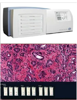
[image:2.595.306.559.235.398.2]Source: https://www.archivesofpathology.org/doi/pdf/10.5858/arpa.2016-007 4-OA
Fig 1. Image analysis using whole slide imaging
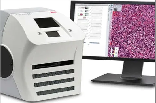
So urce:https://www.researchgate.net/figure/Top-Omnyx-whole-slide-imaging-scanner-Bottom-Omnyx-viewer-and_fig1_279231201
[image:2.595.308.559.424.749.2]The main components of any WSI scanner include: a microscope with objective lenses; a source of light robotics for loading, as well as moving the glass slide around; at least one digital camera to capture the image and a computer and software used to manage, manipulate and view the digital slides. Some advanced scanners come with a dynamic pre-focusing function that makes use of two cameras, one camera is used to focus the image and the other is used for scanning. Having multiple cameras has helped speed up the process of scanning. A limited percentage of scanners further offer dry scanning and oil immersion functions, including the Apiero CS2 scanner that’s designed for hematopathology as well as microbiology. The Apiero CS2 scanner and other similar appliances can traverse numerous glass slides at high speeds of at least 180 mm/s.
[image:3.595.38.288.237.402.2]Source: https://www.leicabiosystems.com/digital-pathology/scan/aperio-cs2/
Fig 3. Apero CS2 scanners used to create high quality slides with the help of Aperio CS2 capture device from a computer
Some scanners are limited in functionality as they are not compatible to wet slides or are unable to scan slides that lack coverslips. WSI gadgets that demand the glass slides to be uploaded in a vertical position and not when lying flat have a lesser ability of accommodating wet slides. Devices such as VisionTek need a glass slide cover interface for the purpose of focusing when scanning slides. More recent WSI devises apply modern optics and elimination methods, including confocal microscopy. Scanning of slides can be done manually or automatically. Many WSI gadgets do batch scanning and random aces processing. Most of the devices present in the market can also read one-dimensional barcodes and two dimensional bar codes. The scanning speeds for WSI gadgets today vary from less than a minute to about three minutes for every slide. The scanning speed is determined by the object magnification and the quantity of Z stacks that have been acquired. Scanning can be done on an entire slide or a specific region of interest on the respective slide. Some WSI scanners can digitize a slide at different z-axes, therefore creating a multi-plane image that has fine focus control that’s equivalent to that of the conventional microscope. The Z stacking technique provides the capability demonstrated above and is appropriate for viewing the cytology slides. Following the collection of digital data through the charge coupled device of a camera, a computer then uses special imaging software to create a virtual slides. The most common methods used to generate a virtual slide include tile based scanning and line based scanning. Tile based scanning has a robotic-controlled and motorized slide stage that’s used to obtain many square image frames that are then Scanning of slides can be done
manually or automatically. Many WSI gadgets do batch scanning and random aces processing. Most of the devices present in the market can als magnification and the quantity of Z stacks that have been acquired. Scanning can be done on an entire slide or a specific region of interest on the respective slide. Some WSI scanners can digitize a slide at different z-axes, therefore creating a multi-plane image that has fine focus control that’s equivalent to that of the conventional microscope. The Z stacking technique provides the capability demonstrated above and is appropriate for viewing the cytology slides. Following the collection of digital data through the charge coupled device of a camera, a computer then uses special imaging software to create a virtual slides. The most common methods used to generate a virtual slide include tile based scanning and line based scanning. Tile based scanning has a robotic-controlled and motorized slide stage that’s used to obtain many square image frames that are then assembled in a mosaic pattern. Every slide has a 2 to 5% overlap because of the overly precise saccadic movement of the slide stage. The digital data captures by the charged couple device are then correlated to one another so as to ensure proper alignment after which the tiles are stitched together to form a vast and seamless image. In line based scanning, a servomotor based slide is used for acquisition of digital images. The servomotor-based slide stage traverses in a jitter free and linear fashion along one axis of acquisition. The slide stage moves numerous times in a sequence along different points of the slide, it then generates a group of images in the form of long and uninterrupted strips. This technique of slide acquisition simplifies the process of image alignment since it reduces the number of lines or tiles and the freedom associated with the lines or tiles.
management, Bashshur et al. made use of storage tiers for managing storage requirements through a model that makes WSI techniques or virtual microscopy more efficient. Bashshur
et al. made use of storage tiers. The top tier served as quick
disk based solution while the lower tier served as an archive where digital files were copied after a specified time period. Slides that are being used for ongoing diagnostic works are usually stored in the first tier while slides that may require a second opinion are placed in the lower tier for long term storage. The storage management variation described by Bashshur et al. also includes a procedure to purge slides, digital slides that are stored in the short term storage expire after a period of time and are then deleted too free space.
Conclusion
When executing pathology from a remote areas, static image systems have been the most used concept in India because its cost effective. However, drawbacks such as lower accuracy of the image and only a limited subsection of a larger field under evaluation can be asses off site by the concerned pathologist. Conventional pathology however, has a higher risk of loss of data through damage of the material carrying a specimen, or the specimen itself as it is physically taken for observation. Static image systems are thus a step in the right direction. Real time systems and virtual pathology, or whole slide imaging are more accurate techniques compared to static image systems, and generate results much faster. However, both are relatively expensive. For developing nations such as India, I recommend the adoption of virtual pathology or WSI which is discussed in detail in this paper. The technique produces composite slide images within an efficient timeline, therefore improving the quality of the outcome from diagnosis. WSI comes in different variations to suit the varying requirements by users, and is further supported by developments in technology such as the development of higher processing speeds, camera lenses with higher resolution and more storage space availed on the cloud. The aforementioned developments satisfy the storage needs, timeline demands, and the image quality demands for pathologists
Conflict of Intrest: None Funding-None
REFERENCES
Abate, Admasu, Mengistu Kifle, Sena Okuboyejo, and Victor Mbarika. 2018. "A mobile-based telepathology system for
a low resource setting in Ethiopia." Applied computing and
informatics, 14, no.2: 186-191.
Bashshur, Rashid L. and Elizabeth, A. Krupinski, Ronald, S., Weinstein, Matthew, R., Dunn and Noura Bashshur. 2017. "The empirical foundations of telepathology: evidence of
feasibility and intermediate effects." Telemedicine and
e-Health, 23, no.3: 155-191.
Chandraratnam, Edward, Leonardo, D., Santos, Shaun Chou, Jun Dai, Juan Luo, Syeda Liza, and Ronald, Y. 2018. Chin. "Parathyroid frozen section interpretation via desktop
telepathology systems: A validation study." Journal of
pathology informatics, 9.
Chordia, Trupti Dinesh, Ashok Vikey, Anuraag, B., Choudhary, Yashpal Samdariya, and Dipti Samdariya Chordia. 2016."Current status and future trends in
telepathology and digital pathology." Journal of oral and
maxillofacial pathology: JOMFP 20, no.2: 178.
Ekong, Donald, Fang Liu, G., Thomas Brown, Arunima Ghosh, and Paul Fontelo, 2017. "Evaluation of android
smartphones for telepathology." Journal of pathology
informatics, 8.
Elizabeth and Arcellana-nuqui, 2016."Telepathology in the
Philippines: A review and future prospects." Acta Medica
Philippina, 50, no. 4: 201-205.
Evans, Andrew, J., Elizabeth, A., Krupinski, Ronald, S.,
Weinstein, and Liron Pantanowitz, 2015. "2014 American
Telemedicine Association clinical guidelines for
telepathology:anotherimportantstepinsupportof increased
adoption of telepathology for patient care." Journal of
pathology informatics, 6.
Farahani, Navid, and Liron Pantanowitz,2015. "Overview of telepathology." Surgical pathology clinics, 8, no.2: 223-231.
Farris, Alton Brad, Cynthia Cohen, Thomas E. Rogers, and
Geoffrey H. Smith, 2017. "Whole slide imaging for
analytical anatomic pathology and telepathology: practical
applications today, promises, and perils." Archives of
pathology and laboratory medicine, 141, no.4 : 542-550.
Fontelo, P., Liu, F. and Yagi, Y. 2015. Evaluation of a
smartphone for telepathology: lessons learned. Journal of
pathology informatics, 6.
Hon, Jane Date, Christine Minerowicz, Sumi Thomas, Nicola Barnard, and Billie Fyfe. 2016. "Distinction of Tissue Marking Dye Colors Using Traditional Glass Slide Micro
scopy, Live Telepathology, and Virtual Microscopy.”|
AmericanJournalofClinical Pathology, 146, no. suppl_1.
Huang, Yingxin, Yan Lei, Qi Wang, Dazhou Li, Lili Ma, Lili
Guo, Minshan Tang et al. 2018. “Telepathology
consultation for frozen section diagnosis in China.”
Diagnostic pathology, 13, no. 1: 29.
Kessler, Stephen E. and Peter F. White, 2015. "Reflectance
confocal microscopy: An overview of technology and
advances in telepathology." Cutis, 95, no.5: E39-E46.
Krupinski, Elizabeth A., Achyut, K., Bhattacharyya, and Ronald S. Weinstein, 2016. "Telepathology and digital
pathology research." In Digital Pathology, pp. 41-54.
Springer, Cham.
Meyer, Julien, and Guy Paré, 2015."Telepathology impacts
and implementation challenges: a scoping review."
1550-1557.
Mpunga, Tharcisse, Bethany, L., Hedt-Gauthier, Neo Tapela, Irenee Nshimiyimana, Gaspard Muvugabigwi, Natalie
Pritchett, Lauren Greenberg et al. 2016. "Implementation
and validation of telepathology triage at cancer referral
center in rural Rwanda." Journal of global oncology, 2, no.
2: 76-82.
Onega, Tracy, Lisa, M., Reisch, Paul, D., Frederick, Berta, M.,
Geller, Heidi D. Nelson, Jason P. Lott, Andrea C. Radick et
al. 2016. "Use of digital whole slide imaging in
dermatopathology." Journal of digital imaging, 29, no.2:
243-253.
Pantanowitz, Liron, Jeffrey McHugh, William Cable, Chengquan Zhao, and Anil V. Parwani, 2015. “Imaging file management to support international telepathology”.
Journal of pathology informatics, 6.
Pare, Guy, Julien Meyer, Marie-Claude Trudel, and Bernard Tetu. 2016. "Impacts of a large decentralized telepathology
network in Canada." Telemedicine and e-Health, 22, no.3:
246-250.
Rubio, Patricio, and César Merino Soto. 2017. “Approaching the oral pathology to primary care dentists through Telepathology in the Health Service of Concepción”.
Siegel, Gabriel, Dan Regelman, Robert Maronpot, Moti Rosenstock, Shim-mo Hayashi, and Abraham Nyska, 2018. "Utilizing novel telepathology system in preclinical studies
and peer review." Journal of toxicologic pathology, 31, no.
4: 315-319.
Vosoughi, Aram, Paul Taylor Smith, Joseph A. Zeitouni, Merce Jorda, Carmen Gomez-Fernandez, Monica
Garcia-Buitrago, Atousa Ordobazari et al. 2018. "Frozen section
evaluation via dynamic real-time nonrobotic telepathology system in a university cancer center by resident/faculty
cooperation team." Human pathology, 78: 144-150.
Weinstein anf Ronald, S. 2015. "Telepathology system
development and implementation." The medicine,
e-health, m-e-health, telemedicine, and telehealth handbook:
Telemedicine and electronic medicine, 1: 577-591.
Zhao, Chengquan, Tao Wu, Xiangdong Ding, Anil V. Parwani,
Hualin Chen, Jeffrey McHugh, Anthony Piccoli et al.
"International telepathology consultation: three years of experience between the University of Pittsburgh Medical
Center and King Med Diagnostics in China." Journal of
pathology informatics, 6. (20