Disrupted flow sensing impairs hydrodynamic performance and increases the
metabolic cost of swimming in the yellowtail kingfish,
Seriola lalandi
Kazutaka Yanase, Neill A. Herbert and John C. Montgomery
10.1242/jeb.082107
There was an error published in J. Exp. Biol.215, 3944-3954.
On p.3947, the π in Eqn3 was incorrectly diplayed as a δ. The equation should have read:
Af= πdw/4.
We apologise to the authors and readers for any inconvenience that this error may have caused.
INTRODUCTION
In contrast to animals with aerial or terrestrial locomotion, aquatic locomotion in fishes is affected to a greater extent by forces, i.e. pressure drag and viscous drag, resulting in severe restrictions on forward speed and energetic performance due to the high density and viscosity of the aquatic medium (Schmidt-Nielsen, 1972; Daniel and Webb, 1987; Fish, 1994). These drags increase with increased swimming speed, and also increase as a result of thrust production through the movements of the trunk and the tail fin (Anderson et al., 2001). Given the metabolic benefits of a lowered cost of locomotion for a given swimming speed, and the complex interaction of boundary layers, drag and thrust production, it seems reasonable that active flow sensing by fish could play a part in improving the efficiency of locomotion. Active flow sensing by the lateral line has been shown to be important in allowing fish to seek out flow refuges generated by obstacles in the flow (Montgomery et al., 2003), and in allowing fish to modify their swimming and save energy in structured turbulent flows (Liao et al., 2003). However, a direct contribution of the lateral line to efficient swimming has not yet been shown for fish swimming under normal conditions.
Two potential roles for a contribution of lateral-line feedback to swimming efficiency have been proposed. Lighthill (Lighthill, 1993) suggested that the lateral-line sensors in the subcerebral canal system of the herring could provide an appropriate feedback signal for controlling yaw by oscillatory neck deflections so as to minimize the effective pressure difference and any associated cross-flow effects
over the head of the fish. It was proposed that swimming clupeid fishes may use this as an ‘active’ mechanism for reduction of hydrodynamic resistance. This theory was supported by analysis of the mechanics of the subcerebral perilymph canal, which crosses the head between the lateral lines of clupeid fishes (Denton and Gray, 1993), and an analysis of the head turning movements in herring and other fishes (Rowe et al., 1993). However, direct experimental evidence for a role of lateral-line feedback in this behaviour was not provided in those studies. A recent study by McHenry et al. (McHenry et al., 2010) in a different species of fish (golden shiner, Notemigonus crysoleucas) concluded from kinematic analysis that ‘flow sensing does not facilitate active drag reduction’, at least with respect to coordinating the motion of the head relative to detected flow signals. However, the golden shiner is not a particularly active species, and this study does not provide any direct measures of swimming efficiency relative to the manipulation of the lateral line.
The second suggested role for lateral-line feedback in the efficient control of swimming came from the detailed measurements of the thin layers of flowing water immediately adjacent to the surface of the swimming fish (i.e. the boundary layers), where influence of viscosity predominates (Anderson et al., 2001). This study observed inflected boundary layers that appeared to be stabilized during the later phases of the undulatory cycle, and suggested that these boundary layer profiles may provide evidence of a contribution of hydrodynamic sensing to the optimization of swimming performance. But, again, this suggestion remains to be directly tested.
SUMMARY
The yellowtail kingfish, Seriola lalandi, shows a distribution of anaerobic and aerobic (red and pink) muscle fibres along the trunk
that is characteristic of active pelagic fishes. The athletic capacity of S. lalandi is also shown by its relative high standard
metabolic rate and optimal (i.e. least cost) swimming speed. To test the hypothesis that lateral line afferent information
contributes to efficient locomotion in an active pelagic species, the swimming performance of S. lalandi was evaluated after
unilateral disruption of trunk superficial neuromasts (SNs). Unilaterally disrupting the SNs of the lateral line impaired both
swimming performance and energetic efficiency. The critical swimming speed (Ucrit; mean ± s.d., N12) for unilaterally
SN-disrupted fish was 2.11±0.96forklengths(FL)s–1, which was significantly slower than the 3.66±0.19FLs–1 U
crit of sham
SN-disrupted fish. The oxygen consumption rate (mgO2kg–1min–1) of the unilaterally SN-disrupted fish in a speed range of
1.0–2.2FLs–1was significantly greater than that of the sham SN-disrupted fish. The least gross cost of transport (GCOT; N6) for
SN-disrupted fish was 0.18±0.06JN–1m–1, which was significantly greater than the 0.11±0.03JN–1m–1GCOT for sham SN-disrupted
fish. The factorial metabolic scope (N6) of the unilaterally SN-disrupted fish (2.87±0.78) was significantly less than that of sham
controls (4.14±0.37). These data show that an intact lateral line is important to the swimming performance and efficiency of carangiform swimmers, but the functional mechanism of this effect remains to be determined.
Key words: lateral line, superficial neuromast, standard metabolic rate, cost of transport, oxygen consumption, critical swimming speed, feedback motor control, kingfish, Seriola lalandi.
Received 5 April 2012; Accepted 22 July 2012
The Journal of Experimental Biology 215, 3944-3954 © 2012. Published by The Company of Biologists Ltd doi:10.1242/jeb.073437
RESEARCH ARTICLE
Disrupted flow sensing impairs hydrodynamic performance and increases the
metabolic cost of swimming in the yellowtail kingfish,
Seriola lalandi
Kazutaka Yanase*, Neill A. Herbert and John C. Montgomery
Leigh Marine Laboratory, University of Auckland, 160 Goat Island Road, Leigh 0985, New Zealand
The lateral line of fish is made up of two submodalities, canal neuromasts and superficial neuromasts (Sns). The canal neuromasts respond less to steady currents and low-frequency flows, and are better suited to encode higher frequency signals (Montgomery et al., 2001). The SNs, in contrast, have anatomically appropriate properties to sense steady currents and low-frequency flows immediately above the surface of the fish body (Coombs and Janssen, 1989; Coombs and Janssen, 1990; Kroese and Schellart, 1992; Montgomery et al., 1994). Therefore, SNs are the most likely submodality to contribute to motor control for sustained or prolonged level of swimming associated with boundary layer flows. Here we experimentally test the hypothesis that the SNs of the trunk lateral line contribute to swimming efficiency. The critical swimming speed, Ucrit, and metabolic cost of locomotion were measured in an active pelagic species, the yellowtail kingfish, Seriola lalandi Valenciennes 1833. The premise that should be supported in the present study is that the yellowtail kingfish is an active pelagic species where active flow sensing for swimming efficiency is likely to be important. The muscle locomotor system in fishes contains two functionally independent components, designated red and white muscle due to their usual colour (Lindsey, 1978). Although the white muscle is faster, more powerful and capable of burst activity, which may be anaerobic, the red muscle is usually slow with low contractile power. The relative development of red and white muscle in different species may be correlated roughly with their mode of life (Videler, 1993; Ellerby and Altringham, 2001). Therefore, we conducted macroscopic examination of locomotor muscle of S. lalandi, by which the active athletic nature of this species was documented based on the relative mass proportion of the red muscle and cross-sectional profiles of muscle fibre types that were found. The constant rhythmic oscillatory tail motion that is usually found in prolonged swimming activity, such as migration, foraging, etc., is predominantly powered by aerobic metabolism (Hudson, 1973; Webb, 1975). Such an aerobically powered mode of swimming demonstrates a linear increase in oscillatory tail-beat frequency with increased swimming speed, resulting in an exponential increase in oxygen demand with increased swimming speed (Lowe et al., 1998; Lowe, 2001; Webber et al., 2001; Steinhausen et al., 2005). Measuring the rate at which oxygen is consumed during locomotion is therefore a direct and non-invasive way to determine the physiological cost of locomotion and is commonly undertaken in accordance with incremental velocity tests, which measure Ucrit (Brett, 1964). These measurements were made for control fish, and fish with a unilateral ablation of the trunk SNs.
The control system for rhythmic animal locomotion, including fish swimming, is formed by sets of neurons in the central nervous system, the so-called central pattern generators (CPGs) (Tytell and Cohen, 2008). The CPGs exhibit certain properties of adaptation and robustness to the environmental changes by generating rhythmic neural output, where the feedback is not essential (Iwasaki and Zheng, 2006). The CPGs, however, receive sensory feedback capable of modulating their rhythmic activity in order to achieve adaptation to environmental changes (Iwasaki and Zheng, 2006). The simplest explanation of the mechanism of CPG activity may be given by the reciprocal inhibition oscillator (Brown, 1911) located in the spinal cord (Friesen, 1994). The CPG consists of two pools of interneurons mutually coupled with inhibitory synaptic connections (Iwasaki and Zheng, 2006). Based on the motor pattern measured using an electromyographic technique by Jayne and Lauder (Jayne and Lauder, 1995), these inhibitory connections in the reciprocal inhibition oscillator generate out-of-phase oscillations in the activities of the two neurons, which drive a pair of muscles
(i.e. anterior and posterior) alternately in sequence down to the tail on an ipsilateral side of the fish body. To ensure stable phase transition in the locomotor cycle to the contralateral side of the fish body, the afferent signals from different (at least two) channels of neurons innervating trunk SNs on the respective right and left sides of the fish body must appropriately be associated with each other in the central nervous system. Because of this characteristic feature of CPG-based control described above, we used unilateral ablation of sensory input from trunk SNs as experimental manipulation on the rationale that asymmetric disruption of sensory input may be particularly effective in demonstration of a sensory feedback component to central pattern generation for locomotion efficiency.
MATERIALS AND METHODS Animals
Ninety-six juvenile S. lalandiwere obtained from the Bream Bay Aquaculture Park, National Institute of Water and Atmospheric Research (NIWA), New Zealand. Fish were held in 3000l tanks at the Leigh Marine Laboratory, and were fed three times a week on chopped fish, such as pilchard. The holding tanks were continuously aerated and flushed with high-quality filtered seawater. All the experiments were conducted under the approval of the University of Auckland Animal Ethics Committee (AEC application number R817).
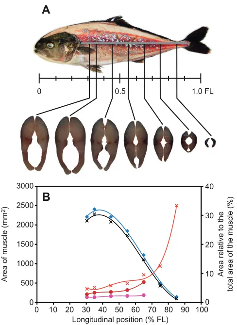
Anatomical analysis of distribution of muscle fibres Fish used for the measurements described in this section were starved for a period of 2days and then euthanized with an overdose of MS-222 (100mg ethyl 4-aminobenzoate per 1l seawater). Five adult fish were selected in order to scale the mass of different muscle fibres. After the wetted mass of the fish was measured, each of the five specimens was submerged in 40°C water for 20min so that the skin could easily be removed from the muscle. The mass of glycolytic (white) and oxidative (red and pink) muscle fibres were dissected separately from the fish body, and was immediately measured using an analytical balance. The mass of the remaining components, including bones, guts and skin, was also measured. The density of the dissected muscle groups containing oxidative (red + pink) and glycolytic (white) muscle was measured using the method of volumetric displacement. Nine adult fish were frozen for analysis of the distribution of the muscle fibres of different type. Each of the frozen specimens was first cut immediately behind the pectoral fin base, on average 30.4±1.8% fork length (FL; mean ± s.d.) from the snout, and then at standardized increments of 10% FL from 35 to 85% FL. Each transverse section was pictured using a digital camera (EX-F1, Casio, Tokyo, Japan), after which the areas of red, pink and white muscle were analysed with ImageJ (v1.43r, National Institutes of Health, MD, USA) and expressed as a percentage of the total muscle cross-sectional area.
Unilateral ablation of the lateral line
width, were also recorded before fish were transferred to a recovery tank. The fish were allowed an additional 3–5days before experimentation and the procedure was deemed successful if fish resumed feeding after a couple of hours. To minimise handling stress, ImageJ was used to estimate the fork length (FL) of control fish according to background grids placed in the working section of the flow chamber before the experimental trial. The body depth and width of the control fish was estimated using linear regressions obtained during preliminary measurements (N60). If the true FL, depth and width measured at the end of the experiment differed from the estimated values, the flow velocity for the calculation of the critical swimming speed was corrected in the post-experimental analysis.
Critical swimming speed test protocol
The critical swimming speed (Ucrit) test (Brett, 1964) has long been used to assess the prolonged swimming capacity of various species. Ucrit is obtained from fishes subjected to incremental changes in speed over time in an experimental flow chamber. It is generally assumed that maximum oxygen uptake occurs at Ucrit(Farrell and Steffensen, 1987; Farrell, 2007) and is thus a convenient way to measure both swimming performance and the maximum aerobic capacity of fish. Several investigators have found that Ucrit overestimates sustained speed because recruitment of the fast-twitch, glycolytic muscle fibres at higher speeds is evident in a change in gait from a continuous tail beat to intermittent bursts of high-frequency tail beats followed by gliding (i.e. burst-and-glide) in the final stages of the Ucrittest (Kolok, 1991; Webb, 1998). To avoid this discrepancy, Drucker (Drucker, 1996) proposed the gait transition speed (e.g. the speed at which pectoral fin locomotion is supplemented by body caudal fin locomotion) as an alternative cut-off swimming speed. Dickson et al. (Dickson et al., 2002) also defined the upper limit of scope for activity as swimming speed at which a continuous tail beat was shifted to a ‘burst-and-glide’ gait three times within 30s. However, it is practically difficult to demonstrate the upper limit of scope for activity by counting how many times such gait shifts are conducted by the fish swimming in the swim tunnel within a certain period of time. Therefore, Ucritis considered to be a relatively robust and reliable metric for comparing the relative aerobic swimming capabilities of a body-caudal-fin swimmer such as S. lalandi(Williams and Brett, 1987; Hartwell and Otto, 1991; Peake and McKinley, 1998; Hogan et al., 2007).
The experiment commenced in late October 2010. At that time, the water temperature in the holding tanks was 18°C. Thirty-six fish were used for the Ucrittest, divided into three groups: the control group (0.275±0.043m FL, N12); a group that received sham treatment for SN disruption (sham SN-ablated fish: 0.277±0.034m FL, N12); and an experimental group that received the treatment for SN disruption (SN-disrupted fish: 0.280±0.027m FL, N12). Six individuals in each group were used for the respirometric measurement, and the other six individuals were used for kinematical measurements (reported separately). The Ucrittest was carried out in a Brett-type swim tunnel (38.4l) with a 0.16⫻0.16⫻0.55m (width
⫻height ⫻length) working section, where the flow was rectified by passing the water through a square mesh filter placed upstream in order to ensure a high degree of homogeneous and unidirectional flow. The downstream end of the working section was closed by a metal grid. During the test, fish swimming behaviour was recorded by two video cameras (EX-F1, Casio, Tokyo, Japan) that were set directly and obliquely above the working section of the respirometry flow chamber. The respirometry flow chamber was submerged in a water bath. The water was pumped from an experimental sump up to the water bath viaan in-line water chiller (HC-1000A, Hailea, Guandong, China). A flushing pump was able to swap the water inside the respirometry flow chamber with water from the temperature controlled water bath. The water flow was generated by a frequency-modulated induction motor that was controlled with a frequency inverter (CFW-10, WEG, Scoresby, VIC, Australia). The flow velocity was controlled using customized software that altered the frequency of the power supply to the motor. The experimental flow system was surrounded by a plastic black-out sheet to prevent the fish from being disturbed. Supplementary fluorescent light was provided in the Ucrittests.
The experimental water temperature was maintained at 18–19°C during the experiment, although water temperature in the holding tanks did increase up to 21°C until the experiment finished in early March 2011. The slight discrepancy in the holding and experimental temperatures over the short period is not likely to have influenced our findings. At the start of the experiment, an individual fish was introduced to the swimming section of the swim flume and left to swim overnight (14–16h) while taking respirometric measurements in a flow set to 0.7forklengths (FL)s–1. Fish were only used once in the experiments to preclude any effect of prior experience (Liao, 2006). Once fish were acclimated to the swim flume, the flow velocity was elevated to 1.0FLs–1. The flow speed, and thus the swimming speed, was increased by 0.3FLs–1every 30min until the fish failed to swim by fatigue, and Ucritwas interpolated from this final level of swimming performance [i.e. incremental velocity test (Brett, 1964; Jones, 1982; Farrell, 2007)]. In the present study, we assumed that fatigue was reached when the accumulated time that the body or fin of fish touched the downstream grid reached 1min.
The absolute value of Ucritin ms–1is dependent upon the length of fish. Therefore, Ucritwas normalized by FL of the fish (Beamish, 1978) and was calculated as follows:
UcritUmax+ (tf/ti)Ui, (1) where Uiis the velocity increment (0.3FLs–1), Umaxis the maximum speed at which the fish was able to complete a 30-min period of swimming, tfis the elapsed time from the velocity increase to fatigue and tiis the time between the velocity increments (Brett, 1964). The presence of the fish in the enclosed chamber results in an increase in water velocity around the fish. Due to this phenomenon, the
[image:4.612.52.288.67.183.2]so-A
B
called solid blocking effect, the flow velocity that the fish was exposed to was corrected in accordance with the equation described by Bell and Terhune (Bell and Terhune, 1970):
UUw(1 + Af/Ac), (2) where Uis corrected velocity, Uwis the flow velocity without fish,
Acis the cross-sectional area of the flow chamber and Afis the cross-sectional area of the fish. Afwas approximated by an ellipse with the maximum depth of the body for the major axis (d) and the maximum width of the body for the minor axis (w), and was described as:
Af dw/4. (3)
Respirometric measurements and analysis
The mass-specific rate of oxygen consumption (MO2) of 18 fish was resolved during Ucrit swimming tests (see above for details on acclimation and flow velocity increments, etc.) to assess the swimming energetics of fish with intact and non-intact lateral lines: control group (0.304±0.022m FL, N6); sham SN-disrupted fish (0.308±0.011m FL, N6) and SN-disrupted fish (0.301±0.008m FL, N6). The respirometer ran with a single measurement cycle consisting of three periods (i.e. flushing, waiting and measuring) according to the protocol of Steinhausen et al. (Steinhausen et al., 2005) and Brown et al. (Brown et al., 2011). During the flushing period, the flushing pump was active to mix the water inside the respirometry flow chamber and to ensure proper flow past the oxygen sensor. The flushing period (2–7min) was changed depending on the fish and swimming speed so that the air saturation level of water would be ~100% at the end of the flushing period. During waiting and measuring periods, the flushing pump was turned off and the respirometry flow chamber was closed by a solenoid valve to prevent water from entering from an ambient tank. Before starting a new measuring period, a waiting period is necessary to account for a lag in the system response, resulting in a non-linear declination change in oxygen saturation of the water inside the respirometry flow chamber over the elapsed time. To avoid any risk of hypoxia developing, the length of the waiting and measuring periods were also modified to ensure air saturation never dipped below 80% saturation by the end of the measuring period. In general, however, measurement cycles were either 10 or 15min, allowing two to three MO2measurements for an individual fish swimming at each flow velocity.
The change in oxygen saturation over time, and hence MO2, was measured at 1Hz with a needle-type oxygen sensor (NTH-PSt1-L5-TS-NS40/1.2-YOP, PreSens Precision, Regensburg, Germany) connected to a Microx-TX3 oxygen meter (PreSens Precision). The entire respirometer was under the control of customized software that not only managed water flow velocity but also initiated the flush, wait and measure cycle and recorded water O2saturation. The software then calculated the rate of change in water saturation per second (slope of the linear regression of oxygen saturation over the elapsed time), and this value was converted to MO2
(mgO2kg–1min–1) (sensuSteffensen, 1989) using the equation:
where sat is the recorded change in the percent of air saturation per second, PO2is the measured partial pressure of oxygen at 100% air saturation, wO2is the capacitance coefficient of oxygen in water at a certain salinity (0.369mgO2l–1kPa at 36p.p.t. for this experiment), Vwis the volume of the respirometer, 60 is the number of seconds in a minute and Mis the mass of the fish.
= Δ × ×β × ×
M P v
M
( sat / 100) 60
, (4)
O2 O wO w
2 2
MO2 was described as a power function of swimming speed (Steinhausen et al., 2005), U(FLs–1) and M(kg) by:
MO2aM–0.2+ bM–0.2Uc. (5)
Oxygen consumption data were fitted to the curve (Eqn5) by the least-square method (generalized reduced gradient nonlinear algorithm, Microsoft Excel Solver, Microsoft, Redmond, WA, USA) in order to estimate the model parameters a, b and c. To account for variations in MO2due to size differences amongst the fish, Clarke and Johnston (Clarke and Johnston, 1999) used the mass-specific exponent of M(0.8–1), thus M–0.2 describes the allometric relationship between the standard metabolic rate (SMR) and mass of 69 species of teleost fish. SMR represents the energy required to maintain basic biological functions independent of activity, digestion or the costs of physiological stressors. The SMR was extrapolated as the oxygen consumption at zero swimming speed (U0) using Eqn5. To compare the cost of swimming as work per metre (WPM; Jm–1), M
O2was recalculated with the units mgO2s–1 and multiplied by a general oxycaloric value of 14.1Jmg–1O
2 (Videler, 1993). The gross cost of transport (GCOT) is the value of WPM that is corrected for size effects by dividing by body weight (N) (Videler, 1993). GCOT (JN–1m–1) at U
optis therefore given by:
GCOT AMRopt(MgUopt)–1, (6) where AMRopt(Js–1) is the active metabolic rate at the optimum swimming speed, Uopt, and g is the acceleration due to gravity (ms–2). After Steinhausen et al. (Steinhausen et al., 2005), using the relationship between U and GCOT with Eqn5 and 6, the optimum swimming speed, Uopt, where WPM is at its minimum, was obtained as:
Uopt[a/b(c– 1)]1/c. (7) The level of metabolism available for locomotion (aerobic metabolic scope for activity) is usually calculated as the difference between SMR and AMRmax. AMRmaxgenerally occurs at Ucrit, and the fractional difference of AMRmaxto SMR rate from this point onwards is referred to as factorial metabolic scope (FMS) (Fry, 1957):
FMS AMRmax/SMR. (8)
Composition of unsteady swimming
The acceleration and deceleration of fish swimming within the test section of the swim flume is a good indicator of steady (aerobic) versus unsteady (anaerobic) activity, the latter being more energetically expensive to maintain (Boisclair and Tang, 1993). The oxygen consumption of unsteady swimming was therefore analysed to address how swimming style might influence the respirometric measures of SN- and non-SN-disrupted fish.
if the rate of longitudinal displacement was within 10% FL (i.e. ±0.1FLs–2).
Statistics
Basic statistics and pairwise tests were performed using the Analysis ToolPak of Microsoft Excel 2010. The statistical program R (ver. 2.12.2, R Development Core Team, Vienna, Austria) was used to run ANCOVA and multiple group comparisons with post hoc analysis. The effect of unilateral disruption on Ucritand respirometric performance was examined further if ANCOVA determined any significant difference in the following pairwise comparisons: (1) between control and SN-disrupted groups and (2) between sham SN-disrupted and SN-disrupted groups. Given statistical significance for the above pairwise comparisons, an insignificant result was desired for the comparison between control and sham SN-disrupted groups in order to support equivalence of the effect of unilateral disruption for control and sham SN-disrupted groups. In this scenario, the significance limit of an F-statistic was adjusted by Bonferroni correction in order to reduce the risk of Type I errors. If not stated otherwise, values are presented as means ± s.d.
RESULTS
Distribution of muscle fibres
The mean mass of the whole lateral muscle was 0.4065±0.0941kg (N5), and this corresponded to 52.3% of the body mass of the fish. The mean mass of oxidative (red + pink) and glycolytic (white) muscle was 0.0291±0.0060kg (N5) and 0.3774±0.0886kg (N5), which corresponded to 7.2 and 92.8% of whole muscle mass, respectively (Table1). Red muscle was concentrated along the lateral line but penetrated the white muscle, and even reached the vertebral column along the horizontal septum (Fig.2A). The change of muscle distribution along the body axis and the proportions of the red and pink muscle to the whole lateral muscle are shown in Fig.2B. The areas of red and white muscle both reached a maximum at 35.0% FL from the snout (red: 80mm2and white: 2282mm2). The area of the pink muscle reached a maximum at 45.0% FL from the snout (48mm2). The proportion of the oxidative red and pink muscle increased posteriorly, but the pink muscle was indistinguishable in most of the cross-sections from 75 to 85% FL from the snout. Oxidative muscle for the 75 and 85% FL sections could possibly contain pink muscle, but the ratio is so small it was deemed insignificant. A sharp increase in the proportion of the area of red muscle in the area of the whole muscle was found in the cross-sections posterior to 65% FL from the snout. The percentage of oxidative red muscle posterior to 65% FL was significantly greater than that in the anterior part (i.e. 30.4–45.0% FL from the snout) (Kruskal–Wallis test, H48.80, d.f.6, P<0.01,
followed by a post hocSteel–Dwass test, P<0.05). In contrast, the proportion of the white muscle in the area of the whole muscle decreased posteriorly.
Critical swimming speed
The mean critical swimming speed (Ucrit) of the unilaterally SN-disrupted fish group was 2.11±0.96FLs–1 (N12), and was significantly slower than the 3.66±0.19FLs–1Ucrit(N12) of sham SN-disrupted fish group (ANCOVA, F1,2131.87, P<0.01; Fig.3). The Ucritof control fish group was 3.89±0.45FLs–1(N12; Fig.3), which was the fastest Ucritof the three. No significant difference in FL was detected between the three groups (ANOVA, F2,330.07,
P>0.05). The Ucritin the control group was negatively correlated with the FL (two-tailed Spearman’s rank correlation, t3.48, d.f.10, P<0.01). It might be expected that ability of the smaller fish to execute burst-and-coast gait swimming would be less restricted by the size of the flow chamber, resulting in a faster Ucrit(Castro-Santos, 2004; Castro-Santos, 2005; Peake and Farrell, 2006; Tudorache et al., 2007). However, the Ucrit, from which this extraneous influence depending on the fish length was removed, was not significantly different between control and sham SN-disrupted fish groups (ANCOVA, F1,213.79, P>0.05).
The mean maximum swimming speed (Umax) was 3.79±0.42FLs–1(N12) for control fish and 3.60±0.21FLs–1(N12) for sham SN-disrupted fish. There was no significant difference between these two Umaxvalues (ANCOVA, F1,212.99, P>0.05). However, the Umax for SN-disrupted fish was lower, at 1.99±0.97FLs–1(N12), significantly slower than that for control (ANCOVA, F1,2133.25, P<0.01) and sham SN-disrupted fish (ANCOVA, F1,2132.20, P<0.01).
Respirometric measurement
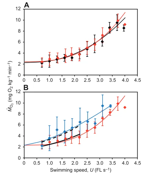
Basic statistics describing the respirometric measures are given in Table2. There was no significant difference in the FL and body mass between control, sham SN-disrupted and SN-disrupted fish groups (ANOVA, FL: F2,150.19, mass: F2,150.41, P>0.05), and so any difference reported herein is not due to body size effects. As shown in Fig.4A, MO2for sham SN-disrupted fish group was almost identical to the control group MO2at all swimming speeds. A significant linear correlation between the logarithm of the MO2
and the swimming speed was found, but there was no significant difference in log MO2between control and sham SN-disrupted fish groups (ANCOVA, F1,1380.27, P>0.05). The relationship between
MO2 and swimming speed (U) was expressed by
MO21.75M–0.2+0.27M–0.2U2.3 (R20.77, F
1,143493.00, P<0.01) for the control group and MO21.91M–0.2+0.16M–0.2U2.7(R20.77,
F1,148500.81, P<0.01) for the sham SN-disrupted fish. The
Table1. Mass of glycolytic white and oxydative (red + pink) muscle and relative percentage of the mass of the yellowtail kingfish Oxidative (red + pink) Whole muscle mass Lost mass (kg) White muscle mass muscle mass (kg) (white + red + pink) Remaining mass after measurement Wet mass (kg) [% relative to [% relative to whole [% relative to (kg) [% relative to [% relative to
Individual FL (kg) whole muscle mass] muscle mass] wet mass] wet mass] wet mass]
1 0.357 0.601 0.247 [92.8] 0.019 [7.2] 0.266 [44.3] 0.325 [54.1] 0.010 [1.6]
2 0.393 0.871 0.422 [93.5] 0.029 [6.5] 0.452 [51.8] 0.403 [46.2] 0.017 [2.0]
3 0.404 0.915 0.481 [93.3] 0.035 [6.7] 0.516 [56.4] 0.378 [41.3] 0.021 [2.3]
4 0.365 0.684 0.341 [92.0] 0.030 [8.0] 0.370 [54.1] 0.296 [43.3] 0.018 [2.6]
5 0.386 0.813 0.395 [92.3] 0.033 [7.7] 0.428 [52.6] 0.379 [46.6] 0.007 [0.8]
Mean 0.381 0.777 0.377 [92.8] 0.029 [7.2] 0.407 [52.3] 0.356 [45.9] 0.014 [1.8]
s.d. 0.020 0.131 0.089 [0.6] 0.006 [0.6] 0.094 [4.6] 0.044 [4.9%] 0.006 [0.7]
intercept of the model curve with the vertical axis (Fig.4A) was determined as the SMR. The estimated SMR values for control and sham SN-disrupted fish groups were 2.16 and 2.32mgO2kg–1min–1, respectively. Because the SN-disrupted fish exhibit a high rise in MO2 between 0.7 and 1.3FLs–1 (Fig.4B), their extrapolated SMR was unreasonably underestimated. Because SN ablation should not affect SMR, it was assumed that the SMR of the disrupted fish was the same as that of the sham SN-disrupted fish group for the model fitting process (Fig.4B). This assumption allowed the MO2 of SN-disrupted fish group to be reasonably described by MO21.88M–0.2+0.97M–0.2U1.4(R20.53,
F1,91103.33, P<0.01). This correction revealed a significant difference in MO2between sham SN-disrupted and SN-disrupted fish groups in the swimming speed range of 1.0–2.2FLs–1 (ANCOVA, F1,6510.71, P<0.01), where 2.2FLs–1was the closest swimming speed to the mean Ucritof SN-disrupted fish group. It is important to note that only one SN-disrupted fish could complete two cycles of the MO2measurement during the 3.4FLs–1velocity swimming, and recorded MO2values statistically similar to those for the other two groups (ANOVA, F2,200.95, P>0.05; Fig.4B):
8.6±1.3mgO2kg–1min–1 (10 measurements) for control fish, 8.3±1.1mgO2kg–1min–1 (11 measurements) for sham SN-disrupted fish and 9.5±0.2mgO2kg–1min–1(two measurements) for SN-disrupted fish.
The mean AMR at Umax (A—M—R–max) was 8.76±1.60mgO2kg–1min–1 for control fish (N6) and 9.23±1.19mgO2kg–1min–1for sham SN-disrupted fish (N6). The A
—M—R–max for SN-disrupted fish was 7.47±3.29mgO2kg–1min–1 (N6). This was the lowest A—M—R–maxof the three groups, but there was no significant difference between the three A—M—R–maxvalues (one-way ANOVA, F2,151.01, P>0.05). The mean FMS (F—MS—) for the SN-disrupted fish (2.87±0.78, N6) was significantly less than that of control fish (4.04±0.75, N6) and sham SN-disrupted fish (4.14±0.37, N6; one-way ANOVA, F2,156.80, P<0.05, followed by a Tukey’s post hocHSD multiple comparison test, P<0.05). There was no significant difference in F—MS— between control and sham SN-disrupted fish (one-way ANOVA, F2,156.80, P<0.01, followed by a Tukey’s post hocHSD multiple comparison test, P>0.05).
The mean GCOT of fish in the control and sham SN-disrupted groups was also almost identical at each swimming speed (Fig.5A) and reflects similarity in the MO2–swim-speed relationship (above). The mean GCOT of SN-disrupted fish, however, was significantly greater than that of sham SN-disrupted fish at swimming speeds from 1.0 to 2.2FLs–1 (ANCOVA, F
1,6514.43, P<0.01; Fig.5B). The Uoptthat was extrapolated using the power model (i.e. Eqns5, 7) was 2.0FLs–1for both control and sham SN-disrupted fish. Using the same approach, a Uoptvalue of 2.7FLs–1was obtained for SN-disrupted fish. The corresponding value of GCOTmin at Uopt (calculated from Eqn6 using the mean mass of each group) was similar between control fish (0.15JN–1m–1) and sham-disrupted fish (0.14JN–1m–1), but was approximately 1.5 times greater for SN-disrupted fish (0.22JN–1m–1).
In addition to the overall power model estimations (above), the mean optimal swimming speed (Uopt) obtained from individual fish was estimated to be 2.25±0.50FLs–1 (N6) for control fish, 2.00±0.70FLs–1 (N6) for sham SN-disrupted fish and 1.70±0.81FLs–1(N6) for SN-disrupted fish. Although the overall difference in Uoptbetween the three groups was not significant (one-way ANOVA, F2,150.96, P>0.05), the mean GCOT at Uopt (G—C—O—Tmin) of SN-disrupted fish (0.18±0.06JN–1m–1, N6) was significantly greater than that of the other two groups (one-way
Area of muscle (mm
2)
Area relative to the
total area of the muscle (%)
Longitudinal position (% FL)
0 10 20 30 40 50 60 70 80 90 1000
10 20 30 40
0 500 1000 1500 2000 2500 3000
0 0.5 1.0 FL
A
[image:7.612.49.293.64.399.2]B
Fig. 2. Distribution of the mean area of red, pink and white muscle and the percent of the area relative to the whole muscle area. (A)Lateral view of the distribution of the red muscle and transverse sections. (B)The absolute area of the white (black crosses) and whole muscle (blue diamonds), and percent of areas of different fibre type relative to the whole muscle area for the oxydative red + pink (red crosses), red (red circles) and pink (pink circles) muscle at different longitudinal positions. The mean area of each muscle type was adjusted by the area versusFL2relationship (ANCOVA) to account for allometry effects. The curves (fourth-order polynomials) are indicated for illustration purposes only. FL, fork length.
Control Sham SN-disrupted SN-disrupted
Critical swimming speed,
Ucrit
(FL
s
–1) 6 5
4
3
2
1
0
[image:7.612.335.564.66.225.2]ANOVA, F2,154.28, P<0.05, followed by a Tukey’s post hocHSD multiple comparison test, P<0.05). The G—C—O—Tminwas comparable between control (0.10±0.03JN–1m–1, N6) and sham SN-disrupted fish (0.11±0.03JN–1m–1, N6; one-way ANOVA, F
2,154.28,
P<0.05, followed by a post hocTukey’s HSD multiple comparison test, P>0.05). Contrary to the overall power model estimate of Uopt
(i.e. 2.7FLs–1), the Uopt for SN-disrupted fish was significantly slower than the Ucritof this group (Friedman test, 27.00, d.f.2,
P<0.05, followed by a post hoc Scheffe’s method for pairwise comparison, P<0.05), but it was not significantly different from the Umaxof this group (Friedman test, 27.00, d.f.2, P<0.05, followed by a post hocScheffe’s method for pairwise comparison, P>0.05). Table2. Basic statistics from the respirometric measurement (mean ± s.d., N6) of the yellowtail kingfish
Control fish Sham SN-disrupted fish SN-disrupted fish
Fork length, FL (m) 0.304±0.022 0.308±0.011 0.301±0.008
Mass, M(kg) 0.347±0.070 0.366±0.051 0.340±0.026
Standard metabolic rate, SMR (mgO2kg–1min–1) 2.16 2.32 2.32
Maximum metabolic rate, AMRmax(mgO2kg–1min–1) 8.76±1.60 9.23±1.19 7.47±3.29 Optimal swimming speed, Uopt(FLs–1) 2.25±0.50 (2.00) 2.00±0.70 (2.00) 1.70±0.81 (2.74) Gross cost of transport, GCOT (JN–1m–1) 0.10±0.03 (0.15) 0.11±0.03 (0.14) 0.18±0.06* (0.22)
Factorial metabolic scope, FMS 4.04±0.75 4.14±0.37 2.87±0.78**
Maximum swimming speed, Umax(FLs–1) 3.79±0.42 3.60±0.21 1.99±0.97**
The SMR of superficial neuromast (SN)-disrupted fish was assumed to be the same as that of sham SN-disrupted fish group. *Significant at P<0.05; **significant at P<0.01.
The values in parentheses represent the estimates based on a power model of mass-specific oxygen consumption (MO2), MO2aM–0.2+bM–0.2Uc, where parameters a, band care estimated from data, and Mis the mean mass of the fish for each group.
A
0 0.5 1.0 1.5 2.0 2.5 3.0 3.5 4.0 4.5
Swimming speed, U (FL s–1)
0 0
0.5 1.0 1.5 2.0 2.5 3.0 3.5 4.0 4.5
MO
2
(mg O
2
kg
–1 min –1) 2
4 6 8 10 12
0 2 4 6 8 10
12
B
[image:8.612.49.568.79.176.2].
Fig. 4. Comparison of mass-specific oxygen consumption (MO2) between fish groups with and without a unilateral ablation of trunk superficial neuromasts (SNs). The plots compare MO2(mean ±s.d. MO2) (A) between control (black) and sham SN-disrupted (red) fish groups and (B) between sham SN-disrupted (red) and SN-disrupted (blue) fish groups. The curves represent the power model MO2aM–0.2+bM–0.2Ucfor (A) control (black) and sham disrupted (red) fish, and (B) sham disrupted (red) and SN-disrupted (in blue) fish. The model parameters a, band care approximated from empirical data obtained from fish with mean body mass of M. The ANCOVA regression models of log(MO2) on the swimming speed (U1.0–2.2FLs–1) for sham disrupted (solid black curve) and SN-disrupted (dashed black curve) fish groups are compared in B. The error bars represent s.d. from a mean of N6. FL, fork length.
0 0.1 0.2 0.3 0.4 0.5
GCOT
(J N
–1 m –1)
0 0.1 0.2 0.3 0.4 0.5
A
B
0 0.5 1.0 1.5 2.0 2.5 3.0 3.5 4.0 4.5
0 0.5 1.0 1.5 2.0 2.5 3.0 3.5 4.0 4.5
Swimming speed, U (FL s–1)
GCOTmin
GCOTmin
Uopt
[image:8.612.328.561.322.607.2]Uopt
[image:8.612.53.288.329.614.2]Composition of unsteady swimming
For both control and sham SN-disrupted fish, the percent frequency of quasi-steady swimming (i.e. steady swimming or accelerations <1.0FLs–2) was nearly twofold greater than the percent frequency of unsteady swimming (i.e. accelerations ≥1.0FLs–2) (two-way ANOVA, F1,5048.07, P<0.01 for control fish and F1,4836.33,
P<0.01 for sham SN-disrupted fish; Fig.6A,B). For SN-disrupted fish, however, quasi-steady and unsteady measures shared approximately equal frequencies (two-way ANOVA, F1,341.79,
P>0.05; Fig.6C). The percentage frequency of all acceleration measures was generally independent of flow speed in all three groups (regression analysis for control fish: F1,280.81, P>0.05; for sham SN-disrupted fish: F1,270.45, P>0.05; for SN-disrupted fish:
F1,200.34, P>0.05). Most importantly, the average percentage frequency of unsteady activity (≥1.0FLs–2) for the SN-disrupted fish was significantly greater than that for the control and sham SN-disrupted fish (two-way ANOVA, F2,664.88, P<0.05; Fig.7A). There were differences between the three groups in terms of quasi-steady swimming (two-way ANOVA, F2,663.86, P<0.05; Fig.7B); the percentage frequency of quasi-steady swimming by SN-disrupted fish was less than that of controls (post hocTukey’s HSD multiple comparison test, P<0.05) but statistically similar to that of sham SN-disrupted fish (post hocTukey’s HSD multiple comparison test, P>0.05). There was, however, no significant difference in the average percentage frequency of deceleration activity (≤–0.1FLs–2) between the three groups (two-way ANOVA, F2,661.37, P>0.05; Fig.7C).
DISCUSSION
Seriola lalandimuscle and its relationship with aerobic
swimming performance
Myotomal muscle is the primary means of propulsion in fishes and, with slow oxidative (red) and fast glycolytic (white) muscle fibres being both anatomically and functionally separated, the proportional mix of muscle type is generally believed to reflect the swimming ecology of different species (Videler, 1993). Fishes power steady undulatory swimming with both red and pink muscle but recruit white glycolytic fibres when fast unsteady movements, such as sprint and burst swimming, are required (Rome et al., 1992; Coughlin and Rome, 1996; Ellerby and Altringham, 2001). Examining the relative proportion of the different muscle fibres thus provides a valuable insight into the routine swimming performance of different species. For instance, constant-swimming pelagic species generally have more slow muscle than benthic species (Videler, 1993; Ellerby and Altringham, 2001), and our analysis of muscle fibre allocations
certainly provides support for S. lalandialso being a fast-swimming cruiser. The percentage cross-sectional area of aerobic (red and pink) muscle in S. lalandiincreased posteriorly, and the percentage for the posterior 65% of the body was significantly greater than the anterior 35%, suggesting that the red and pink muscle plays a very important role in carangiform swimming, where large lateral undulations are restricted to the posterior part of the body. It is probable that the pink muscle of S. lalandiis a fast-twitch muscle with contraction speeds lying somewhere between the performance of red and white fibres (Coughlin et al., 1996).
The total mass ratio of the oxidative red and pink muscle fibres was found to range from 6.5 to 8.0% in the whole muscle of S. lalandi, which, although less than common active pelagic species [e.g. sardine Sardinopus melanostica, 20.7% and chub mackerel Scomber japonicus, 12.0% (Obatake and Heya, 1985)], is considerably greater than that of common demersal species [e.g. yellow sea bream Taivs tumifrons, 2.2% and sillago Sillago japonica, 1.6% (Obatake and Heya, 1985)]. Seriola lalandishows a red muscle mass ratio similar to that of the jack mackerel (Trachurus japonica), the latter having a red muscle fraction range of 7.7–8.6% (Obatake and Heya, 1985; Xu et al., 1993). The fact that both carangids have red muscle ratios in the vicinity of 6.5–8.6% suggests that this ratio is presumably sufficient to support rapid long-term schooling, a characteristic of both species. Examining the shift in muscle type along the length of carangiform and sub-carangiform modes of swimming lends further support to this argument. For example, in S. lalandi, the posterior bias for oxidative red and pink muscle fibres is clearly seen in Fig.2A. The gadoid Merlangius merlangus, a sub-carangiform swimmer that has more body bending and does not solely create thrust from posterior sections, was found to have 2% red muscle at 35% of the body length and 14.3% at 79% of the body length (Greer-Walker and Pull, 1975). Seriola lalandiat similar positions in the present study (Fig.2B) was found to have 5.1% oxidative fibres at 35% FL and 22.9% at 80% FL (if a linear correlation was assumed between the proportions at 75 and 85% FL). Seriola lalanditherefore shows features of a cruising specialist with a carangiform mode of swimming.
The use of fast carangid swimming by S. lalandiis also supported by our respirometry data. Active epipelagic species are generally seen to have high SMR to maintain swimming machinery supporting peak performance at high AMRmax(Benetti et al., 1995). Clark and Seymour (Clark and Seymour, 2006) measured the SMR of 2.1kg S. lalandi at 20 and 25°C and derived values of 1.55 and 3.31mgO2kg–1min–1, respectively. Their 1.55mgO2kg–1min–1 SMR at 20°C corresponds to 2.22mgO2kg–1min–1for a 0.347kg
0 10 20 30 40 50 60 70
1.0 1.3 1.6 1.9 2.2 1.0 1.3 1.6 1.9 2.2
Frequency of unsteady or quasi-steady activity (%)
A
Flow speed (FL s–1)
B
C
[image:9.612.54.563.579.697.2]1.0 1.3 1.6 1.9 2.2
fish if a mass-specific exponent of –0.2 is used (Clarke and Johnston, 1999). The 2.16mgO2kg–1min–1SMR of our 0.347kg control fish at 18°C is therefore close to the measures of Clark and Seymour (Clark and Seymour, 2006). When looking at other pelagic carangids, the chub mackerel Scomber japonicusprovides a good comparison of SMR because it is an epipelagic predator specialized for rapid and efficient swimming (Dickson et al., 2002). Dickson et al. (Dickson et al., 2002) reported an SMR of 2.11mgO2kg–1min–1 for a 0.095kg S. japonicus at 18°C. This measure of SMR is close to that of our control fish, but obviously applies to a smaller fish and is therefore less than that of our S. lalandiif a mass exponent of –0.2 is applied. The Uoptof control and sham treated fish within the current line of work was 2.0FLs–1 at 18°C, which is directly comparable to 2.2FLs–1U
optof S. lalandi at 22°C within the study of Brown et al. (Brown et al., 2011) using the same swim flume and similarly sized fish. However, other more sluggish species such as saithe and whiting are seen to have much lower Uoptvalues in the vicinity of 1.4 and 1.0FLs–1, respectively (Steinhausen et al., 2005). The specialized musculature and relatively high Uoptand SMR of S. lalanditherefore suggest that this species uses relatively faster swimming speed during routine moving and foraging, and it is within these more energy-demanding speeds that the lateral line is likely to play a more substantive role.
Effect of lateral-line disruption on aerobic metabolic capacity and mode of swimming
SN-disrupted fish had a lower swimming performance and swam less efficiently. Not only were they unable to attain high critical swimming speeds (Fig.3), but across a comparable range of swimming up to the Ucrit level of 2.11FLs–1, their rate of O2 consumption per unit swimming speed was greater (Fig.4B). Although MO2was higher for SN-disrupted fish, the drop in Ucrit swimming performance translated into a reduced aerobic scope, implying that fish were possibly forced to recruit anaerobic fibres earlier in the Ucrittest. As illustrated in Fig.5B, some SN-disrupted individuals would have had to swim beyond their Ucrit limit to capitalize on the benefits of Uopt, their least costly swimming speed at 2.74FLs–1, which emphasizes the mismatch in swimming efficiency and performance in this group. Only one SN-disrupted fish could complete two cycles of the MO2measurement at 3.4FLs–1, and showed MO2 measurements similar to those of the fish with intact lateral-line input (Fig.4B). This may result from the stabilized swimming performance by multisensory substitution involving canal neuromasts, which have a higher threshold for hydrodynamic
stimuli (high-frequency flow). The estimated Uoptfor control fish suggests that S. lalandiuse swimming speeds as fast as 2FLs–1for routine swimming, such as during migration and foraging. These considerations taken together suggest that the upper limit of the swimming speed range where trunk SNs are responsible for motor control for swimming efficiency may fall between 2.0 and 3.4FLs–1. The fish with intact lateral lines were able to sustain swimming across an extended range of speeds and were able to capitalize on the benefit of the least costly swimming at Uopt. The results of the present study therefore suggest that the majority of SN-disrupted fish were subject to increased swimming costs and showed poor swimming efficiency and performance across a range of swimming speeds. In relation to the MO2measures, no significant difference in GCOT was detected between fish with and without intact lateral lines when swimming at Uopt. This suggests that SN-disrupted fish are required to take the same period of time to reach the same distance if swimming at Uopt. This is the least costly swimming activity. However, SN-disrupted fish will subsequently consume 1.5 times greater aerobic energy compared with control and sham SN-disrupted fish. The higher GCOT at Uoptfor SN-disrupted fish and shallower U-shaped relationship of GCOT with swimming speed (Fig.5B) suggest that the cost of swimming for this group of fish was considerably high up to Ucrit. That is because the difference between the GCOT at Uoptand Ucritwas negligibly small despite the fact that the GCOT at swimming speeds around Ucritshould arguably be high due to the exponentially increased hydrodynamic resistance that the fish had to overcome. It is essential that, like S. lalandi, fish species leading a pelagic life use the most cost-effective swimming speed (i.e. Uopt) for longer and more directional movement. Therefore, relatively inefficient swimming at Uoptmust have substantial impact on their fitness components, such as survival, growth, reproduction and other relevant responses.
Behavioural aspects of swimming (i.e. steady versus unsteady manoeuvres of swimming; Figs6, 7) analysed in the Ucrit test obviously have important implications for the bioenergetics of swimming S. lalandi. It would appear that the greater G—C—O—T of SN-disrupted fish was attributed to their frequent use of an unsteady swimming gait (i.e. ≥1.0FLs–2; Fig.7A) and other spontaneous activities, such as turns, breaks and fin movements for stability control, that were observed in SN-disrupted fish during intermediate swimming between 1.0 and 1.9FLs–1. However, the percentage frequency of quasi-steady swimming (steady swimming or accelerations <1.0FLs–2) for SN-disrupted fish swimming at 2.2FLs–1was almost exactly the same as that for control and sham
A
B
C
0 10 20 30 40 50 60 70
Frequency of unsteady or quasi-steady activity (%) 1.0 1.3 1.6 1.9 2.2 1.0 1.3 1.6 1.9 2.2
Flow speed (FL s–1)
[image:10.612.70.562.65.184.2]1.0 1.3 1.6 1.9 2.2
SN-disrupted fish swimming at 2.2FLs–1. Nevertheless, the G
—C—O—T of SN-disrupted fish (0.23JN–1m–1) swimming at 2.2FLs–1 was significantly greater than that of control (0.14JN–1m–1) and sham SN-disrupted fish (0.13JN–1m–1) swimming at the same speed. We therefore believe that the relatively high level of inefficient/high-cost swimming seen in the SN-disrupted fish may have contributed to the elevated level of GCOT of this group.
Disrupting the lateral line of S. lalandiis seen to affect the mode of swimming in a way that increases the metabolic costs of travel. This potentially provides evidence for a role of flow-sensing feedback from trunk SNs on both sides of the fish body in swimming efficiency, but the mechanism by which this effect occurs is not yet resolved. One possibility is that SN-disrupted fish experienced an asymmetric disruption of sensory feedback from trunk SNs or asymmetric integration of mechanosensory input within the central nervous system that resulted in unsteady swimming and reduced swimming efficiency (Boisclair and Tang, 1993). We believe that this may have occurred and at least partially explains why Ucrit swimming performance was reduced and O2 consumption at standardized rates of swimming was higher in the SN-disrupted fish. The use of unsteady swimming undoubtedly produced a high-cost relationship between O2consumption and swimming speed in the SN-disrupted fish. Previous investigators have proposed a role for sensory feedback in promoting efficient kinematics (Lighthill, 1993) and boundary layer control (Anderson et al., 2001). However, more detailed studies of kinematics and boundary layer control after sensory manipulation are required to directly test these possibilities. Further work is also required to clarify whether there are other factors, such as changes in swimming behaviour, that contribute to the observed efficiency deficits created by SN ablation.
Previous studies have shown that CPGs are involved in generation of rhythmic neural output for diverse rhythmic activities in various animals, including swimming in fishes (Tytell and Cohen, 2008). It is also known that although CPGs produce a rhythmic motor pattern in the absence of any external cues (i.e. CPG-based control), deafferenated animals were not able to sustain the rhythmic pattern of CPGs for long periods without sensory input, and that in a natural situation, a great number of afferent sensory inputs are involved in the sustained production of the rhythmic motor patterns (Belanger and Orchard, 1992; da Silva and Lange, 2011). Assuming that this is also the case in our S. lalandi, even if lateral-line input from trunk SNs was completely blocked by bilateral SN ablation, the alternative sensory feedback system could compensate for the perturbation that would otherwise be caused by the bilateral SN ablation, by performing a similar function. Therefore, we chose unilateral SN ablation as the experimental manipulation rather than bilateral ablation on the rationale that central pattern generation may be more sensitive to asymmetric disruption of sensory input. It is interesting to note that the observed change in metabolic and cost-of-transport measures is particularly evident at intermediate swimming speeds. One interpretation of these findings is that sensory feedback to modify CPGs for locomotion efficiency is more important over an intermediate swimming range. It is possible that the CPGs are tuned to operate efficiently at higher swimming speeds without sensory feedback from trunk SNs, and that it is at intermediate swimming speeds that active sensory feedback will be more evident.
LIST OF SYMBOLS AND ABBREVIATIONS
a,b,c parameters in a power function of swimming speed and body
mass for oxygen consumption Ac cross-sectional area of the flow chamber Af cross-sectional area of the fish
AMR active metabolic rate
AMRmax maximum active metabolic rate A—M—R–max mean maximum active metabolic rate
AMRopt active metabolic rate at optimal swimming speed CPG central pattern generator
d maximum depth of the fish body FL fork length of the fish
FMS fractional metabolic scop F—MS— mean FMS
GCOT gross cost of transport
GCOTmin minimum GCOT based on a power model of MO2 G—C—O—Tmin mean minimum GCOT
g gravitational acceleration M mass of the fish
MO2 mass-specific oxygen consumption rate PO2 partial pressure of oxygen at 100% air saturation SMR standard metabolic rate
SN superficial neuromasts
tf elapsed time from the velocity increase to fatigue of the fish in the Ucrittest
ti time between the velocity increments in the critical swimming speed test
U flow velocity or swimming speed when the fish held its station relative to the flume
Ucrit critical swimming speed
Ucrit mean critical swimming speed Ui velocity increment in the Ucrittest
Umax maximum swimming speed at which the fish was able to complete 30-min period of swimming in the Ucrittest
Umax mean maximum swimming speed
Uopt optimal swimming speed based on a power model of MO2
Uopt mean optimal swimming speed Uw flow velocity without fish Vw volume of the respirometer WPM cost of swimming as work per metre
wO2 capacitance coefficient of oxygen in water at a certain salinity
sat change in % of air saturation per second
FUNDING
This study was financially supported by the New Zealand Foundation of Research Science and Technology (FRST) under the Fellowship Program jointly
administrated by the Japan Society for Promotion of Science (JSPS).
REFERENCES
Anderson, E. J., McGillis, W. R. and Grosenbaugh, M. A.(2001). The boundary
layer of swimming fish. J. Exp. Biol.204, 81-102.
Beamish, F. W. H. (1978).Swimming capacity. In Fish Physiology, Vol. 7 (ed. W. S.
Hoar and D. J. Randall), pp. 101-187. New York: Academic Press.
Belanger, J. H. and Orchard, I.(1992). The locust ovipositor opener muscle:
proctolinergic central and peripheral neuromodulation in a centrally driven motor system. J. Exp. Biol.174, 343-362.
Bell, W. H. and Terhune, L. D. B.(1970). Water tunnel design for fisheries research.
J. Fish. Res. Board Can. Tech. Rep.195, 1-69.
Benetti, D. D., Brill, R. W. and Kraul, S. A., Jr(1995). The standard metabolic rate of
dolphin fish. J. Fish Biol.46, 987-996.
Boisclair, D. and Tang, M.(1993). Empirical analysis of the influence of swimming
pattern on the net energetic cost of swimming in fishes. J. Fish Biol.42, 169-183.
Brett, J. R.(1964). The respiratory metabolism and swimming performance of young
sockeye salmon. J. Fish. Res. Board Can.21, 1183-1226.
Brown, E. J., Bruce, M., Pether, S. and Herbert, N. A.(2011). Do swimming fish
always grow fast? Investigating the magnitude and physiological basis of exercise-induced growth in juvenile New Zealand yellowtail kingfish, Seriola lalandi. Fish Physiol. Biochem.37, 327-336.
Brown, T. G.(1911). The intrinsic factors in the act of progression in the mammal.
Proc. R. Soc. Lond. B84, 308-319.
Castro-Santos, T.(2004). Quantifying the combined effects of attempt rate and
swimming performance on passage through velocity barriers. Can. J. Fish. Aquat. Sci.61, 1602-1615.
Castro-Santos, T.(2005). Optimal swim speeds for traversing velocity barriers: an
analysis of volitional high-speed swimming behavior of migratory fishes. J. Exp. Biol.
208, 421-432.
Clark, T. D. and Seymour, R. S.(2006). Cardiorespiratory physiology and swimming
energetics of a high-energy-demand teleost, the yellowtail kingfish (Seriola lalandi). J. Exp. Biol.209, 3940-3951.
Clarke, A. and Johnston, N. M.(1999). Scaling of metabolic rate with body mass and
temperature in teleost fish. J. Anim. Ecol.68, 893-905.
Coombs, S. and Janssen, J.(1989). Peripheral processing by the lateral line system