www.impactjournals.com/oncotarget/ Oncotarget, Vol. 7, No. 18
Oncogenic kinase fusions: an evolving arena with innovative
clinical opportunities
Fabrizio Tabbò1,2, Marco Pizzi2,3, Peter W. Kyriakides2, Bruce Ruggeri4 and Giorgio Inghirami1,2,5
1 Department of Molecular Biotechnology and Health Science and Center for Experimental Research and Medical Studies
(CeRMS), University of Torino, Torino, Italy
2 Department of Pathology and Laboratory Medicine, Weill Cornell Medical College, New York, NY, USA
3 General Pathology and Cytopathology Unit, Department of Medicine-DIMED, University of Padova, Padova, Italy 4 Pre-Clinical Discovery Biology, Incyte Corporation, Wilmington, DE, USA
5 Department of Pathology, and NYU Cancer Center, New York University School of Medicine, New York, NY, USA Correspondence to: Giorgio Inghirami, email: ggi9001@med.cornell.edu
Keywords: tyrosine kinase fusions, translocations, signaling pathways, small molecule inhibitors, resistance mechanisms
Received: October 14, 2015 Accepted: January 24, 2016 Published: March 02, 2016
ABSTRACT
Cancer biology relies on intrinsic and extrinsic deregulated pathways, involving a plethora of intra-cellular and extra-cellular components. Tyrosine kinases are frequently deregulated genes, whose aberrant expression is often caused by major cytogenetic events (e.g. chromosomal translocations). The resulting tyrosine kinase fusions (TKFs) prompt the activation of oncogenic pathways, determining the biological and clinical features of the associated tumors. First reported half a century ago, oncogenic TKFs are now found in a large series of hematologic and solid tumors. The molecular basis of TKFs has been thoroughly investigated and tailored therapies against recurrent TKFs have recently been developed. This review illustrates the biology of oncogenic TKFs and their role in solid as well as hematological malignancies. We also address the therapeutic implications of TKFs and the many open issues concerning their clinical impact.
INTRODUCTION
The discovery of Philadelphia chromosome (Ph) in Chronic Myeloid Leukemia (CML) [1], the subsequent
identification of the genes involved in this translocation
[2] and the description of its molecular mechanisms [3,
4] have marked a scientific milestone, which dramatically shaped our understanding of kinase-mediated oncogenesis. Since these findings, many studies have investigated cell transformation via Tyrosine Kinase Fusions (TKFs). A recent appraisal of the literature shows that ~1000 independent studies have been published describing the oncogenic properties of TKFs. Recurrent kinase chimeras are detected in ~2-3% of human tumors and this frequency increases when non-recurrent fusions (4-5%) or cancer-associated abnormal karyotypes (5-7%) are considered [5]. TKFs do not represent the only cancer-associated gene derangement involving Tyrosine Kinases (TKs). During oncogenic transformation, TKs can indeed undergo a
number of molecular impairments, including: (i) gene mutations; (ii) gene amplifications; (iii) chromosomal translocations; and (iv) non-fusion rearrangements (e.g. CRLF2 rearrangements in ALL) [6, 7]. All such Tyrosine Kinase (TK)-associated aberrations induce a huge variety of metabolic and signaling derangements, which ultimately lead to neoplastic transformation.
The widespread occurrence of TKFs in human cancers, their ability to synergistically fire multiple signaling pathways and their overall pivotal role in clinical oncology justify the many recent efforts to identify new anti-TKF target therapies. Of note, similar TKF-related pathways have been reported in different tumors; this
constitutes the rationale for the use of similar therapies
in tumors bearing the same molecular derangements. This approach is well represented by Ph-like leukemias, whose
transcriptomes closely resemble those of conventional
Patient Derived Tumor Xenografts (PDTX) allow the assessment of therapeutic agents targeting common molecular “hubs” [10]. This perspective is in line with the new “basket trial” era, where patients with different tumor types may be grouped together based on their tumors’ mutational profile.
In this review, the role of TKFs in both hematologic and solid neoplasms will be discussed. Special attention will be devoted to the many open issues concerning the treatment of TKF-bearing neoplasms.
STRUCTURES OF TKF
sAND ROLE OF
THE N-TERMINUS DOMAINS
Tyrosine kinases are the most common kinases
and catalyze the transfer of a phosphate group from ATP to tyrosine residues. On a structural level, TK display a variety of modules dictating their cellular localization, function stability and response to therapy [11]. The
enzymatic activity of these proteins relies on their TK
domains (TKD), which are targeted by ATP-competitive and irreversible, covalent inhibitors [12]. Other important regions are the SH2 domains, the extracellular domains and remote allosteric sites, which can also be targeted by neutralizing drugs [13].
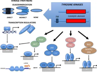
TKD are highly conserved modules, frequently composed by a N-terminal ATP-binding region and by a regulatory C-terminal domain. Upon activation, a cascade of events occurs, including ATP consumption, kinase dimerization, conformational changes, trans-autophosphorylation and oligo-complex formation. TK regulation is disrupted in oncogenic TKFs, which display
constitutive dimerization due to loss of the inhibitory
domains and forced oligodimerization of partner peptides [14]. Both inter- and intra-chromosomal rearrangements can lead to TKFs. Most break-points occur within introns and allow the synthesis of chimeric transcripts carrying the functional kinase domain and its flanking regions (Tables 1 and 2). Translocations involving TK membrane receptors frequently have breakpoints between exons coding for the juxta-membrane region. Though less frequent, the breaks can also split the trans-membrane domain upstream from the GXGXXG-coding exon. If protein products presenting the TK transmembrane domain localize to the cellular membrane, the exclusion of such a domain will localize the TK according to the properties of the translocation partner instead (Figure 1). In both cases, the natural TK ligand-binding site is lost. TKF involving
intra-cytoplasmic kinases are also characterized by the loss of
regulatory regions.
[image:2.612.122.492.383.656.2]Gene fusions typically replace the TK promoter;
Figure 1: Structure and Signaling Transduction Motifs of Tyrosine Kinase Fusions. The constitutive activation of Tyrosine
therefore TKF expression becomes ectopically regulated by the promoter of the partner gene. Partner genes contribute to the oncogenic potential in a number of ways. In most instances, the partner N-terminus region provides dimerization domains, which recruit molecular adaptors and lead to the constitutive trans-phosphorylation and activation of the kinase. TKF partners can also directly activate oncogenic pathways, as observed in the TFG-NTRK1 and TRAF1-ALK fusions, independently activating the NF-kB pathway [15]. We and others have
recently demonstrated that a small subset of systemic
(<5%) and cutaneous (~20%) Anaplastic Large Cell Lymphomas (ALCL) bears ROS1 or TYK2 fusions. In such cases, the TKF N-terminus region comprises a transcription factor (NFkB) or a transcriptional repressor (NCOR2) fused to ROS1 and TYK2, respectively [16]. These TKFs clearly illustrate how the chimera may also influence the transcriptional activity of the partner genes, perturbing transcription promotion or repression. This, in turn, may lead to a widespread derangement of several metabolic and signaling pathways, which ultimately concur to neoplastic transformation [15, 16]. The N-terminus can also contribute to the neoplastic phenotype by preventing protein degradation through the recruitment of heat shock proteins (Hsp70, Hsp90) [17]. This may constitute a valid therapeutic target, as observed with Hsp90 inhibitors for the treatment of NPM-ALK ALCL [16, 17] and FLT3-positive leukemias [18]. Fusion partners can further contribute to neoplastic transformation by directing TKF localization. In T-ALL, for example, NUP214 of the NUP214-ABL orchestrates TKF nuclear pore localization, which is required for neoplastic transformation [19]. Similarly, the ectopic localization of NPM-ALK and FOP-FGFR1 contributes to oncogenesis by impacting centrosome deregulation and chromosomal instability. Lastly, fusion partners can act as dominant negative mutants. By decreasing the access to wild-type TPM3, TPM3-ALK fusions induce changes in the cytoskeleton organization and confer a higher metastatic capacity [20].
TKF IN HUMAN CANCERS
TKFs are found in many human cancers,
although at very different frequencies. Among recurrent translocations, hematologic cancers were first shown to carry rearrangements characterized by TKF expression.
Subsequent studies demonstrated that RET [21], TRK [22] or NTRK1 [23] were frequently translocated in a fraction of thyroid carcinomas, as well as other epithelial and soft tissue neoplasms [24, 25]. This list has grown steadily to >300 oncogenic TKF [5] (http://cgap.nci.nih.gov/ Chromosomes/Mitelman) (Tables 1 and 2).
Are TKF drivers or passengers in human oncogenesis?
There is strong evidence that both TK and TKFs play an oncogenic role in human cancers [26-28]. Nevertheless, TKF-driven transformation requires multiple genomic
defects and the contribution of the host microenvironment
to drive oncogenesis. This paradigm is well represented by Chronic Eosinophilic Leukemia (CEL), where the sustained expression of FIP1L1-PDGFRA is necessary (but insufficient) to sustain eosinophils proliferation [29]. In humans, CEL also requires the overexpression of IL-5 through a single nucleotide polymorphism of IL5RA. The same holds true for systemic mastocytosis (SM), in which the constitutive activation of c-KIT alone is insufficient
to induce the neoplastic phenotype and requires the
stimulation of stem cell factor (SCF) as well [30]. This view is somehow challenged by seminal studies
on other myeloproliferative neoplasms (MPNs), like CML,
which may in fact represent monogenic disorders. The implantation of BCR-ABL (p210) retroviral-transduced marrow precursors into recipient mice can indeed cause rapidly progressing MPNs, which closely resemble human CML [31]. Of note, this scenario is hardly achievable in mouse models, where the TKF is ectopically expressed by means of more conventional molecular strategies [32, 33]. Moreover, Foley et al. have recently shown that the expression of BCR-ABL alone from the knock-in allele is unable to knock-induce leukemia [34], suggestknock-ing
that this TKF is per se insufficient to induce a disease fully recapitulating human CML. This latter hypothesis is consistent with the observation of BCR-ABL-positive hematopoietic cells also in healthy individuals [35], but is challenged by the observation that a fraction of CML patients who have achieved a sustained complete molecular remission (CMR) on imatinib can remain in CMR after drug withdrawal [36]. This latter observation raises the possibility that BCR-ABL signaling may not be the only driving force in some cases of CML. Of note, the concomitant loss of p14 (ARF) [37-39], and the critical role of Bcl-6 [40], miR-29 [41], and Raf1 [42] in CML suggest that the co-occurrence of selective defects and/ or the full activation of selective pathways are required to achieve the complete neoplastic phenotype. This is line with the recent observation that IKF1-mutated Ph-positive high-risk ALL, in which IKF1 and Arf alterations synergistically promote the development of an aggressive lymphoid leukemia, can be effectively treated with retinoid receptor agonists which potentiate the activity of dasatinib in mouse and human BCR-ABL1 ALL [43].
and in normal hematopoietic cells [35, 45]. Taken together,
these observations indicate that TKFs alone are probably
unable to drive T-cell neoplastic transformation completely and require secondary chromosomal abnormalities/TK activating mutations [46].
TKF expression levels may also play a role in tumor development. This is the case of ALK-translocated tumors: high NPM-ALK expression is indeed seen in ALCL, while lower EML4-ALK levels characterize NSCLC. These
differences are mainly due to different transcriptional
activities of the partner genes. Lastly, the regulation of TKF expression may depend on miRNAs/translational regulation and TKF degradation. Overall, these data provide empirical support to the concept of global neutrality, or near-neutrality (i.e. very weak selection
of the mutant clone in the early phases of neoplastic
transformation) for TKFs [47].
Are TKFs required for the maintenance of a neoplastic phenotype?
It is generally believed that TKFs contribute to the maintenance of the neoplastic phenotype. This is well exemplified by the therapeutic success of TK inhibitors (TKi) for the treatment of chronic/indolent hematologic disorders (CML and CEL) [48, 49]. The oncogenic contribution of TKF to tumor maintenance can nevertheless vary according to several variables, including: (i) the properties of each fusion; (ii) the cellular context and the tumor microenvironment; (iii) the disease stage and evolution phase [28, 49]; (iv) the degree of oncogenic addiction to the TKF (e.g. ROS1-positive NSCLCs show better response to Crizotinib than ALK-positive NSLCLs) [50]; (v) the presence of concomitant genomic aberrations [30, 51]. In particular, the occurrence of multiple genetic defects may influence the response to therapy. This is the case of genetically complex disorders (e.g. ALL or end-stage solid cancers), which show a very aggressive clinical behavior and only partial responses to TKi [52]. The same holds true for some cases of ALK+ ALCL. The majority of ALK+ ALCL patients have a fairly good outcome with conventional chemotherapy treatments. However, rapid and even leukemic progression can occur. These are frequently associated with disrupting events, including
TP53 and Blimp1 deletions [53] and c-myc translocation/ amplifications. The acquisition of secondary events may indeed be linked to a more aggressive phenotype and can damper the addiction to ALK signaling [15].
This mechanism has also been reported in CML in blast
crisis [54] and in ALK-positive NSLCLs resistant to Crizotinib. In the first case, BCR-ABL induces an IGF1 autocrine loop via Hck and Stat5b, in the latter case, the IGF-1R signaling synergizes with (and overcome) ALK by engaging the adaptor protein IRS-1 [51]. Moreover, restoration of p16 (INKa) or p14 (ARF) into Ph-positive
leukemic cells (both CML in blast crisis and ALL) is able
to determine cell growth arrest and/or apoptosis suggesting a pathogenic role for these deletions and their co-operating role with BCR-ABL [39].
TKFs in hematopoietic disorders
Among hematologic malignancies, distinct subsets of acute leukemias, MPNs and non-Hodgkin lymphomas harbor specific TKFs (Table 1).
The most frequent TKFs involve ABL-1 and PDFGRa/b, but translocations of ARG, JAK2, NTRK3 and SYK have been reported, as well. Not all partner genes provide oligodimerization domains, as in the case of the FIP1L1-PDGFRa fusion, which leads to the activation of the kinase moiety by disruption of its juxtamembrane regulatory motif [55]. Alternatively, the partners do not interact directly, but engage intermediate proteins leading to an indirect dimerization (e.g. CTCCL-ALK, NUP214-ABL, FOP-FGFR1).
The t(9;22)(q34;q11) translocation juxtaposes
the BCR and ABL1 genes in virtually all cases of CML and in a subset of adult/pediatric ALL. The latter may
represent a de novo disease or, as in many adults, the
blastic transformation of CML. Of note, adult and
(more frequently) pediatric ALL carry other TKFs,
which are associated with a Ph-like phenotype [9]. The oncogenic properties of such fusions have been demonstrated in a large variety of in vitro and in vivo
systems [9, 56, 57]. The second most common TKFs involve PDGFRα, PDGFRβ and FGFR1. Such TKFs characterize a group of hematological disorders, sharing similar clinico-pathological features, such as peripheral blood eosinophilia, clinical presentation as MPN and/or lymphoid tumors, and, at least for PDGFRα and PDGFRβ fusions, good response to Imatinib. For this reason, the 2008 WHO Classification of hematopietic and lymphoid tumors has introduced a separate diagnostic category for
such disorders, referred to as “Myeloid and Lymphoid
Neoplasms with Eosinophilia and Abnormalities of PDGFRα, PDGFRβ or FGFR1” [58].
Among mature T-cell neoplasms, subsets of Peripheral T-cell Lymphomas, not otherwise specified (PTCL, NOS) and Angioimmunoblastic T-cell
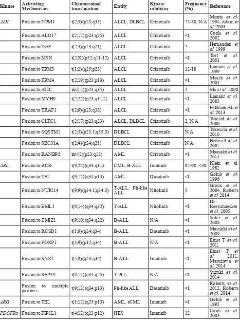
Table 1: Tyrosine kinases fusion in hematological malignancies
Kinase Activating Mechanisms Chromosomal translocation Entity Kinase inhibitor Frequency (%) Reference
ALK Fusion to NPM1 t(2;5)(p23;q35) ALCL, DLBCL Crizotinib 75-80, N/A Morris 1994, Adam et et al. al. 2003 Fusion to ALO17 t(2;17)(p23;q25) ALCL Crizotinib <1 Cools 2002 et al.
Fusion to TGF t(2;3)(p23;q21) ALCL Crizotinib 2 Hernandez al. 1999 et
Fusion to MSN t(2;X)(p32;q11-12) ALCL Crizotinib <1 Tort 2001 et al.
Fusion to TPM3 t(1;2)(q25;p23) ALCL Crizotinib 12-18 Lamant 1999 et al.
Fusion to TPM4 t(2;19)(p23;p13) ALCL Crizotinib <1 Meech 2001 et al. Fusion to ATIC inv( 2)(p23;q35) ALCL Crizotinib 2 Ma et al. 2000 Fusion to MYH9 t(2;22)(p23;q11.2) ALCL Crizotinib <1 Lamant 2003 et al.
Fusion to TRAF1 t(2;9)(p23;q33) ALCL Crizotinib <1 Feldman AL et al. 2013
Fusion to CLTC1 t(2;17)(p23;q23) ALCL, DLBCL Crizotinib 2, N/A Touriol 2000 et al.
Fusion to SQSTM1 t(2;5)(p23.1;q35.3) DLBCL Crizotinib N/A Takeuchi et al. 2010
Fusion to SEC31A t(2;4)(p24;q21) DLBCL Crizotinib N/A Bedwell 2007 et al.
Fusion to RANBP2 inv(2)(p23;q13) AML Crizotinib <1 Maesako et al. 2014
ABL Fusion to BCR t(9;22)(q34;q11) CML, B-ALL Imatinib 85-90, <30 Klein et al. 1982
Fusion to TEL t(9;12)(q34;p13) AML Dasatinib <1 Golub 1996 et al.
Fusion to NUP214 t(9;9)(q34.1;q34.3) T-ALL, Ph-like ALL Nitolinib 5 Graux 2004, Roberts et al.
et al 2014
Fusion to EML1 t(9;14)(q34;q32) T-ALL Nitolinib <1 De Keersmaecker et al. 2005
Fusion to ZMIZ1 t(9;10)(q34;q22) B-ALL N/A <1 Soler 2008 et al.
Fusion to RCSD1 t(1;9)(q24;q34) B-ALL Dasatinib <1 Mustjoki 2009 et al.
Fusion to FOXP1 t(3;9)(p12;q34) B-ALL N/A <1 Ernst T 2011 et al.
Fusion to SNX2 t(5;9)(q23;q34) B-ALL Imatinib <1
Ernst T et al. 2011, Masuzawa et al. 2014 Fusion to SEPT9 t(9;17)(q34;q25) T-PLL N/A <1 Suzuki 2014 et al.
Fusion to multiple
partners t(9;12)(q34;p13) Ph-like ALL Dasatinib <1
Roberts et al.
2012, Roberts
et al. 2014
ARG Fusion to TEL t(1;12)(q25;p13) AML, aCML Imatinib <1 Golub 1995 et al.
Fusion to BCR t(4;22)(q12;q11.2) CEL, T-ALL, CML-like MPN Imatinib <1
Baxter et al.
2002, Yigit
et al. 2015,
Cluzeau et al.
2015
Fusion to TNKS2 t(4;10)(q12;q23.3) MPN eosinophilia w/ Imatinib <1 Chalmer et al. 2014
Fusion to STRN t(2;4)(p22;q12) MPN eosinophilia w/ Imatinib <1 Curtis 2007 et al.
Fusion to ETV6 t(4;12)(q23;p12) MPN eosinophilia w/ Imatinib <1 Curtis 2007 et al.
Fusion to KIF5B t(4;10)(q12;p11) MPN eosinophilia w/ Imatinib <1 Score 2006 et al.
Fusion to CDK5RAP2 ins(9;4)(q33;q12q25) CEL Imatinib <1 Walz 2006 et al.
PDGFRb Fusion to TEL t(5;12)(q33;p13) CMML Imatinib 4 Golub 1994 et al.
Fusion to HIP1 t(5;7)(q33;q11) CMML Imatinib 4 Ross1998 et al.
Fusion to Rabaptin5 t(5;17)(q33;p13) CMML Imatinib <1 Magnussonal. 2001 et
Fusion to H4(D10S170) t(5;10)(q33;q11-q21) aCML Imatinib <1 Kulkarni et al. 2000
Fusion to CEV14 t(5;14)(q33;q32) AML Imatinib <1 Abe 1997 et al.
Fusion to Myomegalin t(1;5)(q23;q33) Eosinophilia Imatinib <1 Wilkinson al. 2003 et
Fusion to ATF71P t(5;12)(q23;p13) Ph-like ALL Imatinib <1 Kobyashi al. 2015 et
Fusion to EBF1 t(5;5)(q33.1;q33.3) Ph-like ALL Dasatinib <1 Roberts2012, Roberts et al.
et al. 2014
FGFR1 Fusion to multiple partners t(2;8)(q12;p11) EMS, AML None <1 Etienne 2007 et al.
Fusion to FOP t(6; 8)(q27;p11) MPN None <1 Lee et al. 2014
Fusion to SQSTM1 t(5;8)(q35;p11) AML <1 Nakamura Y et al. 2014
FGFR3 Fusion to TEL t(4;12)(p16;p13) PTCL Fiin23, NVP-BGJ398 <1 Maeda 2005 et al.
Fusion to IGH t(4;14) (p16; q32) CLL <1 Geller 2014 et al.
Fusion to TIF1 t(7;8)(q34;p11) MDS, AML CLL, Fiin23, NVP-BGJ398 <1 Maeda 2005 et al.
JAK2 Fusion to TEL t(9;12)(p24;p13) ALL, CML-like Ruxolitinib <5 Lacronique al. 1997 et
Fusion to OFD1 t(X;9)(p22;p24) ALL Jak2 inhibitors <1 Yano 2015 et al.
Fusion to SPAG9 t(9;17)(p24;q21) ALL Jak2 inhibitors <1 Kavamura M et al 2015
Fusion to PAX5 t(9;9)(p13;p24) ALL Jak2 inhibitors <1 Nebral K et al. 2009
Fusion to BCR t(9;22)(p24;q11.2) aCML Ruxolitinib <5 Griesingeral. 2005 et
Fusion to multiple
partners t(9;12)(p24;p13) Ph-like ALL Jak2 inhibitors <1
Roberts et al.
2012, Roberts
et al. 2014
involving TYK2 and ROS-1 in CD30-positive cutaneous lymphoproliferative disorders [64] and systemic ALK-negative ALCL [16] have been previously described. Interestingly, in addition to NPM-1, NFkB2 and NCOR2
are partners of these new TKFs and contribute to the neoplastic phenotype [16]. Finally, precursor T-cell neoplasms (i.e. T-cell acute lymphoblastic lymphoma [T-ALL]) may also harbor TKFs, which usually involve
the ABL gene. Some of such translocations are also observed in B-ALL (e.g. ABL-NUP214), while others seem to be specific of T-ALL (ABL-EML1) [65] (Table 1). Interestingly, several lymphoproliferative disorders also display TK oncogenic mutations, further demonstrating the tumorigenic role of deregulated kinase signaling in hematopoietic disorders [66, 67].
TKFs are less frequent in mature non-Hodgkin B-cell neoplasms, with the notable exception of ALK-positive diffuse large B-cell lymphoma, in which the TKFs bear distinct oncogenic and clinico-pathological features [68] (Table 1).
TKFs in solid tumors
In solid tumors, aberrant TK activity results from
diverse mechanisms, including: (i) point mutations; (ii) gene amplifications; (iii) gene overexpression; and (iv) chromosomal translocations [51, 69, 70]. Abnormal
TK activity can also result from de novo transcription
initiation (ATI) sites, which lead to the synthesis of truncated intracellular TKDs (e.g. ALKATI in melanoma or
ERBB4ATI in ALCL) [71, 72].
In recent years, a wide array of recurrent
translocations has been reported in solid tumors (Table
2) [14, 28]. In a systematic survey of nearly 7000
samples from the Cancer Genome Atlas, Stransky et al.
have depicted an extremely variegated translocation landscape in solid tumors [5], which can display heterogeneous chromosomal translocations, with specific malignancies (such as sarcomas) being characterized by a high percentage of non-recurrent translocations and/or infrequent but recurrent gene fusions.
SYK Fusion to TEL t(9;12)(q22;p12) MDS Imatinib <1 Kanie 2004 et al.
Fusion to ITK t(5;9)(q33;q22) PTCL-NOS, AITL None 17, <1
Streubel B
et al 2006, Attygale et al 2013
TRKC Fusion to TEL t(12;15)(p13;q25) AML and fibrosarcome None <1 Dobus 2001 et al.
FLT3 Fusion to ETV6 t(12;13)(p13;q12) HES Sunitinib, Midostaurin,
Lestaurtinib <1 Vu et al. 2006 ETV6 Fusion to TEL t(4;12)(p16;p13) PTCL-NOS Ruxolitinib <1 Yagasaki 2001 et al.
CSF1R Fusion to SSBP2 t(5;5)(q14;q33) Ph-like ALL Dasatinib <1 Roberts 2014 et al.
CRLF2 Fusion to IGH t(X;14)(p22;q32)/t(Y;14)(p11;q32) Ph-like ALL Jak2 inhibitors <5
Mullighan
et al. 2009, Roberts et al.
2014
ROS1 Fusion to NFKB2 t(6;10)(q22;q24) ALCL Ros1 inhibitors <1 Crescenzo al. 2015 et
Fusion to NCOR2 t(6;12)(q22;q24) ALCL Ros1 inhibitors <1 Crescenzo al. 2015 et
TYK2 Fusion to NFKB2 t(19;10)(p13;q24) ALCL Tyk2 inhibitors <1 Crescenzo et al. 2015
Fusion to NPM1 t(19;5)(p13;q35) LPDs Tyk2 inhibitors <1 Velusamy al. 2014 et
LYN Fusion to NCOR t(8;17)(q13;p11) ALL NA <1 Yano 2015 et al.
Abbreviation:
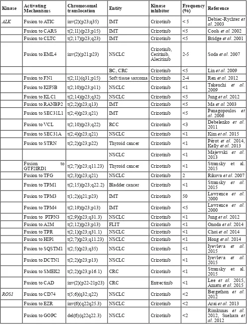
Table 2: Tyrosine kinases fusions human in solid tumors
Kinase Activating Mechanisms Chromosomal translocation Entity Kinase inhibitor Frequency (%) Reference
ALK Fusion to ATIC inv(2)(p23;q35) IMT Crizotinib < 5 Debiec-Rychter al. 2003 et Fusion to CARS t(2;11)(p23;p15) IMT Crizotinib <5 Cools et al. 2002
Fusion to CLTC t(2;17)(p23;q23) IMT Crizotinib <5 Bridge et al. 2001
Fusion to EML4 inv(2)(p21;p23) NSCLC Crizotinib, Ceritinib,
Alecitinib 2-5 Soda et al. 2007
BC, CRC Crizotinib <5 Lin et al. 2009
Fusion to FN1 t(2;11)(q31;p15) Soft tissue sarcoma Crizotinib 2-4 Ren et al. 2012 Fusion to KIF5B t(2;10)(p23;p11) NSCLC Crizotinib <1 Takeuchi 2009 et al. Fusion to KLC1 t(2;14)(p23;q32) NSCLC Crizotinib <5 Jung et al. 2012 Fusion to RANBP2 t(2;2)(p23;q13) IMT Crizotinib <5 Ma et al. 2003 Fusion to SEC31L1 t(2;4)(p23;q21) IMT Crizotinib <5 Panagopoulos al. 2006 et
Fusion to VCL t(2;10)(p23;q22) RCC Crizotinib <3 Debelenko 2011 et al.
Fusion to SEC31A t(2;4)(p23;q21) NSCLC Crizotinib <1 Kim et al. 2015 Fusion to STRN t(2;2)(p23;p22) Thyroid cancer Crizotinib <1 Pérot Kelly et al.et al. 2013 2014,
NSCLC Crizotinib <1 Majewski 2013 et al. Fusion to
GTF2IRD1 t(2;7)(p23;q11.23) Thyroid cancer Crizotinib <1 Stransky et al. 2015
Fusion to TFG t(2;3)(p23;q21) NSCLC Crizotinib 2 Rikova et al. 2007
Fusion to TPM1 t(2;15)(p23;q22.2) Bladder cancer Crizotinib <1 Stransky 2015 et al.
Fusion to TPM3 t(1;2)(q21;p23) IMT Crizotinib 50 Lawrence 2000 et al.
Fusion to TPM4 t(2;19)(p23;p13) IMT Crizotinib <5 Lawrence 2000 et al.
Fusion to PTPN3 t(2;9)(p23;q31.3) NSCLC Crizotinib <1 Jung et al. 2012
Fusion to A2M t(2;12)(p23;p13) FLIT Crizotinib <1 Onoda et al. 2014 Fusion to TPR t(2;1)(p23;q31.1) NSCLC Crizotinib <1 Choi et al. 2014 Fusion to HIP1 t(2;7)(p23;q11.23) NSCLC Crizotinib <1 Hong et al. 2014
Fusion to SQSTM1 t(2;5)(p23;q35) NSCLC Crizotinib <1 Iyevleva 2015 et al.
Fusion to DCTN1 t(2;2)(p23;p13) NSCLC Crizotinib <1 Iyevleva 2015 et al.
Fusion to SMEK2 t(2;2)(p23;p16.1) CRC Crizotinib <1 Stransky et al. 2015
Fusion to CAD inv(2)(p22-21p23) CRC Entrectinib <1 Lee Amatu et al. et al. 2015, 2015
ROS1 Fusion to CD74 t(5;6)(q32;q22) NSCLC Crizotinib <2 Bergethon 2012 et al.
Fusion to EZR inv(6)(q22q25.3) NSCLC Crizotinib <2 Arai et al. 2013
CCA Crizotinib <1 Gu et al. 2011 Ovarian Cancer Crizotinib <1 Birch et al. 2011 Fusion to LRIG3 t(6;12)(q22;q14.1) NSCLC Crizotinib <2 Takeuchi 2012 et al.
Fusion to SDC4 t(6;20)(q22;q12) NSCLC Crizotinib <2 Davies 2012, Takeuchi et al. et al. 2012
Fusion to SLC34A2 t(4;6)(q15.2;q22) NSCLC Crizotinib <2 Davies et al. 2012
Gastric cancer Crizotinib <1 Lee et al. 2013
Fusion to TPM3 t(1;6)(q21.2;q22) NSCLC Crizotinib <2 Takeuchi 2012 et al.
Fusion to TFG t(6;3)(q22.1;q12.2) IMT Crizotinib <1 Yamamoto 2015 et al.
RET Fusion to CCDC6 inv10(q11;q21) NSCLC Cabozantinib, Vandetanib <2 Wang et al. 2012
Thyroid cancer Cabozantinib, Vandetanib <2 Celestino 2012 et al.
Fusion to KIF5B inv(10)(p11;q11) NSCLC Cabozantinib, Vandetanib <2 Ju et al. 2012
Fusion to NCOA4 inv(10)(q11;q11) Thyroid cancer Cabozantinib, Vandetanib <2 Rui et al. 2012
Fusion to
PRKAR1A t(10;17)(q11.2;q23) Thyroid cancer Cabozantinib, Vandetanib <2 Rui et al. 2012
Fusion to ACBD5 inv(10)(p12.1;q11.2) Thyroid cancer Cabozantinib, Vandetanib <1 Hamatani 2014 et al.
BRAF Fusion to KIAA1549 t(7;7)(q34;q34) Brain tumors BRAF/MEK inhibitors <1 Tian et al. 2011
Fusion to FAM131B t(7;7)(q34;q34) Brain tumors BRAF/MEK inhibitors <1 Cin et al. 2011
Fusion to CEP89 t(7;19)(q34;q13) Melanoma BRAF/MEK inhibitors < 5 Wiesner 2014 et al.
Fusion to LSM14A t(7;19)(q34;q13) Melanoma BRAF/MEK inhibitors < 5 Wiesner 2014 et al.
FGFR1 Fusion to TACC1 t(8;8)(p11.23;p11.22) GBM FGFR inhibitor <3 Singh et al. 2012
Fusion to BAG4 t(8;8)(p11.23;p11.23) NSCLC FGFR inhibitor <1 Rui et al. 2014
FGFR2 Fusion to BICC1 t(10;10)(q26;q21.1) CCA FGFR inhibitor <1 Yi-Mi et al. 2013
Fusion to
KIAA1967 t(10;8)(q26;p21.3) NSCLC FGFR inhibitor <1 Yi-Mi et al. 2013
Fusion to PPHLN1 t(10;12)(q26;q12) CCA FGFR inhibitor 45 Sia et al. 2015
FGFR3 Fusion to TACC3 del4(p16;p16) GBM FGFR inhibitor <3 Singh Bao et al. et al.2014 2012,
Bladder cancer FGFR inhibitor <2 Williams 2013 et al.
NSCLC FGFR inhibitor <2 Rui et al. 2014
To date, fusions involving 14 different TKs
have been described in solid tumors (Table 1). ALK is translocated in several tumors, including lung, colorectal, breast, renal, thyroid carcinomas and soft tissue tumors.
NPC FGFR inhibitor <3 Yuan et al. 2014
Cervical cancer FGFR inhibitor <1 Carneiro 2015 et al.
NTRK1 Fusion to TPM3 t(1;3)(q21;q11) Thyroid cancer TRKA inhibitor 7-8 Beimfohr 1999 et al.
CRC TRKA inhibitor <1 Creancier 2015 et al.
HGG TRKA inhibitor <1 Wu et al. 2014
Fusion to TPR inv1(q23;q21) Thyroid cancer TRKA inhibitor <1 Greco et al. 1999
CRC TRKA inhibitor <1 Creancier 2015 et al.
Fusion to MPRIP t(1;17)(q21;p11) NSCLC TRKA inhibitor <5 Vaishanvi 2013 et al.
Fusion to CD74 t(1;5)(q21;q32) NSCLC TRKA inhibitor <5 Vaishanvi 2013 et al.
Fusion to
RABGAP1L t(1;1)(q21;q25.1) CCA TRKA inhibitor <1 Ross et al. 2014 Fusion to SQSTM1 t(1;5)(q21;q35) NSCLC Entrectinib <1 Farago et al. 2015
Fusion to LMNA t(1;1)(q21;q22) Soft tissue sarcoma LOXO-101 <1 Doebele 2015 et al.
CRC Entrectinib <1 Sartore-Bianchi al. 2015 et
NTRK2 Fusion to VCL t(9;10)(q22.1;q22) HGG TRKA inhibitor <1 Wu et al. 2014
Fusion to AGBL4 t(9;1)(q22.1;p33) HGG TRKA inhibitor <1 Wu et al. 2014
NTRK3 Fusion to ETV6 t(12;15)(p13;q25) Thyroid cancer TRKA inhibitor 2-14 Ricarte-Filho 2013 et al.
CFS TRKA inhibitor <1 Knezevih 1998 et al.
IMT TRKA inhibitor <1 Yamamoto 2015 et al.
GIST TRKA inhibitor <1 Brenca et al. 2015
MASC TRKA inhibitor <1 Skalovà 2015 et al.
HGG TRKA inhibitor <1 Wu et al. 2014
Fusion to BTBD1 t(15;15)(p24;q25) HGG TRKA inhibitor <1 Wu et al. 2014
AKT2 Fusion to BCAM t(19;19)(q13.2;q13.3) HGSC AKT2 inhibitor 10 Kannan et al. 2015
PRKACA Fusion to DNAJB1 t(19;19)(p13.1;p13.2) FL-HCC PKA inhibitors 100 Honeyman 2014 et al.
PRKD1 Fusion to ARID1A t(14;1)(q12;p36) Salivary tumor gland PRKD1 inhibitor <3 Weinreb 2014 et al. MET Fusion to PTPRZ1 t(7;7)(q31.2;q31.3) GBM MET inhibitor 15 Bao et al. 2014
PIK3CA Fusion to TBL1XR1 t(3;3)(q26.3;q26.32) BC PIK3CA inhibitor <1 Stransky et al. 2015
Abbreviation:
To date, fusions involving 14 different TKs have been described in solid tumors (Table 1). ALK is translocated in several tumors, including lung, colorectal, breast, renal, thyroid carcinomas and soft tissue tumors.
Fusion partners include EML4, TFG, KIF5B, KLC1 and STRN [28]. Rare translocations involving SMEK2 (rectal adenocarcinoma) or GTF2IRD1 (thyroid carcinoma) have also been reported.
Next generation sequencing (NGS) analyses have highlighted new patterns of translocations, defining specific subgroups of common solid tumors (BRAF in melanoma; FGFR1, FGFR2, FGFR3 in lung cancer;
NTRK in thyroid cancer), and in clinically unmet patients (BRAF and FGFR1 translocations in pediatric low-grade glioma; NTRK1 and NTRK2 in high-grade gliomas, and cholangiocarcinoma, Table 1). Lastly, MET is involved in chromosomal translocations of secondary glioblastomas
and papillary renal carcinomas and PI3KCA translocations
are observed in breast and prostate carcinomas.
The transforming activity of TKF in solid cancers has been extensively characterized through pre-clinical models [73], proving that classical MEK-ERK and PI3K-AKT pathways are critical players in sustaining the transformed phenotype and in mediating resistance to targeted therapies [74]. TKF dependency thus provides a molecular explanation for the “oncogene addiction” of different TKF-driven tumors [75].
Mechanistically, multiple causes of chromosomal
translocations have been predicted: (i) exposure to toxic agents (probably the major initiating event); (ii) exposure to ionizing radiation (e.g. in papillary thyroid carcinoma); (iii) oxidative stress and chemotherapeutic agents (e.g. topoisomerase inhibitors).
DISCOVERY AND DETECTION OF TK
FUSIONS
The oncogenic role of TKFs has prompted huge
efforts to improve their detection in cancer patients to
better guide patient-directed treatment strategies [76]. Affinity purification coupled with mass spectrometry (AP-MS) is a powerful tool to characterize aberrant gene products and protein-protein interactions (PPIs). AP-MS has been successfully used to discover novel TKFs in NSCLC (ALK and ROS1) [77] and rare TKFs in other tumor histotypes [78]. AP-MS could also define activating mutations linked to TKi-resistant phenotypes [79]. Furthermore, proteomic patterns distinguish normal from pathological tissues and correctly classify primary/ metastatic lesions, allowing tailored treatments and a better stratification of NSCLC [80].
The introduction of NGS-based approaches has rapidly enriched the overall knowledge of TKF, giving
a better representation of their distribution in human
cancers. In hematologic malignancies, NGS studies of Ph-like ALL have detected TKFs in >90% of patients [8].
Technically, both RNAseq and Whole Exome Sequence (WES) made these achievements possible. In some cases an exon capture specific for TK genes followed by massive parallel sequencing was applied [81-84]. Since these analyses can be performed on formalin-fixed
paraffin-embedded samples, they can be applied to routine clinical
practice. The possibility to test archived material will also allow for the characterization of the clonal evolution of TKF-bearing tumors, with consequent implementation of cancer tailored therapies.
Clinical diagnostic tests: tumor fingerprints
Clinically, TKF in human hematological cancers can be detected by an array of techniques. Genomic/ conventional cytogenetics (CG) or FISH are commonly used for the detection of BCR-ALB translocations. Negative samples (<5% of cases) frequently carry variant translocations or cryptic rearrangements, which can occasionally be detected by FISH. Both approaches are also used to monitor drug responses and disease evolution. FISH and RT-PCR have shown higher sensitivity at diagnosis and better MRD detection than CG. In particular, qRT-PCR has become the gold standard for the diagnosis of MRD [49]. FuseFISH probe-based approaches for the detection of fusion transcripts have also recently emerged [85], but their usefulness in clinical practice is uncertain.
Resistance to TKi treatments is currently detected by RT-PCR coupled with DNA Sanger sequencing, but new methods have been developed. Massive parallel sequencing is rapidly replacing the above reported strategies, as it can also identify subclonal populations and characterize tumor clonal heterogeneity [86]. Analogous approaches have now been extended to the detection of actionable mutations, copy number variations, and gene rearrangement in clinical cancer specimens.
In solid tumors, TKFs are clinically identified by several techniques, some of which are performed as companion tests to select the best TKi agent. In 2013, the USA and China’s FDA approved FISH- and IHC-based diagnostic tests to identify ALK fusions or ALK protein
expression in NSCLC. Similar strategies were proposed to
assess ROS1 and RET fusions [87]. Alternatively, a
NGS-based detection kit for RET-rearrangements has been used to enroll patients in NSLCL clinical trials (Eisai sponsored trial; NCT01829217). It is conceivable that screening approaches to detect simultaneously multiple fusions will be implemented in the clinical arena [88].
Since different TKFs can be present in distinct
cancer subsets, multi-targeted platforms have emerged as powerful tools to screen diagnostic samples. As multi-gene screening methods are entering into routine diagnostic procedures, new technical and therapeutic questions need to be addressed. Indeed, the greater efficacy of
TKi compared to conventional treatments needs to be
Similarly, patients treated with multiple rounds of chemotherapeutic regimens need to be evaluated for the
compliance to selective TKi, as limited data are available
to justify their usage.
TUMOR-ASSOCIATED TK TRANSLOCATIONS: CURRENT THERAPEUTIC STRATEGIES
The discovery of recurrent tumor-associated TK translocations has brought new strategies for the treatment of hematologic and non-hematologic tumors. In the last
decades, TKi have revolutionized the treatment of CML,
Ph-positive B-ALL [89] and NSCLC carrying ALK or ROS-1 gene rearrangements [14, 90].
TKi-based therapies for the treatment of hematological tumors
BCR-ABL1 gene rearrangements are reported in all cases of CML and in about 30% of adult B-ALL. The fusion gene usually results from the t(9;22)(q34;q11.2) translocation, but 5-10% of cases bear variants and/ or cryptic translocations [2]. BCR-ABL1 oncogenic properties are due to the constitutive activation of ABL1 TK [91, 92] and the loss of ABL-1 N-terminus regulatory domain.
Clinically, Imatinib has revolutionized the
prognosis and outcome of CML, converting a highly lethal hematological neoplasm to a chronic and curable disease [49]. Complete cytogenetic responses (CCyR) are indeed reported in 87% of Imatinib-treated patients, with a progression-free survival (PFS) of 93% [93]. After 2 years of sustained CCyR, the life expectancy for CML patients is comparable to that of the general population [94]. Despite
these results, several studies pinpointed a subset of cases
with primary/secondary resistance to Imatinib, which can be overcome by new generation TKi (i.e. Nilotinib, Dasatinib and Bosutinib) with improved clinical efficacy [95]. These lines of evidence led the European Leukemia Net to recommend Imatinib, Nilotinib and/or Dasatinib as first line drugs for the treatment of CML. More aggressive therapeutic approaches (e.g. hematopoietic stem cell
transplant) are considered only in very selected cases
(e.g. refractory/relapsing cases unresponsive to TKi or accelerated/blast phase CML) [49].
In CML patients, resistance to TKi can result
from two major mechanisms: (i) occurrence of TKi-resistant BCR-ABL1 mutations; (ii) enhancement of ABL1-independent pro-survival pathways. BCR-ABL1-dependent resistance has been overcome by third generation TKi that also target the most common ABL1 mutations (e.g. BCR-ABL1T315I). The most promising
therapeutic agents are the conventional TKi, Ponatinib, and the BCR-ABL1 switch control inhibitor, DCC-2036 [96]. Notably, good results with Ponatinib have been
obtained even in accelerated and blast phase CML [97], while the distinct action of DCC-2036 may overcome pre-existing mutations, including T315I [96]. Pemovska et al.
have recently re-proposed the utility of Axitinib, an anti-VEGFR molecule approved for renal cell carcinoma, to counteract the resistance phenotype of T315I gatekeeper CML. Indeed, Axitinib tightly binds the T315I mutated kinase and inhibits leukemic cell growth, showing a better toxicity profile than other new-generation drugs, such as Ponatinib [98].
BCR-ABL1-independent resistance is mainly due to the activation of alternative signaling pathways promoting neoplastic cell survival [99]. Such pathways can be either extrinsic (i.e. activated by the marrow microenvironment) or intrinsic (i.e. due to microenvironment-independent mechanisms). Both intrinsic and extrinsic pathways lead to the activation of STAT3, through its phosphorylation at Tyr-705 [100]. Combined inhibition of STAT via BP-5-087, a potent and selective STAT3 SH2 domain inhibitor and BCR-ABL1 leads to synthetic lethality in resistant CML and constitutes a very promising therapeutic approach for refractory/relapsing CML [100]. Another therapeutic opportunity is represented by glitazones, anti-diabetic drugs that down-regulate STAT5 expression by inhibiting the peroxisome proliferator-activated receptor-γ (PPARγ). Recent data show that their use may lead to the complete eradication of leukemic stem pools, thus greatly improving the survival of CML patients in sustained CMR [101].
Unlike what has been observed in CML, the use of single-agent TKi in Ph-positive ALL has not produced sustained clinical responses. This is probably due to the rapid development of TK mutations, which are reported in >80% of adult positive ALLs at relapse [102]. Ph-positive ALL and CML also greatly differ in terms of genomic backgrounds, which may influence the response to TKi. For instance, INK4-ARF deletions, which often occur in acute Ph-positive ALL with aggressive clinical course, facilitate the emergence of BCR-ABL drug-resistant clones. This molecular event is not observed in indolent CML or in CML in blast crisis [103].
These data provide the rationale for the use of multi-modal regimens for the treatment of Ph-positive ALL,
based on a combination of intensive chemotherapy and
TKi. Very encouraging results have been first obtained in pediatric trials (COG, EsPhALL and SHOP-2005 trials) [104, 105] and, more recently, in adult patients [106]. In
and adult Ph-positive ALL is still under evaluation, but preliminary results indicate their safety and efficacy when used with high dose chemotherapy and/or ASCT [107]. Encouraging data have also been obtained with Dasatinib and corticosteroid, which proves the potential efficacy of chemo-free protocols [108]. Finally, the association of TKi and bispecific T-cell engaging antibodies (BiTE) or chimeric antigen receptors T-cells (CARTs) represents a promising strategy for the treatment of Ph-positive ALL [109].
TKI-based therapies for the treatment of solid tumors
The introduction of TKi for the treatment of ALK-, ROS1- and RET-translocated NSCLC [14, 110, 111] has led to an astonishing improvement of patients’ outcome and survival. More recently, fusions involving NTRK1 and FGFR1/2/3 have been reported (Table 1), thus enlarging the spectrum of promising therapies for NSCLC (i.e. Crizotinib, Ceritinib, Alecitinib for ALK-positive NSCLC; Crizotinib for ROS1-positive NSCLC; Cabozantinib and Vandetanib for RET-positive NSCLC).
Crizotinib represents the paradigm of efficient translation of molecular knowledge to the bedside. Initially developed as a c-Met inhibitor, Crizotinib showed clinical activity on advanced ALK-positive NSCLC in both the PROFILE 1001 (phase I) and PROFILE 1014 (phase III) clinical trials. Of note, in such tumors, Crizotinib displayed even greater efficacy than standard chemotherapy [112]. Similar results have been observed in ROS1- or RET-positive NSCLCs, with good responses to Crizotinib in ROS1-positive cases [14] and to Cabozantinib in Crizotinib-refractory cells [113]. The application of TKi has recently expanded to RET-mutated medullary thyroid carcinoma, RET-translocated NSCLC and other solid tumors (NCT02183870, NCT0194502, NCT01823068, NCT01639508). More recently, inhibitors with broad spectrum of activities were tested (RXDX-101), providing novel strategies to target different molecular defects (NTRK1/2/3, ROS1 and ALK translocation) shared by wide range of solid tumors (NCT02568267, NCT02097810).
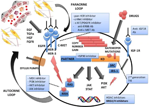
Despite these outstanding results, TKi often lose their efficacy due to the development of treatment-related resistance [114]. Much like hematologic neoplasms,
primary and secondary mechanisms determine resistance
to TKi in solid tumors. Primary resistance is linked to the tridimensional structures of the protein that confer TKi-resistance [115], whereas secondary TKi-resistance mainly consists of gene mutations, bypass mechanisms, and/ or gene amplifications overcoming the effect of TKi at therapeutic levels.
As for target mutations, different genetic alterations associated with resistance have been described for
ALK-positive NSCLC: i) L1169M within the “gatekeeper” domain (the most frequent); ii) L1152R, associated with EGFR signaling activation; iii) G1202R and S1206Y within the ATP-binding pocket, leading to a decreased Crizotinib affinity [116]. The most frequently reported bypass mechanisms associated with secondary resistance to ALK inhibition are: i) EGFR/KRAS mutations [117] and Tyrosine Kinase Receptor (ERBB, c-MET, etc.) activation triggered by paracrine stimuli [118, 119]; ii) c-KIT amplification, requiring SCF [120], IGF-1R up-regulation [51] and recruitment of P2Y receptors [121]. Other resistance mechanisms include impaired drug influx (OCT-1), increased drug efflux (MDR1), or microenvironment-mediated resistance [122].
Different strategies have been proposed to overcome TKi acquired resistance, including the development of next-generation drugs, such as the ALK-inhibitors LDK378-Ceritinib and AP26113-Brigatinib (for
ALK-positive NSCLC resistant to Crizotinib), Lecitinib (for
ALK-positive NSCLC carrying the L1169M mutation) and PF06463922 (a dual ALK/ROS1 inhibitor with a good CNS penetration profile) [123]. Additionally, Hsp-90 inhibitors (AUY922-Ganetespib) are currently under evaluation in phase I/II clinical trials. Other emerging strategies include the administration of combined therapies to by-pass the “addiction” to single TKFs (e.g., combined inhibition of HER family [Dacomitinib], c-KIT [Dasatinib], src [Saracatinib], MEK [AZD6244], and IGF-1R [Linsitinib]). The re-timing of drug administration
should also be considered, as recently demonstrated by
the case of a Crizotinib-resistant ALK-positive NSCLC, displaying good response to this TKi after a withdrawal period [124]. Similar results have been obtained in an EGFR-mutated NSCLC, which regained sensitivity to EGFR-inhibitors upon therapy discontinuation [125]. Lastly, immunotherapeutic strategies (i.e. vaccination against over-expressed oncogenic antigens) may represent
an effective approach for solid tumors, as recently reported
in ALK-positive disorders [59, 126].
A final major roadblock concerning TKi is the poor
response of central nervous system (CNS) metastases to
therapy. This drawback may be overcome by different strategies: (i) combined therapies with drugs favoring TKi CNS accumulation, as recently attempted through the co-administration of Vandetanib and Everolimus (an immunosuppressive drug also modulating the P-glycoprotein efflux activity) [127]; and (ii) usage of single compound with higher brain penetrance, such as Ceritinib compared to Crizotinib.
FUTURE PERSPECTIVES
relapses [128] (Figure 2). Possible solutions for this limitation are: (i) the development of more potent drugs with limited side effects; (ii) the co-administration of TKi and chemotherapeutic agents [129], immune-based protocols [130] and/or bone marrow transplant [131]; and (iii) further research on treatment timing and dosage.
Of note, a growing number of protocols combining chemotherapy and TKi are currently being tested in both TKF-induced hematological and solid tumors [107, 132] [133], and several trials are evaluating the efficacy of new TKi either alone or in combination with more conventional therapies (NCT02269085; NCT02427620; NCT02315768; NCT01606878; NCT02134912; NCT02511184).
Preclinical studies have also demonstrated a substantial
advantage of combining classical chemotherapy with TKi: Das and colleagues showed a synergistic effect of Crizotinib and Temozolomide in FIG-ROS1-positive glioblastoma patient-derived cells, paving a route to improve clinical outcome [134]. Similarly, Appelmann
and colleagues evaluated the possibility of combining Dasatinib, Ruxolitinib and Dexamethasone to prolong remission and avoid major toxicities in a mouse model of Ph-positive ALL [135].
One of the most challenging issues on the combined
use of TKi and chemotherapeutics concerns the optimal
doses and schedules to use. Toward this end, mathematical models might improve the clinical success, limiting unacceptable side effects and TKi/conventional therapy-resistance [136]. This approach also needs dedicated trials, the cooperative agreement of pharmaceutical companies and new regulations on intellectual and commercial issues. Moreover, we need to develop biological and
mathematical models to predict cancer evolution,
[image:14.612.65.546.298.643.2]capable of predicting cancer evolutionary trajectories based on pre-treatment diversity. Lastly, we foresee that these predictions need to be adjusted in each individual patient by integrating longitudinally acquired molecular readouts representative of the global cancer landscape
Figure 2: Mechanisms of Intrinsic and/or Acquired Resistances to Tyrosine Kinase Exposure. Multiple mechanisms are
(i.e. comprehensive genomic data [WES] of multiple sites, liquid biopsies, etc.).
Other questions regarding TKi concern the risks and benefits of second/third generation drugs. Thus, the use of first generation compounds remain a valuable option in many settings, including highly targeted diseases and the management of third world populations. The increasing
health costs, the uncontrolled fee schedules in many
industrialized countries, the increasing co-payer costs and the accessibility of generic TKi represent very tangible issues which may be solved only via international based supports and regulations. Finally, the need of cutting health system costs poses unprecedented challenges to molecular medicine programs. In fact, the success of such
therapies depends on the characterization of individual
cancers (both primary and recurrent/metastatic lesions).
These analyses have to be performed by a pool of medical
centers following certified programs, with consequent huge health costs.
These practical problems are paralleled by a series
of scientific and methodological challenges. Although the genomic characterization of each cancer has originally prompted the use of selective compounds, it is now clear that this approach may not be sufficient. Indeed, favorable responses to selective drugs are not strictly linked to defined genetic defects. Thus, functional tests (e.g. comprehensive phosphorylation maps) should be performed together with genomic analyses. As an
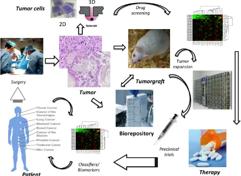
[image:15.612.66.555.247.604.2]alternative, in vitro preclinical tests on patient-derived cell lines or PDTX may be introduced (Figure 3).
Figure 3: Comprehensive Management of Tyrosine Kinase-driven Cancers. Molecular and functional characterization of
human cancers in combination with drug screening tests on primary patient tumor samples is expected to drive precise target therapies. It is anticipated that tumor samples will be used to generated 2D/3D and well as PDTX models. In vitro models from primary samples or from
Of note, in vitro tests with patient-derived tissue samples may allow personalized pharmacologic interventions without the limits related to the use of immortalized cell lines or other tumor models. Patient-derived cancer models are indeed pivotal for drug discovery, for the characterization of cancer biology and for the assessment of personalized therapies [137-139]. Several cancer PDTX models have been developed, but only handful of them carries TKFs [15, 140-142]; thus, a more systematic effort in generating “ad hoc” models is highly recommended.
We anticipate that in vitro primary cell lines
will allow for the rapid testing of drug libraries, with subsequent validation in more complex models (e.g. PDTX). It is also desirable that new clinical trials will include parallel co-clinical mouse PDTX-trials, which will establish the efficacy of such models. The molecular stratification of these models will finally provide new cancer classifiers with possible diagnostic/prognostic implications (Figure 3).
The considerable therapeutic progress against TKF-driven tumors may also prove beneficial for the treatment of those translocation-negative neoplasms, which nonetheless carry defects of TKF-related signaling pathways. As previously highlighted, TKFs indeed represent only one of the several mechanisms leading to TK dysfunction, and other TK impairments (i.e. gene mutations/amplifications or non-fusion rearrangements) lead to phenotypes closely resembling those of TKF-driven neoplasms. This is, for instance, the case of subsets of ALK-negative ALCL, carrying JAK1 and STAT3 activating mutations, which induce transcriptional signatures akin to those of ALK-positive ALCL [16]. Moreover, it is now recognized that activating mutations can synergize with TFKs and lead to full neoplastic transformation [8]. This molecular evidence constitutes
the rationale for the administration of TKi compounds in
specific subsets of both TKF-negative and TFK-positive tumors. Further studies and clinical trials will help clarify this captivating opportunity.
In conclusion, it is desirable that the academic
community promotes dedicated programs on TKi and TKF-bearing cancers, also encouraging the support
of pharmaceutical companies and the distribution of
compounds under compassionate use. This approach, together with the creation of open-access clinical databases, will greatly contribute to the understanding of cancer-related TKFs and to a better management of (TKF-positive and TKF-negative) cancer patients.
ACKNOWLEDGMENTS
GI is supported by Italian Association for Cancer
Research (AIRC) (5x1000 No. 10007); ImmOnc (BIO F.E.S.R. 2007/13, Asse 1 ‘Ricerca e innovazione’ della LR 34/2004) the Oncology Program of Compagnia di San
Paolo, Torino and Ricerca Finalizzata, Ministero della Salute.
Authors’ contributors
FT, MP, PK, BR and GI researched and reviewed the existing literature and editedwrote the manuscript.
CONFLICTS OF INTERESTS
We declare no competing interests.
REFERENCES
1. Nowell PC. A minute chromosome in human chronic granulocitic leukemia. Science. 1960; 132:1497.
2. Rowley JD. Letter: A new consistent chromosomal abnormality in chronic myelogenous leukaemia identified by quinacrine fluorescence and Giemsa staining. Nature. 1973; 243(5405):290-293.
3. Heisterkamp N, Stam K, Groffen J, de Klein A and Grosveld G. Structural organization of the bcr gene and its role in the Ph’ translocation. Nature. 1985; 315(6022):758-761.
4. Heisterkamp N, Stephenson JR, Groffen J, Hansen PF, de Klein A, Bartram CR and Grosveld G. Localization of the c-ab1 oncogene adjacent to a translocation break point in chronic myelocytic leukaemia. Nature. 1983; 306(5940):239-242.
5. Stransky N, Cerami E, Schalm S, Kim JL and Lengauer C. The landscape of kinase fusions in cancer. Nat Commun. 2014; 5:4846.
6. Mullighan CG, Collins-Underwood JR, Phillips LA, Loudin MG, Liu W, Zhang J, Ma J, Coustan-Smith E, Harvey RC, Willman CL, Mikhail FM, Meyer J, Carroll AJ, Williams RT, Cheng J, Heerema NA, et al. Rearrangement of CRLF2 in B-progenitor- and Down syndrome-associated acute lymphoblastic leukemia. Nature genetics. 2009; 41(11):1243-1246.
7. Hantschel O. Structure, regulation, signaling, and targeting of abl kinases in cancer. Genes & cancer. 2012; 3(5-6):436-446. doi: 10.1177/1947601912458584.
8. Roberts KG, Li Y, Payne-Turner D, Harvey RC, Yang YL, Pei D, McCastlain K, Ding L, Lu C, Song G, Ma J, Becksfort J, Rusch M, Chen SC, Easton J, Cheng J, et al. Targetable kinase-activating lesions in Ph-like acute lymphoblastic leukemia. N Engl J Med. 2014; 371(11):1005-1015.
10. Valent P. Targeting the JAK2-STAT5 pathway in CML. Blood. 2014; 124(9):1386-1388.
11. Medves S and Demoulin JB. Tyrosine kinase gene fusions in cancer: translating mechanisms into targeted therapies. J Cell Mol Med. 2012; 16(2):237-248.
12. Tan L, Wang J, Tanizaki J, Huang Z, Aref AR, Rusan M, Zhu SJ, Zhang Y, Ercan D, Liao RG, Capelletti M, Zhou W, Hur W, Kim N, Sim T, Gaudet S, et al. Development of covalent inhibitors that can overcome resistance to first-generation FGFR kinase inhibitors. Proc Natl Acad Sci U S A. 2014; 111(45):E4869-4877.
13. Cowan-Jacob SW, Mobitz H and Fabbro D. Structural biology contributions to tyrosine kinase drug discovery. Curr Opin Cell Biol. 2009; 21(2):280-287.
14. Shaw AT and Engelman JA. Ceritinib in ALK-rearranged non-small-cell lung cancer. N Engl J Med. 2014; 370(26):2537-2539.
15. Abate F, Todaro M, van der Krogt JA, Boi M, Landra I, Machiorlatti R, Tabbo F, Messana K, Abele C, Barreca A, Novero D, Gaudiano M, Aliberti S, Di Giacomo F, Tousseyn T, Lasorsa E, et al. A novel patient-derived tumorgraft model with TRAF1-ALK anaplastic large-cell lymphoma translocation. Leukemia. 2015; 29(6):1390-401. 16. Crescenzo R, Abate F, Lasorsa E, Tabbo F, Gaudiano M,
Chiesa N, Di Giacomo F, Spaccarotella E, Barbarossa L, Ercole E, Todaro M, Boi M, Acquaviva A, Ficarra E, Novero D, Rinaldi A, et al. Convergent Mutations and Kinase Fusions Lead to Oncogenic STAT3 Activation in Anaplastic Large Cell Lymphoma. Cancer Cell. 2015; 27(4):516-532.
17. Bonvini P, Gastaldi T, Falini B and Rosolen A. Nucleophosmin-anaplastic lymphoma kinase (NPM-ALK), a novel Hsp90-client tyrosine kinase: down-regulation of NPM-ALK expression and tyrosine phosphorylation in ALK(+) CD30(+) lymphoma cells by the Hsp90 antagonist 17-allylamino,17-demethoxygeldanamycin. Cancer Res. 2002; 62(5):1559-1566.
18. Yao Q, Nishiuchi R, Li Q, Kumar AR, Hudson WA and Kersey JH. FLT3 expressing leukemias are selectively
sensitive to inhibitors of the molecular chaperone heat
shock protein 90 through destabilization of signal transduction-associated kinases. Clinical cancer research. 2003; 9(12):4483-4493.
19. De Keersmaecker K, Rocnik JL, Bernad R, Lee BH, Leeman D, Gielen O, Verachtert H, Folens C, Munck S, Marynen P, Fornerod M, Gilliland DG and Cools J. Kinase activation and transformation by NUP214-ABL1 is dependent on the context of the nuclear pore. Mol Cell. 2008; 31(1):134-142.
20. Armstrong F, Lamant L, Hieblot C, Delsol G and Touriol C. TPM3-ALK expression induces changes in cytoskeleton organisation and confers higher metastatic capacities than other ALK fusion proteins. Eur J Cancer. 2007; 43(4):640-646.
21. Grieco M, Santoro M, Berlingieri MT, Melillo RM, Donghi R, Bongarzone I, Pierotti MA, Della Porta G, Fusco A and Vecchio G. PTC is a novel rearranged form of the ret proto-oncogene and is frequently detected in vivo in human
thyroid papillary carcinomas. Cell. 1990; 60(4):557-563. 22. Greco A, Pierotti MA, Bongarzone I, Pagliardini S, Lanzi
C and Della Porta G. TRK-T1 is a novel oncogene formed by the fusion of TPR and TRK genes in human papillary thyroid carcinomas. Oncogene. 1992; 7(2):237-242. 23. Butti MG, Bongarzone I, Ferraresi G, Mondellini P,
Borrello MG and Pierotti MA. A sequence analysis of the genomic regions involved in the rearrangements between TPM3 and NTRK1 genes producing TRK oncogenes in papillary thyroid carcinomas. Genomics. 1995; 28(1):15-24. 24. Tognon C, Knezevich SR, Huntsman D, Roskelley CD,
Melnyk N, Mathers JA, Becker L, Carneiro F, MacPherson N, Horsman D, Poremba C and Sorensen PH. Expression of the ETV6-NTRK3 gene fusion as a primary event in human secretory breast carcinoma. Cancer Cell. 2002; 2(5):367-376.
25. Knezevich SR, McFadden DE, Tao W, Lim JF and Sorensen PH. A novel ETV6-NTRK3 gene fusion in congenital fibrosarcoma. Nature genetics. 1998; 18(2):184-187. 26. Blume-Jensen P and Hunter T. Oncogenic kinase signalling.
Nature. 2001; 411(6835):355-365.
27. Krause DS and Van Etten RA. Tyrosine kinases as targets for cancer therapy. N Engl J Med. 2005; 353(2):172-187. 28. Shaw AT, Hsu PP, Awad MM and Engelman JA. Tyrosine
kinase gene rearrangements in epithelial malignancies. Nature reviews Cancer. 2013; 13(11):772-787.
29. Buitenhuis M, Verhagen LP, Cools J and Coffer PJ. Molecular mechanisms underlying FIP1L1-PDGFRA-mediated myeloproliferation. Cancer Res. 2007; 67(8):3759-3766.
30. Yamada Y, Sanchez-Aguilera A, Brandt EB, McBride M, Al-Moamen NJ, Finkelman FD, Williams DA, Cancelas JA and Rothenberg ME. FIP1L1/PDGFRalpha synergizes with
SCF to induce systemic mastocytosis in a murine model of
chronic eosinophilic leukemia/hypereosinophilic syndrome. Blood. 2008; 112(6):2500-2507.
31. Daley GQ, Van Etten RA and Baltimore D. Induction of chronic myelogenous leukemia in mice by the P210bcr/ abl gene of the Philadelphia chromosome. Science. 1990; 247(4944):824-830.
32. Elefanty AG, Hariharan IK and Cory S. bcr-abl, the
hallmark of chronic myeloid leukaemia in man, induces
multiple haemopoietic neoplasms in mice. EMBO J. 1990; 9(4):1069-1078.
33. Van Etten RA. Models of chronic myeloid leukemia. Curr Oncol Rep. 2001; 3(3):228-237.
5(1):51-60.
35. Ismail SI, Naffa RG, Yousef AM and Ghanim MT. Incidence of bcrabl fusion transcripts in healthy individuals. Mol Med Rep. 2014; 9(4):1271-1276.
36. Mahon FX, Rea D, Guilhot J, Guilhot F, Huguet F, Nicolini F, Legros L, Charbonnier A, Guerci A, Varet B, Etienne G, Reiffers J, Rousselot P and Intergroupe Francais des Leucemies Myeloides C. Discontinuation of imatinib in patients with chronic myeloid leukaemia who have
maintained complete molecular remission for at least 2
years: the prospective, multicentre Stop Imatinib (STIM) trial. Lancet Oncol. 2010; 11(11):1029-1035.
37. Williams RT and Sherr CJ. The ARF tumor suppressor in acute leukemias: insights from mouse models of Bcr-Abl-induced acute lymphoblastic leukemia. Adv Exp Med Biol. 2007; 604:107-114.
38. Iacobucci I, Ferrari A, Lonetti A, Papayannidis C, Paoloni F, Trino S, Storlazzi CT, Ottaviani E, Cattina F, Impera L, Abbenante MC, Vignetti M, Vitale A, Potenza L, Paolini S, Soverini S, et al. CDKN2A/B alterations impair prognosis in adult BCR-ABL1-positive acute lymphoblastic leukemia patients. Clinical cancer research. 2011; 17(23):7413-7423. 39. Bai Y, Lu Z, Lin Y, Sun B, Wang S and Wang G.
Restoration of INK4a/ARF gene inhibits cell growth and cooperates with imatinib mesylate in Philadelphia chromosome-positive leukemias. Oncology research. 2013; 21(1):23-31.
40. Hurtz C, Hatzi K, Cerchietti L, Braig M, Park E, Kim YM, Herzog S, Ramezani-Rad P, Jumaa H, Muller MC, Hofmann WK, Hochhaus A, Ye BH, Agarwal A, Druker BJ, Shah NP, et al. BCL6-mediated repression of p53 is
critical for leukemia stem cell survival in chronic myeloid
leukemia. J Exp Med. 2011; 208(11):2163-2174.
41. Lee TY, Ezelle HJ, Venkataraman T, Lapidus RG, Scheibner KA and Hassel BA. Regulation of human RNase-L by the miR-29 family reveals a novel oncogenic role in chronic myelogenous leukemia. J Interferon Cytokine Res. 2013; 33(1):34-42.
42. Albers C, Illert AL, Miething C, Leischner H, Thiede M, Peschel C and Duyster J. An RNAi-based system for loss-of-function analysis identifies Raf1 as a crucial mediator of BCR-ABL-driven leukemogenesis. Blood. 2011; 118(8):2200-2210.
43. Churchman ML, Low J, Qu C, Paietta EM, Kasper LH, Chang Y, Payne-Turner D, Althoff MJ, Song G, Chen SC, Ma J, Rusch M, McGoldrick D, Edmonson M, Gupta P, Wang YD, et al. Efficacy of Retinoids in IKZF1-Mutated BCR-ABL1 Acute Lymphoblastic Leukemia. Cancer Cell. 2015; 28(3):343-356.
44. Zhang Q, Wei F, Wang HY, Liu X, Roy D, Xiong QB, Jiang S, Medvec A, Danet-Desnoyers G, Watt C, Tomczak E, Kalos M, Riley JL and Wasik MA. The potent oncogene NPM-ALK mediates malignant transformation of normal human CD4(+) T lymphocytes. Am J Pathol. 2013; 183(6):1971-1980.
45. Maes B, Vanhentenrijk V, Wlodarska I, Cools J, Peeters B, Marynen P and de Wolf-Peeters C. The NPM-ALK and the ATIC-ALK fusion genes can be detected in non-neoplastic cells. Am J Pathol. 2001; 158(6):2185-2193.
46. Boland JM, Jang JS, Li J, Lee AM, Wampfler JA, Erickson-Johnson MR, Soares I, Yang P, Jen J, Oliveira AM and Yi ES. MET and EGFR mutations identified in ALK-rearranged pulmonary adenocarcinoma: molecular analysis of 25 ALK-positive cases. J Thorac Oncol. 2013; 8(5):574-581.
47. Jankovic GM, Pavlovic M, Vukomanovic DJ, Colovic MD and Lazarevic V. The fundamental prevalence of chronic myeloid leukemia-generating clonogenic cells in the light of the neutrality theory of evolution. Blood Cells Mol Dis. 2001; 27(5):913-917.
48. Baccarani M, Cilloni D, Rondoni M, Ottaviani E, Messa F, Merante S, Tiribelli M, Buccisano F, Testoni N, Gottardi E, de Vivo A, Giugliano E, Iacobucci I, Paolini S, Soverini S, Rosti G, et al. The efficacy of imatinib mesylate in patients with FIP1L1-PDGFRalpha-positive hypereosinophilic syndrome. Results of a multicenter prospective study. Haematologica. 2007; 92(9):1173-1179.
49. Baccarani M, Deininger MW, Rosti G, Hochhaus A, Soverini S, Apperley JF, Cervantes F, Clark RE, Cortes JE, Guilhot F, Hjorth-Hansen H, Hughes TP, Kantarjian HM, Kim DW, Larson RA, Lipton JH, et al. European LeukemiaNet recommendations for the management of chronic myeloid leukemia: 2013. Blood. 2013; 122(6):872-884.
50. Camidge DR, Bang YJ, Kwak EL, Iafrate AJ, Varella-Garcia M, Fox SB, Riely GJ, Solomon B, Ou SH, Kim DW, Salgia R, Fidias P, Engelman JA, Gandhi L, Janne PA, Costa DB, et al. Activity and safety of crizotinib in patients with ALK-positive non-small-cell lung cancer: updated results from a phase 1 study. Lancet Oncol. 2012; 13(10):1011-1019.
51. Lovly CM, McDonald NT, Chen H, Ortiz-Cuaran S, Heukamp LC, Yan Y, Florin A, Ozretic L, Lim D, Wang L, Chen Z, Chen X, Lu P, Paik PK, Shen R, Jin H, et al. Rationale for co-targeting IGF-1R and ALK in ALK fusion-positive lung cancer. Nature medicine. 2014; 20(9):1027-1034.
52. Ottmann OG and Wassmann B. Treatment of Philadelphia chromosome-positive acute lymphoblastic leukemia. Hematology / the Education Program of the American Society of Hematology American Society of Hematology Education Program. 2005:118-122.
53. Boi M, Rinaldi A, Kwee I, Bonetti P, Todaro M, Tabbo F, Piva R, Rancoita PM, Matolcsy A, Timar B, Tousseyn T, Rodriguez-Pinilla SM, Piris MA, Bea S, Campo E, Bhagat G, et al. PRDM1/BLIMP1 is commonly inactivated in anaplastic large T-cell lymphoma. Blood. 2013; 122(15):2683-2693.