Original Article
Electron microscopic features of brain edema in rodent
cerebral malaria in relation to glial fibrillary acidic
protein expression
Sumate Ampawong1, Urai Chaisri1, Parnpen Viriyavejakul1, Apichart Nontprasert2, Georges E Grau3, Emsri Pongponratn1
1Department of Tropical Pathology, Faculty of Tropical Medicine, Mahidol University, 420/6, Ratchawithi Road, Ratchathewi, Bangkok, 10400, Thailand; 2Department of Clinical Tropical Medicine, Faculty of Tropical Medicine, Mahidol University, 420/6, Ratchawithi Road, Ratchathewi, Bangkok, 10400, Thailand; 3Department of Pathol-ogy, Faculty of Medicine and Bosch Institute, University of Sydney, 92-94, Parramatta Road, Camperdown, NSW, 2050, Australia
Received March 3, 2014; Accepted April 4, 2014; Epub April 15, 2014; Published May 1, 2014
Abstract: The mechanisms leading to cerebral malaria (CM) are not completely understood. Brain edema has been suggested as having an important role in experimental CM. In this study, CBA/CaH mice were infected with Plas-modium berghei ANKA blood-stage and when typical symptoms of CM developed on day 7, brain tissues were processed for electron-microscopic and immunohistochemical studies. The study demonstrated ultrastructural
hall-marks of cerebral edema by perivascular edema and astroglial dilatation confirming existing evidence of vasogenic
and cytogenic edema. This correlates closely with the clinical features of CM. An adaptive response of astrocytic
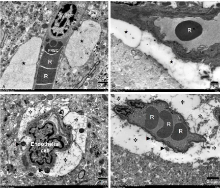
ac-tivity, represented by increasing glial fibrillary acidic protein (GFAP) expression in the perivascular area and increas -ing numbers of large astrocyte clusters were predominately found in the CM mice. The presence of multivesicular and lamellar bodies indicates the severity of cerebral damage in experimental CM. Congestion of the microvessels with occluded white blood cells (WBCs), parasitized red blood cells (PRBCs) and platelets is also a crucial covariate role for CM pathogenesis.
Keywords: Electron microscope, brain edema, rodent cerebral malaria, glial fibrillary acidic protein (GFAP)
Introduction
The most widely studied murine cerebral malar-ia (CM) model is Plasmodium berghei ANKA (PbA) in CBA or C57BL/6 mice [1]; it results in cerebral syndromes seven days post infection [2]. The pathogenic mechanisms underlining the occurrence of cerebral lesions are still incompletely understood, but may result from accompanying microvessel obstruction and
inflammation [3]. A fatal outcome generally
depends on sequestration of activated blood cells particularly monocytes and macrophages, parasitized erythrocytes, and platelets in the cerebral vessels [1] consequence of increased pinocytotic activity occurring in the endothelial cells, associated with degenerative changes in the basement membrane and perivascular astrocyte swelling and contributes to the
appearance of a perivascular edema [4]. The mouse model of CM, in which cerebral edema appears to play an important role, bears more resemblance to the CM observed in African chil-dren than that in South East Asian adults [5].
Maegraith and Fletcher demonstrated exces -sive movement of water and proteins into the brain of P. berghei-infected rodents [6]. Damage to the blood brain barrier (BBB) in P. berghei -infected mice was detected, which led to endo-thelial lesions, edema, and hemorrhage [4, 7]. The two main types of brain edema are cyto-genic and vasocyto-genic [8]. Vasocyto-genic edema
involves accumulation of excess fluid in the
extracellular space of the brain parenchyma because of a leaky BBB [9]. Cytogenic edema
consists of intracellular fluid accumulation that
Brain edema in experimental cerebral malaria
such as hypoxia [10]. Both cytogenic and vaso-genic edema are predominant features of experimental CM [4], however evidence of these in experimental CM has yet not been demonstrated by quantitative electron micro-scopic study.
Astrocytic swelling is also seen in vasogenic edema. One function of this swelling appears to be uptake of the extravasated plasma protein [11]. Perivascular astrocyte swelling contrib-utes to the appearance of a cytogenic edema seen in reactive astrogliosis, in which the cell
cytoplasm is packed with glial fibrillary acidic protein (GFAP) [12]. It is believed that the astro -cyte is the major cell type showing swelling after ischemia and trauma [13]. Astrocyte swelling may be an important early event pre-disposing the brain to further damage, because of the impairment of protective homeostatic mechanisms [14].
Considering the evidence of a causal
relation-ship between brain edema and GFAP expres -sion in experimental CM, this study set out to compare CM-susceptible mice; CBA/CaH mice (CM) and CM-non susceptible mice; BALB/cA mice (non-CM) that inoculated with P. berghei
ANKA (PbA). This study was designed to com-pare the evidence of astrogliosis and
perivas-cular GFAP expression between CM and
non-CM mice using immunohistochemical study.
Fine morphological structure in the perivascu -lar space and within vessels of CM and non-CM mice was also evaluated using an
ultrastruc-tural study. The findings of this study demon -strated the correlation between the evidence of
astrogliosis and GFAP expression on CM and
explained the pathogenic role of cytogenic and vasogenic brain edema together with other related ultrastructural changes in an experi-mental CM model.
Materials and methods
Animals and parasites
Animal studies were conducted in accord with guidelines under of the Australian Code of Practice for the Care and Use of Animals for
Scientific Purposes, and were approved by the
University of Sydney Animal Ethics Committee. 14 female CBA/CaH mice (CM) and 15 female BALB/c mice (non CM) (8-10 weeks old) were used in this study, 3 of each were used as con-trols and 11 CBA/CaH (CM) and 12 BALB/c
(non CM) that were infected with P. berghei
ANKA by intraperitoneal injection of 106 para-sitized erythrocytes, as described in Grau et al. [15]. The mice were housed in individual venti-lation cages and fed ad libitum in the laborato-ry animal facility of the Department of Pathology, University of Sydney. The mice were screened daily for neurological manifestations.
Specimen processing
Mice were euthanized with an over dose
inhala-tion of Isoflurane® on 7days post inoculation, when the mice showed terminal-stage of severe cerebral complications, including convulsions, paralysis, and coma [16]. A median cranial inci-sion of the brain was done. All specimens were
fixed in 10% neutral buffer formalin for 8-h, at 4°C [17]. Fixed specimens were dehydrated and infiltrated using standard tissue process
-ing. The tissues were embedded in paraffin and
sectioned at 5 µm. The continuous tissue
sec-tions were mounted on SuperFrost Plus slides (Menzel GmbH & Co KG, SF41296PL) for the
immunohistochemistry study.
Immunohistochemistry for GFAP
Heat-induced antigen retrieval with citrate buf-fer, pH 6 was used to unmask the antigen.
Endogenous peroxidase was quenched with 1%
v/v hydrogen peroxide in methanol after sec-tions were cooled. Secsec-tions were washed with
0.2% v/v Tween in Tris buffered saline (TBS) and blocked with 10% w/v skimmed milk for 20
min. Sections were incubated for 30 min at
room temperature with 1:40 rabbit anti-GFAP
(Biogenex, San Ramon, CA, USA) diluted in TBS
with 1% v/v normal goat serum (NGS, Vector,
USA, S1000). The sections were washed in TBS and incubated for 30 min with 1:200 biotinyl-ated goat anti-rabbit antibodies (Vector, USA,
BA1000) in 1% NGS/TBS at room temperature.
The slides were washed, incubated with avidin biotin peroxidase complex (ABC Vectastain, Vector, USA, PK4000) in TBS for 30 min at room temperature, and visualized with diamino-benzidine (DAB, DAKO, K3468). Slides were counterstained with hematoxylin before perma-nent mounting with Vectamount (Vector, USA).
Quantitative immunohistochemistry analysis
In each group, multiple random fields were examined for a total of 100 microvessels. From
pixel resolution (at 400X) were acquired with a light microscope (BX51, Olympus®) and digital camera (DP70, Olympus®). GFAP expression, microvessel labeling, was then analyzed by semi-quantitative digitalized image analysis
using analySIS FIVE, Olympus® as described by Kaczmarek et al. and Ampawong et al. [18, 19]. Color images were adjusted, turning the color of non-interesting area to white by replace color mode. Adjusted images were converted to grey-scale images. Then, the area of positive
reac-tion was estimated by the number of black pix-els. Thus, the area fraction of the positive reaction was determined as the percentage of black pixels in the binary image.
All of the obvious astrocytic cells were included for counting. The measurement was applied to all areas to determine the number of astrocytic
clusters that could be classified as small (astro -cytic number < 5 cells/cluster) and large
[image:3.612.91.522.68.576.2]clus-ter (astrocytic number ≥ 5 cells/clusclus-ter).
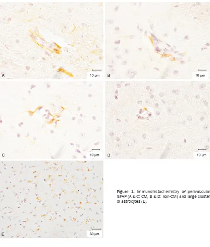
Figure 1. Immunohistochemistry of perivascular
Brain edema in experimental cerebral malaria
Electron microscopy
After euthanizing the mice with an overdose
inhalation of Isoflurane® on day 7 post inocula-tion, specimens were collected from four areas of the brain: cerebellum, cerebrum, brainstem, and the remaining area which composed of midbrain or colliculum hippocampus, and
dien-cephalons. Tissues were then fixed in 2% glu -taraldehyde in 0.1 M phosphate buffer, pH 7.4,
post-fixation with 1% osmium tetroxide in 0.1 M
phosphate buffer, pH 7.4, dehydrated in a
grad-ed ethanol series, infiltratgrad-ed with propylene
oxide, and embedded in Spurr’s epoxy resin (TABB Laboratories, Reading, UK). Thin sec-tions were cut with glass knives on an ultrami-crotome. Copper grids (200-mesh squares)
(Agar Scientific, Stansted, UK) were used to col -lect the thin sections, which were stained with
uranyl acetate and lead citrate before electron microscopic examination [20].
Quantitative ultrastructural analysis
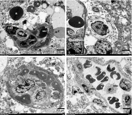
Qualitative examinations of pathologic features from these samples were performed as described in Pongponratn et al. [20]. In the peri-vascular space, the presence of periperi-vascular edema, astroglial dilatation, and multivesicular and lamellar bodies in the astrocytes were examined. Within the vessels, the nature and severity of the following changes were recorded by counting the vessels showing sequestered white blood cells (WBCs), parasitized red blood cells (PRBCs), and platelets. Endothelial cells were assessed for pseudopodia formation, cell swelling, vacuolation, changes in organelle structure, and intercellular bridges. These
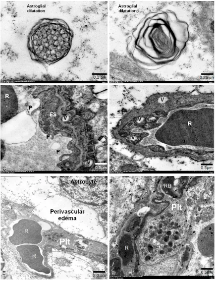
fea-Figure 2. Electron micrographs of perivascular edema and astroglial dilatation. Perivascular edema (★), A: CM and
[image:4.612.90.524.70.442.2]tures were examined in four different areas of the brain per group. Electron micrographs were taken of relevant areas.
Statistical analysis
Data analysis used IBM® SPSS® statistical
soft-ware version 20. To determine the level of GFAP
expression in each group, Kruskal-Wallis test was used. Pearson’s correlation test was used to determine any correlations among variables of interest. Chi-square test with Yates’
correc-tion and Fisher’s exact test were used to com -pare between four different brain areas of CM-susceptible mice (CM) and CM-non suscep-tible mice (non-CM).
Results
GFAP
The perivascular GFAP expression pattern in
CM mice (Figure 1A and 1C) was significantly
higher than in non-CM mice (Figure 1B and 1D) (13.50 ± 1.57 and 9.75 ± 1.00, respectively) and the number of small astrogliosis clusters were identical in both groups (5.0 ± 4.00 and 5.1 ± 3.00). However the number of large astro-gliosis clusters (Figure 1E) in the CM mice was
significantly higher than in the non-CM mice
(6.2 ± 2.00 and 4.2 ± 3.50). There was a
posi-tive correlation between perivascular GFAP
and 0.020, respectively. Perivascular edema in
all investigated areas was significantly more
pronounced in CM than non-CM mice (Table 1),
while astroglial dilatation was only significantly
higher in cerebrum and brain stem (Table 2). There was a positive correlation between the presence of perivascular space and astroglial
dilatation to CM (Pearson’s correlation coeffi -cient 0.344 and 0.108: p-value 0.000 and 0.017 respectively).
The fine morphological structure of secondary
lysosome which may indicate severity brain
damage could be identified by multivesicular
and lamellar bodies in their advanced stages (Figure 3A and 3B). The numbers of secondary lysosomes among the groups of CM and
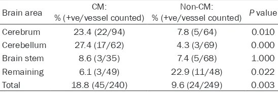
non-CM were significantly different. Multivesicular bodies in the non-CM mice (27.3%; 68/249) were significantly higher than in the CM mice (17.5%; 42/240), in contrast with, number of lamellar bodies, which was significantly greater in the CM mice (18.8%; 45/240) than the non-CM mice (9.6%; 24/249). The features of the
secondary lysosomes in each area of the brain are detailed in Tables 3 and 4. There is a posi-tive correlation between the presence of
lamel-lar bodies to CM (Pearson’s correlation coeffi -cient 0.131: p-value 0.004), while there was a negative correlation between the presence of multivesicular bodies and CM (Pearson’s
[image:5.612.91.368.96.188.2]corre-lation coefficient -0.117: p-value 0.009). Table 1. Presence of perivascular edema in four different brain
areas comparing CM and non-CM groups
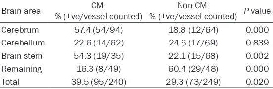
Brain area % (+ve/vessel counted)CM: % (+ve/vessel counted)Non-CM: P value
Cerebrum 42.6 (40/94) 31.3 (20/64) 0.049
Cerebellum 69.4 (43/62) 15.9 (11/69) 0.000
Brain stem 65.7 (23/35) 17.6 (12/68) 0.000
Remaining 49.0 (24/49) 18.8 (9/48) 0.000
Total 54.2 (130/240) 20.8 (52/249) 0.000
Table 2. Presence of astroglial dilatation in four different brain areas comparing CM and non-CM groups
Brain area % (+ve/vessel counted)CM: % (+ve/vessel counted)Non-CM: P value
Cerebrum 57.4 (54/94) 18.8 (12/64) 0.000
Cerebellum 22.6 (14/62) 24.6 (17/69) 0.839 Brain stem 54.3 (19/35) 22.1 (15/68) 0.002
Remaining 16.3 (8/49) 60.4 (29/48) 0.000
Total 39.5 (95/240) 29.3 (73/249) 0.020
expression and the presence of large astrogliosis clusters to CM with Pearson’s correlation
coef-ficient 0.239 and 0.312 at p -val-ues 0.001 and 0.004.
Fine morphology in the perivas-cular space
Brain edema can be identified by
perivascular space and astrogli-al dilatation (Figure 2) which were frequently observed in this study. The results showed that perivascular space and astrogli-al dilatation in the brains of CM
mice (54.2%; 130/240 vessels, 39.5%; 95/240 vessels, respec
-tively) were significantly higher
than in those from non-CM mice
[image:5.612.91.367.235.325.2]Brain edema in experimental cerebral malaria
Figure 3. Fine morphological structure of secondary lysosome. A: A multivesicular body is round to oval contain -ing more small vesicles and limited by a double membrane. B: A lamellar body is round to oval contain-ing lots of
thin electron dense lamellae and limited by a double membrane. Fine morphological structure of endothelial cell
changes: C & D: (P = pseudopodia formation, R = erythrocyte, V = vacuolation, ES = endothelial cell swelling). Fine morphological structure of platelets in brain microvessel: E & F: E: The vascular occluded by red blood cells and
Fine morphology within vessels
The cerebral endothelial cell changes in the PbA model included pseudopodia formation, endothelial cell swelling and vacuolization (Figure 3C and 3D). There was no significant difference between CM (23.8%; 57/240, 17.1%; 41/240, 14.2%; 34/240, respectively) and non-CM mice (28.5%; 71/249, 20.5%; 51/249, 20.9%; 34/249, respectively). The presence of
those endothelial cell changes in each brain area was also identical in both groups.
Despite the rare evidence of platelets in the examined brains, the presence of platelets in the brain microvessels (Figure 3E and 3F) in
the CM group (6.7%; 16/240) showed a signifi -cantly higher trend than in the non-CM mice
(1.6%; 4/249). The significantly higher numbers
of platelets were found in the cerebrums of the CM mice. There was a positive correlation between the presence of platelets and CM
(Pearson’s correlation coefficient 0.128: p -val-ue 0.005).
The presence of PRBCs within cerebral
microvessels quantified by electron microscopy showed that non-CM mice (32.1%; 80/249) exhibited significantly higher levels than CM mice (15.8%; 38/240), and this corresponded
with percentage of parasitemia on day 6 post infection, when non-CM and CM mice were
(especially WBCs, PRBCs, and platelets) adher-ence to the vascular endothelium, ischemia and hypoxia, loss of vascular cell integrity, hem-orrhage, neuronal damage, edema at the termi-nal stage, focal demyelination, astrocyte response (redistribution, astrogliosis, activa-tion), microglia and perivascular macrophage response. Electron and light microscopic stud-ies have shown that both infected groups are characterized by widespread cerebral endothe-lial activation, such as swelling, pseudopodia and vacuolization, together with lesions rang-ing from isolated damage to necrosis.
The role of cerebral edema in the pathogenesis of CM has been debated for decades, and sev-eral techniques has been used, including Evans
blue dye leakage, MRI (water diffusion coeffi -cient, imaging, perfusion, angiography,
spec-troscopy), CT scan, fine morphological studies
(perivascular vacuolization, astrocytic swell-ing), immunohistopathological studies (macro-phage associated with perivascular edema; CD68, tight junction associated proteins; occlu-din, clauocclu-din, JAM, ZO-1, ZO-2, ZO-3, reactive
gliosis and GFAP) [4, 12, 20, 25-31]. This study
determines of perivascular spaces and astro-glial dilatation and their correlation with the severity of brain edema. The results show that
brain edema was significantly greater in CM
[image:7.612.92.368.98.188.2]than in non-CM mice. The distribution of brain edema was observed throughout the brain, Table 3. Presence of multivesicular bodies in 4 different parts
comparing CM and non-CM groups
Brain area % (+ve/vessel counted)CM: % (+ve/vessel counted)Non-CM: P value
Cerebrum 25.5 (24/94) 17.2 (11/64) 0.246
Cerebellum 21.0 (13/62) 18.8 (13/69) 0.828
Brain stem 8.6 (3/35) 22.1 (15/68) 0.106
Remaining 4.1 (2/49) 60.4 (29/48) 0.000
Total 17.5 (42/240) 27.3 (68/249) 0.006
Table 4. Presence of lamellar bodies in 4 different parts compar-ing CM and non-CM groups
Brain area % (+ve/vessel counted)CM: % (+ve/vessel counted)Non-CM: P value
Cerebrum 23.4 (22/94) 7.8 (5/64) 0.010
Cerebellum 27.4 (17/62) 4.3 (3/69) 0.000
Brain stem 8.6 (3/35) 7.4 (5/68) 1.000
Remaining 6.1 (3/49) 22.9 (11/48) 0.022
Total 18.8 (45/240) 9.6 (24/249) 0.003
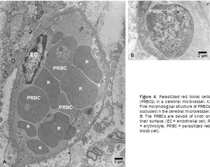
20.35% and 11.68%, respective -ly. PRBCs are devoid of knobs on their surfaces (Figure 4). The dis-tribution of PRBCs in all parts of the brain showed a higher trend among all mice of non-CM group. White blood cell accumulation in the cerebral microvessels (Figure 5) was quantified by electron
microscopy, it was found that, the presence of WBC per vessel was no identical in both CM and non-CM mice, without any inter-action to other cells.
Discussion
[image:7.612.91.365.233.325.2]Brain edema in experimental cerebral malaria
particularly the cerebrum, brain stem, and
dien-cephalon, in both infected groups. Fine mor -phological analysis of brain edema regarding the perivascular space and astroglial dilatation
confirmed the presence of vasogenic and cyto -genic edema. This study also found a positive correlation between the presence of perivascu-lar edema and astroglial dilatation to CM. These data supports the claim that both cytogenic and vasogenic edema are crucial features in experimental CM.
Cytotoxic edema involves an influx of extracel -lular water into the intracel-lular compartment leading to cell swelling and irreversible cell damage. It results from anoxic depolarization subsequent to the failure of Na+/K+ ATPases to
maintain membrane potential after ATP loss [32-34]. It is not considered responsible for brain swelling, because it does not lead to a change in total water content. It is
character-ized by reduced apparent diffusion coefficient
(ADC) value because of con-strained diffusive motion in the extracellular compartment and by
impaired energetic metabolism and reduced pH [35]. Vasogenic edema is characterized by expansion of the extracellular compartment after BBB breakdown and relocation of intra-vascular water into the extraintra-vascular
compart-ment. Inflammation is one of the possible
mechanisms at the origin of BBB disruption. The result of Penet et al. demonstrate that BBB lesions, brain swelling, and ventricular enlarge-ment play key roles in the developenlarge-ment of the
cerebral syndrome and confirm the existence of
a vasogenic edema [4].
An important role of astrocytes in the normal central nervous system (CNS) is to induce and maintain BBB properties in the vascular endo-thelium [36]. Astrocytes also make close con-tact with neuronal synapses and are thought to be intimately involved in maintaining acid-base, electrolyte and neurotransmitter balance [37, 38]. Reactive astrogliosis, which includes an increase in astrocyte size, is actually a hyper-trophy of the cells involving increased synthesis
of glial filaments and other intracellular constit
-Figure 4. Parasitized red blood cells (PRBCs) in a cerebral microvessel. A:
Fine morphological structure of PRBCs
[image:8.612.92.522.71.413.2]uents associated with increase in size and is usually seen as a delayed response (1-5 days) to injury [14]. This study demonstrates that CM
mice had higher GFAP expression in the peri -vascular area and more large astrocyte clus-ters than in non-CM mice. Moreover, the
corre-lation between perivascular GFAP expression
and the presence of large astrogliosis clusters to CM was positive. These may be an adaptive response of the astrocytes in pathogenesis of CM.
Astroglial swelling during experimental isch-emia and trauma in animals has been studied in considerable detail, and is recognized by a pale and watery cytoplasm under electron microscopic analysis [12]. These correspond to
the fine morphological features of astroglial
dilatation, multivesicular bodies, and lamellar bodies in the astrocyte, which demonstrated that the degenerative level of astrocytes was
significantly higher in CM than non-CM mice.
The accumulation of P. berghei-infected red blood cells in the brain is crucial for the devel-opment of CM in C57BL/6 mice, susceptible strain for CM [39]. PRBCs also accumulate in the brains of PbA-infected CBA/CaH mice, although this is less marked than in other murine models [2]. Therefore PRBCs them-selves are an important factor for the cascade of pathological processes in CM.
[image:9.612.90.523.71.451.2]In addition to PRBC and leukocyte accumula-tion in the brain microvasculature, platelets
Figure 5. Fine morphological structure of white blood cells (WBCs) accumulation in cerebral microvessel. A: The
Brain edema in experimental cerebral malaria
also seem to contribute to neurovascular lesions in murine CM. Wassmer et al. reviewed four lines of evidence supporting platelet involvement in murine CM [40]. (i) electron microscopic analysis when of CM disclosed platelets in the lumen of brain venules, between sequestered monocytes and PRBCs (ii) plate-lets sequestered in the brain during CM but not in non-CM mice (iii) in vivo treatment with a monoclonal antibody to leukocyte function-associated antigen-1 (which is expressed on platelets) selectively abrogates the cerebral sequestration of platelets, and this correlates with prevention of CM, (iv) malaria-infected
ani-mals rendered thrombocytopenic are signifi -cantly protected against CM. In this study,
although fine morphological analysis showed
that the accumulation of platelets in the brain
microvessels is significantly more important in
CM than in non-CM mice, interestingly the occurrence of platelets in the brain
microves-sels was quite rare, at 16/240 and 4/249 (+ve/
vessels) in CM and non-CM mice, respectively. A positive correlation between the presence of platelets and CM was found. This supports the idea that platelets play an important role in experimental CM, by acting as effectors of neu-rovascular lesions as previously described [40, 41].
In conclusion, transmission electron microsco-py and immunohistochemistry analysis of the rodent malaria model revealed a pathological alteration particularly brain edema. Brain ede-
ma and GFAP expression correlate closely with
rodent cerebral malaria model. Perivascular space and astroglial dilatation are evidence
confirming of vasogenic edema and cytogenic
edema respectively. The presences of lamellar bodies in the astrocyte demonstrate the degen-erative level of astrocytes. All morphological studies further support the prominent role for CM pathogenesis giving a better understanding of the relationship between brain edema, astro-cytic activity, and CM.
Acknowledgements
This study was supported by Research Grant
from the Faculty of Tropical Medicine, Mahidol University, Fiscal Year 2014.
Disclosure of conflict of interest
None.
Address correspondence to: Dr. Emsri Pongponratn,
Department of Tropical Pathology, Faculty of Tropical
Medicine, Mahidol University, 420/6, Ratchawithi Road, Ratchathewi, Bangkok, 10400, Thailand. Tel:
(662) 3069184; Fax: (662) 3069184; E-mail: emsri. pon@mahidol.ac.th
References
[1] Hunt NH, Grau GE. Cytokine: accelerators and brakes in the pathogenesis of cerebral malar-ia. Trends Immunol 2003; 24: 491-499. [2] Lou J, Lucas R and Grau GE. Pathogenesis of
cerebral malaria: recent experimental data and possible applications for humans. Clin Mi-crobiol Rev 2001; 14: 810-820.
[3] Medana IM, Hien TT, Day NP, Phu NH, Mai NT, Chu’ong LV, Chau TT, Taylor A, Salahifar H,
Stocker R, Smythe G, Turner GD, Farrar J, White NJ and Hunt NH. The clinical significance of cerebrospinal fluid levels of kynurenine
pathway metabolites and lactate in severe ma-laria. J Infect Dis 2002; 185: 650-656. [4] Penet MF, Viola A, Confort-Gouny S, Le Fur Y,
Duhamel G, Kober F, Ibarrola D, Izquierdo M,
Coltel N, Gharib B, Grau GE, Cozzone PJ. Neu-robiology of disease. Imaging experimental
ce-rebral malaria in vivo: significant role of isch -emic brain edema. J Neurosci 2005; 10: 7352-7358.
[5] Sanni LA. Hypothesis: The role of cerebral ede-ma in the pathogenesis of cerebral ede-malaria. Redox Report 2001; 6: 137-142.
[6] Maegraith B and Fletcher A. The pathogenesis
of mammalian malaria. Adv Parasitol 1972; 10: 49-75.
[7] Thumwood CM, Hunt NH, Clark IA and Cowden WB. Breakdown of the blood-brain barrier in murine cerebral malaria. Parasitology 1988; 96: 579-589.
[8] Klatzo I. Neuropathological aspects of brain edema. J Neuropathol Exp Neurol 1967; 26: 1-14.
[9] Pfister HW, Feiden W and Einhaupl KM. Spect -rum of complications during bacterial meningi-tis in adults. Results of a prospective clinical study. Arch Neurol 1993; 50: 575-581. [10] Papadopoulos MC, Krishna S, Verkman AS.
Re-search seminar: Aquaporin water channels and brain edema. Mount Sinai J Med 2002; 69: 242-248.
[11] Trachtenberg MC. Brain cell responses to ex-tracellular protein. In: Grossman RG, Gilden-berg PL, editors. Head Injury: Basic and clinical aspects. New York: Raven press; 1982. pp: 169-178.
trau-matic brain injury: a light and electron micro-scopic study in rats. J Neurotrauma 1994; 11: 289-301.
[13] Kimelberg HK. Current concepts of brain ede-ma. Review of laboratory investigations. J Neu-rosurg 1995; 83: 1051-1059.
[14] Kimelberg HKN. Astrocytic responses to cen-tral nervous system trauma. In: Salzman SK,
Faden AI, editors. The Neurobiology of Central
Nervous System Trauma. New York: Oxford Uni-versity Press; 1994. pp: 193-208.
[15] Grau GE, Piguet PF, Engers HD, Louis JA, Vas
-salli P and Lambert PH. L3T4+ T lymphocytes
play a major role in the pathogenesis of murine cerebral malaria. J Immunol 1986; 137: 2348-2354.
[16] Medana IM, Chaudhri G, Chan-Ling T and Hunt NH. Central nervous system in cerebral malar-ia: ‘Innocent bystander’ or active participant in the induction of immunopathology? Immunol Cell Biol 2001; 79: 101-120.
[17] Presnell JK, Schreibman MP. Fixative. Huma -son‘s Animal Tissue Techniques. Baltimore: Johns Hopkins University Press, 1997.
[18] Ampawong S, Combes V, Hunt NH, Radford J, Chan-Ling T, Pongponratn E and Grau GE. Quantitation of brain edema and localisation of aquaporin 4 expression in relation to sus-ceptibility to experimental cerebral malaria. Int J Clin Exp Pathol 2011; 4: 566-574.
[19] Kaczmarek E, Gorna A, Majewski P. The
tech-niques of image analysis for quantification im -munohistochemistry. Anal Acad Med Bial 2004; 49: 155-158.
[20] Pongponratn E, Turner GD, Day NP, Phu NH, Simpson JA, Stepniewska K, Mai NT,
Viriyave-jakul P, Looareesuwan S, Hien TT, Ferguson DJ
and White NJ. An ultrastructural study of the brain in fatal Plasmodium falciparum malaria. Am J Trop Med Hyg 2003; 69: 345-359. [21] Grau GE, Fajardo LF, Piguet PF, Allet B, Lam
-bert PH, Vassalli P. Tumor necrosis factor (ca-chectin) as an essential mediator in murine cerebral malaria. Science 1987; 237: 1210-1212.
[22] Hearn J, Rayment N, Landon DN, Katz DR and de Souza JB. Immunopathology of cerebral malaria: morphological evidence of parasite sequestration in murine brain microvascula-ture. Infect Immun 2000; 68: 5364-5376. [23] Hunt NH, Golenser J, Chan-Ling T, Parekh S,
Rae C, Potter S, Medana IM, Miu J and Ball HJ. Immunopathogenesis of cerebral malaria. Int J Parasitol 2006; 36: 569-582.
[24] Lackner P, Beer R, Helbok R, Broessner G, Engelhardt K, Brenneis C, Schmutzhard E and Pfaller K. Scanning electron microscopy of the neuropathology of murine cerebral malaria. Malar J 2006; 5: 116.
[25] Ebisu T, Naruse S, Horikawa Y. Discrimination between different types of white matter edema with diffusion-weighted MR imaging. J Magn Res Imag 1993; 3: 863-868.
[26] Jennings VM, Actor JK, Lal AA, Hunter RL.
Cyto-kine profile suggesting that murine cerebral
malaria is an encephalitis. Infect Immun 1997; 65: 4883-4887.
[27] Looareesuwan S, Wilairatana P, Krishna S, Kendall B, Vannaphan S, Viravan C and White NJ. Magnetic resonance imaging of the brain in patients with cerebral malaria. Clin Infect Dis 1995; 21: 300-309.
[28] Maneerat Y, Viriyavejakul P, Punpoowong B, Jones M, Wilairatana P, Pongponratn E, Turner GD, Udomsangpetch R. Inducible nitric oxide synthase expression is increased in the brain in fatal cerebral malaria. Histopathology 2000; 37: 269-277.
[29] Medana IM and Turner GD. Human cerebral malaria and the blood-brain barrier. Int J Para-sitol 2006; 36: 555-568.
[30] Neill AL and Hunt NH. Pathology of fatal and resolving Plasmodium berghei cerebral malar-ia in mice. Parasitology 1992; 105: 165-175. [31] Patankar TF, Karnad DR, Shetty PG, Desai AP
and Prasad SR. Adult cerebral malaria:
prog-nostic importance of imaging findings and cor
-relation with postmortem findings. Radiology
2002; 224: 811-816.
[32] Ayata C, Ropper AH. Ischaemic brain edema. J Clin Neurosci 2002; 9: 113-124.
[33] Harris NG, Zilkha E, Houseman J, Symms MR, Obrenovitch TP, Williams SR. The relationship
between the apparent diffusion coefficient
measured by magnetic resonance imaging,
an-oxic depolarization, and glutamate efflux dur -ing experimental cerebral ischemia. J Cereb
Blood Flow Metab 2000; 20: 28-36.
[34] Latour LL, Svoboda K, Mitra PP and Sotak CH. Time-dependent diffusion of water in a biologi-cal model system. Proc Natl Acad Sci U S A 1994; 91: 1229-1233.
[35] van der Toorn A, Sykova E, Dijkhuizen RM, Vorisek I, Vargova L, Skobisova E, van Look-eren Campagne M, Reese T and Nicolay K. Dy-namic changes in water ADC, energy metabo-lism, extracellular space volume, and tortuosity in neonatal rat brain during global ischemia. Magn Reson Med 1996; 36: 52-60.
[36] Brightman MW. The anatomic basis of the blood-brain barrier. In: Neuwelt EA, editors. Im-plications of the Blood-Brain Barrier and its Manipulation. New York: Plenum Co.; 1989. pp: 53-78.
Brain edema in experimental cerebral malaria
[38] Walz W. Mechanism of rapid K(+)-induced
swelling of mouse astrocytes. Neurosci Lett 1992; 135: 243-246.
[39] Baptista FG, Pamplona A, Pena AC, Mota MM,
Pied S and Vigario AM. Accumulation of Plas-modium berghei-infected red blood cells in the brain is crucial for the development of cerebral malaria in mice. Infect Immun 2010; 78: 4033-4039.
[40] Wassmer SC, Combes V and Grau GE. Patho-physiology of cerebral malaria: role of host cells in the modulation of cytoadhesion. Ann N Y Acad Sci 2003; 992: 30-38.