INTRODUCTION
The teleost escape behavior is a unique vertebrate model in that the individual neurons that trigger the behavior, as well as their afferent inputs and outputs, have been well characterized both morphologically and electrophysiologically (Faber and Korn, 1978). Furthermore the activation of the network leads to a quantifiable behavior, commonly known as the startle response or as the C-start, because of the shape of the body following contraction of the trunk muscles on one side of the body (Nissanov et al., 1990). These properties enable functional correlation of the network with the behavior it mediates, thereby allowing a myriad of questions related to sensory motor integration and synaptic physiology to be addressed (Korn and Faber, 2005).
In particular, a pair of bilateral reticulospinal neurons, the Mauthner (M-) cells, initiates and controls these C-start escapes from aversive sound, mechanosensory, tactile or visual stimuli (Chang et al., 1987; Eaton et al., 1991; Foreman and Eaton, 1993; Korn and Faber, 1975; Lin et al., 1983; Liu and Fetcho, 1999; Preuss et al., 2006; Weiss et al., 2006; Zottoli and Faber, 1979; Zottoli et al., 1987; Zottoli and Van Horne, 1983). The precision and speed of this behavior requires the M-cells to rapidly integrate sensory information from the numerous sensory afferents that terminate along its two primary dendrites. Furthermore, the aversive nature of auditory evoked escape responses implies that the M-cells are capable of locating the sound source (Blaxter et al., 1981; Foreman and Eaton, 1993; Preuss and Faber, 2003). It has been suggested that integration of different components of the octavo-lateralis sensory inputs along the M-cell lateral dendrite or at the level of presynaptic inhibitory networks may be necessary for the encoding
and execution of directional escape behaviors (Eaton et al., 1995; Eaton et al., 1997; Eaton and Popper, 1995; Weiss et al., 2008). However, the exact mechanism that determines this response has not yet been determined.
Recent behavioral studies have implicated the lateral line (LL) system in the initiation of the acoustic startle response, specifically in its initial directionality, reflecting which of the bilateral M-cells is activated (Mirjany et al., 2011). The neuromast receptors of the LL are specialized to detect displacements in the local hydrodynamic field (Bleckmann, 2008) and hence can locate nearby sound fields. These neuromasts are innervated by two main nerve afferents, the posterior LL nerve (pLLn), which innervates the neuromasts on the trunk of the fish, and the anterior LL nerve (aLLn), which innervates the neuromasts on the head. Transection of the pLLn has been shown to have little effect on the escape response, in particular on its directionality (Mirjany et al., 2011). This is presumably because stimulus inputs from the pLLn, would have too long a latency to influence the escape decision, given that the M-cell computational time window is restricted to 2–3ms (Korn and Faber, 1975; Preuss and Faber, 2003; Weiss et al., 2006; Zottoli, 1977) for abrupt sound stimuli. In contrast, as the aLLn innervates the neuromasts that cover the head of the fish, an input from this nerve would be expected to have a shorter conduction time, and hence could be capable of influencing the escape decision. However, to date there is no evidence that the aLLn afferents make direct contact with the M-cell. Considering that all sensory afferents studied thus far project to one of the two primary dendrites of the M-cell, it is probable that the cell also receives projections from the aLLn.
The Journal of Experimental Biology 214, 3368-3377 © 2011. Published by The Company of Biologists Ltd doi:10.1242/jeb.056226
RESEARCH ARTICLE
Characteristics of the anterior lateral line nerve input to the Mauthner cell
Mana Mirjany and Donald S. Faber*
Albert Einstein College of Medicine, Dominick P. Purport Department of Neuroscience, 1410 Pelham Parkway S., Room 429, Bronx, NY 10461, USA
*Author for correspondence (donald.faber@einstein.yu.edu)
Accepted 5 July 2011
SUMMARY
The goldfish Mauthner (M-) cells, a bilateral pair of reticulospinal neurons, initiate the auditory evoked escape behavior of teleosts. In an open field the fish reliably turns away from the sound source. This implies that the M-cells are capable of a decision-making process that requires the two cells to receive differential directional inputs. Recent studies have indicated that the lateral line (LL) system is necessary in the initial directionality of the escape. This information is thought to be transmitted to the M-cell by the anterior branch of the lateral line nerve (aLLn), which has a shorter conduction time then the posterior branch. This study is the first attempt to characterize the inputs from the aLLn to the M-cell. M-cell intracellular responses to aLLn stimulation indicate a fast monosynaptic input (0.80±0.03ms) that has a small amplitude averaging 5.85±0.42mV. This input is bilateral and has a significantly longer latency and smaller amplitude in the contralateral M-cell. Superimposed on the evoked excitatory postsynaptic potential (EPSP) is a shunting inhibition with a delay of 1ms, which is characteristic of other sensory inputs to the M-cell. Pharmacological manipulation and 50Hz stimulation reveal a component of the evoked EPSP that is electrotonic, a property favoring speed of transmission. In addition, this input is localized to the lateral dendrite proximal to the inputs from the inner ear. The short latency of these inputs and their proximity to the posterior eighth nerve afferents indicate a crucial role for the aLLn in influencing the excitability and directionality of the M-cell.
This study examines whether the M-cell receives inputs from the aLLn afferents, and whether the properties of the responses to these inputs are consistent with a role in the initiation of the C-start. We found that electrical stimulation of this nerve produces a mixed chemical and electronic postsynaptic potential (PSP) in the proximal soma-dendritic region of the M-cell. The short latency of these monosynaptic inputs and their proximity to the posterior eighth nerve afferents may be crucial in influencing the excitability of the M-cell and its directionality. To this end the interaction of postsynaptic potentials evoked by these inputs with those produced by sound pulses and mediated by posterior eighth nerve afferents was examined.
MATERIALS AND METHODS Animals and preparations
Goldfish [Carassius auratis (Linnaeus 1758)], body length 10–15cm) were obtained from EECHO Systems (North Kansas City, MO, USA) and Hunting Creek Fisheries (Thurmond, MD, USA) and housed in our holding tanks for 2weeks to adjust to the laboratory conditions and to ameliorate any physiological stresses incurred during transportation. They were maintained in a controlled photoperiod of 12h:12h light:dark at 18°C in recirculating, aerated water conditioned with NovAqua (0.13mll–1; Novalek, Hayward, CA, USA), Coppersafe (0.4mll–1; St John Laboratories, Harbor City, CA, USA), Instant Ocean (70mgl–1; Aquarium Systems, Mentor, OH, USA), Aquarium Salt (190mgl–1; Jungle Laboratories, Cibolo, TX, USA) and Proper pH (350mgl–1; Aquarium Pharmaceuticals, Chalfont, PA, USA) (Preuss and Faber, 2003). All experiments were conducted in concordance with policies established by the Albert Einstein College of Medicine IACUC.
Prior to electrophysiological recordings, the fish were initially anesthetized with 100mgl–13-aminobenzoic acid ethyl ester (MS-222; Sigma, St Louis, MO, USA) and then further anesthetized in ice water. Fish were then connected to a mouthpiece in the recording chamber and respirated continuously with aerated conditioned water containing 20mgl–1 MS-222. It has been reported that at this concentration of MS-222, there are minimal effects on the spontaneous firing of aLLn afferents of fish (Palmer and Mensinger, 2004). The respirated water was maintained between 10 and 15°C using a Delta Star Chiller (Aqua Logic, San Diego, CA, USA). The posterior spinal cord was exposed and the M-cells were activated antidromically through bipolar stimulation of the spinal cord to determine the minimal stimulus intensity that produced the stereotypic movement of the operculum and mouth associated with M-cell activation. The fish were
then immobilized with intramuscular injection of d-tubocurarine (1mgg–1body mass; Sigma). The midbrain was exposed and the cerebellum lifted for intracellular recording of the M-cell as previously described (Faber and Korn, 1978). M-cells were identified on the basis of their characteristic location, 400m lateral to the midline and 1500m below the surface of the medulla, and the signature large extracellular antidromic field potentials generated at the spike initiation zone in the axon cap of the cell.
M-cell intracellular responses to sound pips as well as to anterior lateral line nerve (orthodromic) and spinal cord (antidromic) stimulation were recorded from the soma (50m lateral to the axon cap) and distally along the lateral dendrite of the cell up to 400m from the axon cap, using 4–7M microelectrodes filled with 5moll–1potassium acetate. The membrane potential as well as the amplitude of the antidromic spike were monitored throughout the experiments as a means of verifying recording conditions and the recording position in the cell. Owing to the passive cable properties of the M-cell lateral dendrite, antidromically activated spike amplitudes decrease in the proximal–distal direction along the dendrite, making it possible to estimate the recording position with a high degree of accuracy. For all experiments reported, the resting membrane potential did not vary by more than 5mV in a given recording session and it was between –75 and –83mV in both the somatic and dendritic recordings.
Pharmacology
The brain was continually superfused with normal fish saline (inmmoll–1: 124.0 NaCl, 5.1 KCl, 2.8 NaH
2PO4·H2O, 0.9 MgSO4, 1.6 CaCl2·2H2O, 5.6 glucose and 20.0 HEPES, pH7.2), which was maintained at the same temperature as the aerated perfusion water. To determine the contribution of the N-methyl-D-aspartate (NMDA)
and a-amino-3-hydroxy-5-methyl-4-isoxazolepropionic acid (AMPA) receptors to the response of the Mauthner cell to aLLn stimulation, the glutamatergic receptors were blocked by the addition of antagonists to the saline solution. Specifically NMDA receptors were blocked with 200moll–1DL-2-amino-5-phosphonovaleric acid (APV; Sigma and Tocris, Ellisville, MO, USA) and AMPA receptors were blocked with 200moll–1 cyano-7-nitroquinoxaline-2,3-dione disodium (CNQX; Sigma, Tocris).
aLL and auditory stimuli
In goldfish there are two branches of the aLLn (dorsal and ventral) that innervate both superficial and canal neuromasts on the head
aLL nerve
VL CB
VL Vi
V cranial nerve
Stim. electrode
[image:2.612.46.351.556.744.2]A
B
(Puzdrowski, 1989). The nerve was stimulated at the point where both branches meet just before entering the medulla. This required a further exposure of the cranium anteriorly; then, slightly lifting the optic tectum revealed the aLLn branches as they merge (Fig.1). In some experiments it was necessary to pull back the anterior eighth nerve and the utricular bundle to gain better access to the aLLn. There was no apparent difference in the recordings obtained using either dissection. The nerve was stimulated using a bipolar electrode. Sound pip stimuli were generated in air using a pair of computer speakers positioned above and to the right of the fish’s head. The sinusoidal waves of 200Hz were produced by a digital waveform generator (Model 39, Wavetech Ltd, Norwich, Norfolk, UK) and amplified with an audio power amplifier (Servo 120; Samson, Syosset, NY, USA). Because the recordings were made with the fish out of the water, these sound pips generated only pressure waves that were transmitted viavibration of the Weberian ossicles of the swimbladder to the inner ear. Therefore, irrespective of their location both M-cells received the same input.
Data analysis
All recordings were done in current clamp using an Axoclamp-2B (Axon Instruments, Foster City, CA, USA). The data were recorded with an Apple Mac Pro using Trace Analyzer 3 acquisition software developed in-house (TA3) at a sampling interval of 20–40s. Data analysis was done with both IGOR Pro (WaveMetrics, Inc., Lake Oswego, OR, USA) and TA3. Statistical significance was calculated using Prism 5 software (GraphPad Software, Inc., La Jolla, CA, USA). All data are reported as means ± s.e.m. P-values were obtained using a one-way ANOVA unless otherwise stated. Nis the number of experimental animals.
RESULTS
Postsynaptic potentials evoked in M-cell by aLLn stimulation The left aLL nerve was always the one stimulated and responses were recorded from either the ipsilateral or contralateral M-cell (Fig.2A). Intracellular recordings from the M-cells revealed that electrical stimulation of the aLLn triggers a complex depolarizing potential in the ipsilateral cell and a smaller response in the contralateral one. Fig.2B shows a typical example where the response to stimulation of the left aLLn was initially recorded in the ipsilateral M-cell 75m from the axon cap in the soma (Fig.2B) and then from the contralateral M-cell (Fig.2C). The amplitude of the antidromic action potential was at least 30mV in both cells, confirming that the recordings were both made in the soma (the difference in spike heights is within experimental variability). The onset latency of the ipsilateral postsynaptic potentials (0.80±0.03ms; N21) is indicative of a monosynaptic input. We observed a significantly longer delay in the cell contralateral to the stimulated nerve (1.52±0.16ms; N4; P<0.0001, unpaired t-test, t7.18, d.f.24; Fig.2D). The amplitude of these PSPs was relatively small, averaging 5.85±0.42mV ipsilaterally, and it was significantly smaller, 2.12±0.28mV, contralaterally (P0.0011, unpaired t-test, t3.73, d.f.24; Fig.2E).
Increasing the stimulus strength had little effect on the response latency but increased the amplitude of the PSP to a maximum of 10mV. This increase in amplitude is presumably due to the recruitment of additional afferent fibers with higher firing thresholds, however, even the maximal PSPs failed to depolarize the M-cell to threshold. Additional peaks with incremental delays in multiples of ~0.5ms after the initial peak may be an indication of the presence of different fiber groups with different conduction
2 ms 2 ms
10 mV
AD
spike PSP
Vipsi Vcontra
A
B
C
0 2 4 6
0 0.5 1 1.5 2
P
S
P l
a
tency (m
s
)
***
**
Ipsi Contra
D
E
Ipsi Contra
P
S
P
a
mplit
u
de (mV)
Intracellular recordingsat the soma
aLL nerve
stimulation
2 mV
PSP
Fig.2. Monosynaptic inputs from the anterior lateral line. (A)Monosynaptic postsynaptic potentials (PSPs) were recorded in the ipsilateral (Vipsi) and
velocities or polysynaptic inputs from the nerve. Alternatively, the relatively constant delay of 0.5ms between these additional peaks may be an indication of an induced synchronization of tonically active afferent fibers. The EPSP rise time, measured from 10 to 90% of the initial peak of the response was 0.44±0.05ms in the ipsilateral and 0.47±0.07 in the contralateral M-cell, indicative of fast synaptic transmission, similar to that mediated through mixed electrical and chemical synapses established on the lateral dendrite by club endings of the posterior eighth nerve (Lin and Faber, 1988).
It is possible that some of the synaptic potentials recorded result from current spread to neighboring cranial nerves. The anterior lateral line nerve enters the brain ventrally and just posteriorly to the optic tectum. This location is also the entry point for cranial nerves V (trigeminal nerve) and VII (facial and motor nerve) that is underneath the anterior lateral line nerve (Puzdrowski, 1989; McCormick and Hernandez, 1996). However, even at lower stimulus intensities the initial electrical peak of the response was present. Furthermore, in a few experiments in which the electrode was positioned on cranial nerve V the evoked PSPs had different waveforms, namely a longer latency response with a broader peak. These peaks were eliminated after application of APV and CNQX to block NMPA and AMDA receptors, respectively. Therefore although it remains a possibility that the later component of the PSP may be mixed with potentials from the neighboring nerves, the initial electrical peak is probably generated by stimulation of the aLLn. Furthermore, the lateral dendrite seems to specifically receive stato-acoustic inputs. Therefore, it is more likely for non-stato-acoustic signals to be located on the ventral dendrite, where visual and somatosensory inputs are received.
aLLn excitatory input is mixed electrical and chemical The short latency of the aLLn ipsilateral input suggest that these afferent synapses could be electrical, chemical or both, i.e. mixed. We, therefore, first investigated whether the aLL-triggered EPSPs were sensitive to blockers of glutamatergic transmission, specifically AMPA and NMDA antagonists, which are effective in the M-cell system (Wolszon et al., 1997). In the experiment shown in Fig.3A, application of 200m of the NMDA receptor antagonist APV resulted in a slight reduction in the amplitude and number of peaks of the later components of the EPSP without affecting the initial peak of the response. This initial component also seems to be unaffected by superfusion with a cocktail of 100m APV and 100m CNQX, to block both NMDA and AMPA receptors. However, the later components were significantly reduced, indicating a contribution of AMPA receptors to the control response. The same results were obtained in a similar experiment, shown in Fig.3B, in which we subtracted the EPSP after drug application from the control. The differential signal is the glutamatergic EPSP. The signal that remains after drug application is the initial peak that is presumably electrotonic. The effects of drug treatment on six such experiments were quantified. The mean amplitude of the initial peak (P1; 3.53±0.38mV) was not significantly affected after superfusion of APV alone (4.05±0.33) or in conjunction with CNQX (3.17±0.43). The second peak of the response (P2; 3.90±0.46mV) was also not significantly reduced after APV (3.16±0.47). However, in combination with CNQX this second chemical peak was significantly reduced to 1.40±0.24mV (P0.0005, unpaired t-test). The mean ratios of P1 and P2 after APV alone and APV and CNQX are given in Fig.3C.
5 ms 2 ms
2 ms 0.25 Hz 50 Hz 0.25 Hz
1.2
0.8
0.4
0
Fr
a
ction (dr
u
g/control)
P1 (e) P2 (c)
A
B
C
D
2 mV
2 mV
1 mV
CTL
CTL
APV
APV APV + CNQX
APV + CNQX
APV + CNQX
e c
Fig.3. High frequency stimulation and pharmacological manipulations reveal two components in the aLLn EPSP.
These pharmacological results were confirmed when the aLLn was tetanized with high frequency stimulation at 50Hz. Fig.3D shows superimposed traces from an experiment where the initial electrotonic peak remained after stimulation at 50Hz, but the second
peak was reduced significantly. The second peak recovered once stimulation was resumed at 0.25Hz, and it was presumably mediated chemically. We also applied 50Hz stimulation in four of the six pharmacology experiments described above and found that the initial peak that remained after APV and CNQX was not affected at 50Hz, confirming that this initial phase is electrotonic.
Spatial distributions of the aLLn EPSPs
In order to determine the spatial distribution of the aLLn inputs, M-cell responses evoked by aLLn stimulation were recorded at successive points along the soma and lateral dendrite (Fig.4A) starting at 100m lateral to the axon cap. The M-cell soma and dendrite are passive and, accordingly, EPSP amplitudes are maximal at the locus of presynaptic input and decline in amplitude with increasing distance from that source. The amplitude of the antidromically evoked action potential that is generated in the soma and conducts passively along the dendrites, has been shown to decline steadily towards the distal tip of the lateral dendrite and therefore serves as a good indication of the recording site (Faber and Korn, 1978; Preuss and Faber, 2003; Szabo et al., 2006). EPSP amplitudes evoked by sound pips were also recorded as a confirmation of our location on the dendrite, because these inputs have been previously shown to arise from the large myelinated club endings that terminate at the distal portion of the lateral dendrite, i.e. 200–400m distal to the axon cap (Szabo et al., 2006).
Fig.4B shows results from a single experiment where recordings were made at three successive sites, 100, 200 and 300m, from the axon cap. Antidromic action potentials are biggest at the most proximal location (1) and decrease in amplitude in more distal recordings (2 and 3). The same amplitude profile is seen for the aLLn EPSP, that is, it is also largest most proximally. In contrast, pip EPSPs are maximal at the most distal recording site (3) and their amplitudes diminish towards the soma. The averages from 12 similar experiments are plotted in Fig.4C. We were able to record successfully up to 200m from all 12 cells, 300m in 10 cells and 400m in three cells. The peak amplitudes of the antidromic action potential, aLLn and pip responses were normalized with respect to the response recorded in the soma. The antidromic spike amplitude decayed steadily to 33% of its original value as the recording site shifted distally along the dendrite. The mean aLLn EPSP was largest in the region encompassing 100–200m distal, and decreased laterally to ~65% of its peak amplitude at 300m and 50% at 400m lateral to the soma, suggesting these afferents terminate on the soma and proximal region of the lateral dendrite. As expected from previous reports the pip EPSP amplitudes were maximal at 400m lateral to the axon cap (Szabo et al., 2006). We did not map out the EPSPs evoked in the contralateral M-cell after stimulation of the left aLLn as closely, but in two experiments we observed similar spatial profiles. In other words the amplitude of these EPSPs, although much smaller, were also maximal near the soma and decreased laterally towards the proximal tip of the dendrite.
Inhibitory potentials evoked by aLLn stimulation We examined whether the aLLn afferents also project to a population of commissural inhibitory glycinergic interneurons [passive hyperpolarizing potential (PHP) neurons], which bilaterally inhibit the M-cells both chemically and electrotonically. The collective firing of these interneurons following afferent input produces an extrinsic hyperpolarizing potential (EHP) that can be measured as a positive extracellular field potential in the axon cap (Fig.5A) (Furshpan and Furukawa, 1962; Furukawa and Furshpan, 1963; Zottoli and Faber, 1980). These field potentials are also apparent
2 ms 2 ms 2 ms
2 mV
AD
aLL PSP
Pip PSP 1
2
3
4 mV
10 mV
0 50 100 150 200
100 200 300 400
Norm
a
lized
a
mplit
u
de
100 150 200
50
0
Pip
AD
Distance from axon cap (µM) aLL
B
C
pVIIIn
1
3 2
A
Recording sites AD
aLLn
pVIIIn
after antidromic activation of the M-cell, activated in this case by PHP neurons that are part of the collateral inhibitory pathway (Furshpan and Furukawa, 1962; Furukawa and Furshpan, 1963; Zottoli and Faber, 1980). Fig.5B shows average traces (N4) from an experiment where the EHPs were initially recorded in the ipsilateral M-cell axon cap (left panel) and then in the contralateral axon cap (right panel) of the same fish.
The traces in Fig.5B illustrate the antidromic action potential, a negative field of approximately 10mV in the ipsilateral M-cell and 8mV in the contralateral cell. The comparable size of these fields is an indication that the potentials were recorded in the same region of the axon caps of the two cells. The small EHPs following the negative antidromic spikes may be due to fatigue or an indication that the M-cell input to some of these interneurons was blocked by excessive administration of the cholinergic blocker, which may have leaked into the brain.
Regardless, EHPs were observed following sound pips (Fig.5C) and aLLn stimulation (Fig.5D) in the same experiment. The latencies of ipsi- and contralateral EHPs evoked by aLLn stimulation were not significantly different, with means of 1.13±0.11ms measured at the axon cap of the ipsilateral cells and 1.05±0.10ms at the contralateral cell (Fig.5E). These latency values are consistent with an extra synaptic delay, as compared with the monosynaptic EPSPs, as would
be expected with the interposition of an interneuron in the feedforward inhibitory pathway. These EHPs had comparable mean amplitudes at the axon caps of both cells, averaging 2.93±0.2mV in the ipsilateral side and 3.13±0.77mV in the contralateral side (Fig.5F). Thus, although excitation of the M-cells by the aLLn is asymmetric, the electrical inhibition is, on average, bilaterally symmetrical.
The inhibitory postsynaptic conductance change that is evoked by the firing of these PHP glycinergic interneurons is not readily detected as a frank potential change because the resting membrane potential of the M-cell is equal to the inhibitory postsynaptic potential (IPSP) equilibrium potential (Furukawa and Furshpan, 1963). Therefore, an indirect method of calculating the inhibitory conductance change involves measuring the shunt of an antidromic action potential superimposed during the evoked PSP (Fig.6A). In this example the onset and amplitudes of the inhibitory conductance changes are the same in both ipsilateral and contralateral M-cells (Fig.6B). The onset corresponds to the peak of the ipsilateral PSP but it precedes the peak of the contralateral EPSP (Fig.6C). On average, the inhibitory postsynaptic conductance change (Gipsp) was 0.27±0.03Gm for the ipsilateral cell (N8) and 0.18±0.02Gmfor the contralateral cell (N3) where Gmis the input conductance, which has been calculated to be approximately 6.08⫻10–6S for the M-cell
2 mV
+
-
-Vipsi Vcontra
A
EHPs recorded at the axon cap5 mV
B
AD
Pip
C
2 ms
2 mV
aLL
D
EHP
a
mplit
u
de
(mV)
EHP
l
a
tency
(m
s
)
0 1 2 3 4
0 0.5 1 1.5
Ipsi Contra Ipsi Contra
E
F
(unpaired t-test, P0.102). Therefore, if we take into account the inhibitory conductance change, the net excitation in the contralateral M-cell is even smaller than the means we calculated. Furthermore, the inhibitory shunt lasts longer than excitation, indicating a narrow time window for the latter.
Linear summation of sensory inputs along the M-cell lateral dendrite
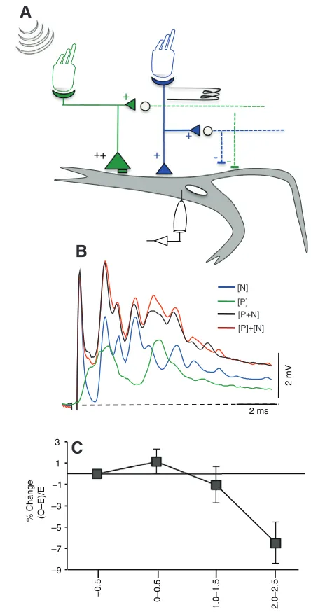
Cable models of distributed synaptic inputs to dendrites suggest supralinear summation when inputs are stimulated centripetally, i.e. distally first and then proximally (Rall, 1964). As the results suggest that aLLn input is proximal to posterior eighth nerve input, these two inputs were paired with different time delays. Both the pip and aLLn EPSPs were subthreshold and failed to trigger the M-cell. The M-cell generally requires over 15mV depolarization before it fires. Pip sounds were paired with aLLn stimulation at intervals ranging from –0.5 to 2.5ms and the data were grouped into four main intervals where the aLLn began, –0.5, 0–0.5, 1.0–1.5 and 2–2.5ms, after the dendritic pip response (Fig.7). Peak amplitudes were compared between these paired responses with those generated by calculating the algebraic sum of the two PSPs evoked alone.
Fig.7B shows average traces from an experiment where there was a 0.5ms delay in the centripetal direction between the onsets of two inputs. The amplitude of the evoked peak is essentially the same as that of the calculated peak, suggesting linear summation. Average peak amplitudes for eight experiments at four different time points indicate a linear mode of summation when the more distal pip inputs are triggered at 0.5ms intervals before the more proximal aLLn inputs (Fig.7C). With a 0.5ms delay there is an average increase of 2.05±1.1% in the amplitude of the evoked response in comparison to the calculated response. This, however, lacks statistical significance. With increasing delay between the pip and aLLn, the summation becomes sublinear, which may reflect the shunt of the aLLn EPSP by inhibition associated with the pip response.
DISCUSSION
Successful escapes rely on integration across multiple sensory modalities. Unraveling the computational mechanism that underlies multisensory integration by the M-cell requires a firm understanding of the properties of each sensory input. This information then lays a strong foundation for a multisensory perspective. This study is the first to demonstrate the existence of monosynaptic connections between aLLn afferents and the M-cell. Stimulation of the distal and ventral branches of the aLLn near their entry to the midbrain evokes a complex PSP that is localized to the soma and proximal lateral dendrite and has both electrotonic and chemical components, as with the input from the posterior eighth nerve, which terminates more distally (Lin and Faber, 1988). We propose that the speed and soma–dendritic location of this input could be an important feature in the gain control of M-cell responses, particularly in relation to directionality.
Pharmacological evidence for mixed synaptic contacts near the soma
Two lines of evidence indicate that the initial peak of the aLLn response has an electrical component. One comes from the pharmacological profile of the PSP. Two glutamate receptor antagonists, APV and CNQX, were used to block excitatory chemical synaptic transmission; the former had no effect and the latter effectively blocked all but the initial depolarizing component (Fig.3A–C). This result also indicates the chemical component of the EPSP is mediated by glutamate receptors of the AMPA subtype. Ineffectiveness of APV at resting potential does not rule out an NMDA receptor-mediated component, although earlier studies indicate the NMDA receptors in this neuron are functional at resting potential (Wolszon et al., 1997). Furthermore, immunohistochemical evidence indicates that NMDA receptors are restricted to the distal lateral dendrite (Sur et al., 1995). Additional confirmation that the first peak is electrotonic comes from the demonstration that it persists during high frequency stimulation, while the later components are depressed (Fig.3D). In this respect, the aLLn inputs seem to differ considerably from those of the pLLn, which have been reported to be only chemical in nature, and mainly inhibitory (Korn and Faber, 1975).
Morphological studies of the lateral dendrite show the presence of mixed junctions, i.e. electrical and chemical, not only at the distal portion of the dendrite where the club endings of the posterior eighth nerve terminate, but also more proximal on the dendrite, close to the soma (Flores et al., 2008; Nakajima, 1974). The sources of these contacts have not been confirmed. When the aLLn was stimulated, responses were maximal in a 100m region that spans the soma and initial segment of the lateral dendrite (Fig.4). This region is closer to the soma than the region where auditory inputs from the posterior nerve are maximal (Fig.4) The spread of the aLLn inputs
V V1
2 ms
10 mV
0.06 Gm
2 ms PSP
Gipsp
Ipsi
Contra
A
B
C
Control AD
Shunted AD
over this region suggests that they are segregated at this postsynaptic region and that their fibers may be responsible for some of the mixed synaptic contacts reported. This region is analogous to the site where pLLn inputs have been reported to be maximal. However, the pLLn was reported to be only chemical in nature (Korn and Faber, 1975).
Chemical and electrical inhibition is bilateral
The aLLn afferents project bilaterally to commissural inhibitory interneurons that chemically inhibit both M-cells. This chemical inhibition is preceded by an ephaptic inhibition via the same interneurons. All sensory afferents studied so far inhibit the M-cell in the same manner when activated. The IPSP can be dissected by measuring the shunt of the antidromic spike height when it is paired with the aLLn. The IPSP starts within 1ms of the onset of excitation and lasts for more than 10ms, as determined by varying the interval between the two stimuli (Fig.4). However, the electrical inhibition follows the EPSP with little to no delay (Fig.5). Stimulation of the nerve produces the same inhibitory conductance change in both the ipsilateral and contralateral cell (Fig.4B). This is in contrast to the excitatory input, which although also bilateral, is much smaller and slower in the contralateral cell. This inhibitory conductance change acts to shunt the recorded EPSP response, and it appears to be the dominant input to the contralateral cell. This observed asymmetry might have a substantial influence on the response threshold, and hence determining which M-cell fires first. This argument is based upon the supposition that the aLLn afferents can be activated unilaterally by naturalistic stimuli, which does not seem to be the case for the pLLn.
Fast monosynaptic excitatory inputs project proximal to inner ear inputs
Korn and Faber showed that electrical stimulation of the pLL nerve just below the operculum evokes a small PSP in the M-cell with latencies between 0.8 and 2.9ms, which is longer than the delay after stimulation of the ipsilateral aLLn, where latencies range between 0.5 and 1.04ms (Korn and Faber, 1975). Considering that afferents of both branches of the nerve innervate functionally similar receptors, this discrepancy is certainly a reflection of the longer conduction distance of the posterior nerve. Schellart and Kroese have reported that the conduction velocity and diameter of the pLLn fibers are not homogeneous and rather depend on the location of the neuromasts that they innervate (Schellart and Kroese, 2002). This velocity compensation is presumably to ensure inputs that are generated towards the tail of the fish remain relevant for central processing of LL information. With this consideration, they report an estimated delay of at least 4ms from hair cell transduction to stimulus detection in the hindbrain. This may be too long to effectively influence evoked responses in the M-cell, which has a shorter processing time (Weiss et al., 2006; Zottoli, 1977). However, because the neuromast receptors on the head are closer to the aLLn, this delay is expected to be shorter.
Although the latency of aLLn-evoked PSPs is indicative of monosynaptic inputs, it is still longer than that of PSPs evoked by stimulation of the posterior eighth nerve (Lin and Faber, 1988). This difference might reflect the fact that the aLLn contains smaller caliber axons than the large (up to 15m diameter) axons in the posterior eighth nerve that terminate distally as large myelinated club endings (Nakajima, 1974). Indeed these large fibers are required for sufficient depolarization of the low resistance M-cell to threshold. Posterior eighth nerve EPSP amplitudes can reach up to 20mV in the distal lateral dendrite, whereas the maximum aLLn EPSP amplitudes observed with electrical stimulation were at most 10mV. Similar to the pLLn, stimulation of the aLLn alone was not sufficient to fire an action potential in the M-cell. Although these inputs from the aLLn are smaller than those of the posterior eighth nerve, if they occur at the right latency, they can contribute to the threshold. Indeed following LL elimination, there are fewer early C-starts in adult
–9 –7 –5 –3
–1 1
3
–0.5
0–0.5
1.0–1.5 2.0–2.5
[P] [P+N] [P]+[N] [N]
2 ms
2 mV
A
B
C
% Ch
a
nge
(O–E)/E
Fig. 7. (A)Schematic of interaction paradigm. M-cell inputs along the lateral dendrite were stimulated centripetally at different time intervals. Distal eighth nerve inputs (green large triangles) were evoked by stimulation of the primary hair cells by sound pips generated with a loud speaker in air, whereas the proximal aLLn inputs (blue small triangles) were stimulated electrically at the nerve afferents. Open circles represent inhibitory interneurons that are excited by M-cell afferents and project bilaterally to both cells. Recordings were made in the ipsilateral M-cell soma.
[image:8.612.51.273.65.502.2]goldfish (Mirjany et al., 2011). Furthermore, in larval herring and zebrafish, where the M-cell presumably has a lower firing threshold, escapes can be evoked through the lateral line (Blaxter and Fuiman, 1989; Blaxter and Fuiman, 1990; Blaxter et al., 1981).
Inputs from the aLL appear less synchronous, with multiple brief peaks and an underlying depolarizing envelope. This apparent asynchrony could be a reflection of polysynaptic inputs or a heterogeneous population of afferents with mixed conduction velocities. Indeed it has been reported that afferent fibers that innervate canal neuromasts have a shorter conduction time than those innervating the superficial neuromasts (Schellart and Kroese, 2002). The fast rise time (approximately 0.5ms) of the initial peak is, however, indicative of fast synaptic potentials mediated by electrically coupled synapses as they are comparable to the coupling potentials reported for the single club-ending fibers of the posterior eighth nerve (Lin and Faber, 1988; Szabo et al., 2006).
Even though aLLn EPSPs were subthreshold in the adult goldfish, their fast electrical component and proximity to the soma may render them effective in modulating the responsiveness of the M-cell. Indeed the M-cell lateral dendrite contains voltage-sensitive K+ channels that facilitate complex computations and nonlinear interactions (Faber and Korn, 1986; Faber et al., 1991; London and Hausser, 2005; Magee, 2000). An inward K+rectifier is thought to increase the short space constant of the M-cell, so that close to threshold, EPSPs generated distally will be transmitted to the soma with less decrement than when the cell is at resting membrane potential. This may also facilitate integration of secondary somatic inputs with longer latencies or smaller amplitudes. This nonlinear property of the dendrite ensures that the cell can maintain a low input resistance while still being capable of reliably triggering a high threshold, rapid response. Also, Rall postulated that with passive dendrites sequential activation of inputs in a distal to proximal direction could yield supralinear summation (Rall, 1964). We investigated the potential for this latter type of nonlinearity by concurrently stimulating distal posterior eighth nerve inputs and proximal aLLn inputs through sound pressure pulses and electrical stimulation, respectively (Fig.7), using relatively weak PSPs. When these two inputs were triggered with a delay of less than 1ms, summation was linear and remained below threshold. However, if somatic inputs arrive later than 1.5ms from those further out on the dendrite, the inhibitory conductance resulting from the distal afferents produces an effective shunt of the more proximal signal. Thus, we found no evidence for supralinearity. This mechanism might occur in the absence of feedforward inhibition, but the prevalence of that inhibitory process instead suggests it contributes to linear summation, as well as to limiting the temporal window of integration. This lack of supralinearity may be due to the short membrane time constant of the M-cell (Rall, 1964), which is 0.3–0.4ms compared with approximately 25ms or more in central neurons, such as CA3 pyramidal neurons (Johnston, 1981). An alternative explanation is that the postulated nonlinearity depends upon synaptic conductance changes and the dominant synaptic input is electrotonic. A better test of Rall’s hypothesis might be to block glycinergic inhibition with strychnine, as was done with the pLLn; however, that manipulation is likely to expose complex polysynaptic inputs (Faber and Korn, 1986; Faber et al., 1991; Korn and Faber, 1975).
Behavioral consequences of aLLn inputs to the M-cell Most startle events are multisensory and depend on precise timing between different inputs for the effective execution of the behavior.
It is most probable that directional decisions by the M-cell are the result of a combination of inputs, consisting, at least, of those of the posterior eighth nerve branches and the lateral line nerves, converging onto the M-cell or associated inhibitory neurons within the time interval allotted for sensory processing, which is roughly 2ms (Eaton et al., 1995; Mirjany et al., 2011). To this end the large electrical inputs from the posterior eighth nerve are required for threshold and speed of transmission. However, current evidence suggests that they may not, by themselves, carry directionally relevant information, because they are activated by pressure waves that lack a directional component. Indeed latencies and amplitudes of pip-evoked EPSPs are the same in both M-cells regardless of the source of the pressure wave (unpublished observations). Other inputs from the inner ear, i.e. vestibular inputs, chemically synapse at the distal tip of the dendrite and are thought to act locally in modulation of M-cell excitability rather than contribute directly to the behavioral threshold (Szabo et al., 2007).
Recent behavioral studies have shown that the initial direction of the escape requires an intact lateral line system. However, escapes can still occur in its absence, confirming the requirement for multisensory inputs with the inner ear inputs setting the behavioral threshold. Therefore, the LL system is capable of both detecting the location of the stimulus and ensuring that the appropriate cell reaches threshold before the other cell. Furthermore, this has to occur with sufficient lead-time so that the inhibitory circuit can prevent the other cell from firing. There studies implicate the aLL in encoding directional responses when the fish is in the open field, as transection of the pLL did not reduce directionality to the same extent as complete LL elimination did. Indeed, the short latency responses we observed after aLLn stimulation provide support for PSPs evoked through these inputs having behavioral relevance. However, both afferents convey information from the same receptor subtypes and it would seem that for a natural startle response, detection of fluid distortion in any quadrant might be important from a directionality perspective. The central nervous system presumably also uses spatial and temporal information from the neuromasts that cover the entire body to obtain a three-dimensional representation of the outside world. Indeed, to account for the position-dependent delays of receptor signals further out on the body, there is a compensatory gradient of fiber conduction rates (Schellart and Kroese, 2002). Therefore, it would be expected that both anterior and posterior lateral line inputs should occur temporally close together. Furthermore, it appears that both branches are localized to the same region of the dendrite, a feature that may enhance functional integration.
Korn and Faber reported longer latencies ranges of between 0.8 and 2.9ms for PSPs evoked by stimulating the posterior branch (Korn and Faber, 1975). PSP latencies for the anterior branch are smaller, ranging between 0.8 and 1.5ms. Conduction delays can certainly account for the limited contribution of the posterior lateral line with the stimulus parameters used.
If this is the main difference, then the pLL can be a contributing factor to directionality when the aversive stimulus is more graded. The M-cell processing times for such stimuli are extended and hence temporal delays may be less significant (Weiss et al., 2006).
1963; Coombs et al., 1988), such that the afferents of the anterior branch of the lateral line convey a higher proportion of acceleration-detecting fibers. Furthermore, it has been reported that the conduction rates of fibers innervating the canal vary from those innervating superficial neuromasts. This may also add to the discrepancies between PSP onsets following anterior versusposterior LL stimulation.
In conclusion, we have established that, consistent with behavioral studies, the aLLn input to the M-cell has physiological characteristics consistent with this modality being able to influence both the threshold and directionality of the acoustically evoked C-start. This result raises the possibility of investigating the contribution of the components of the octavolateralis system to M-cell activation using a combination of behaviorally relevant stimuli, including one capable of triggering directional responses.
LIST OF ABBREVIATIONS aLLn anterior LL nerve
AMPA a-amino-3-hydroxy-5-methyl-4-isoxazolepropionic acid APV DL-2-amino-5-phosphonovaleric acid
CNQX cyano-7-nitroquinoxaline-2,3-dione disodium EHP extrinsic hyperpolarizing potential
EPSP excitatory postsynaptic potential IPSP inhibitory postsynaptic potential
LL lateral line
NMDA N-methyl-D-aspartate
PHP passive hyperpolarizing potential pLLn posterior LL nerve
PSP postsynaptic potential
REFERENCES
Blaxter, J. H. S. and Fuiman, L. A.(1989). Function of the free neuromasts of marine teleost larvae. In The Mechanosensory Latera Line, Neurobiology and Evolution (ed. S. Coombs, P. Gorner and H. Munz), pp. 481-499. New York: Springer-Verlag. Blaxter, J. H. S. and Fuiman, L. A.(1990). The role of the sensory systems of herring
larvae in evading predatory fishes. J. Mar. Biol. Assoc. UK61, 851-869. Blaxter, J. H. S., Gray, J. A. B. and Denton, E. J.(1981). Sound and Startle
responses in herring shoals. J. Mar. Biol. Assoc. UK61, 851-869.
Bleckmann, H.(1993). Role of the lateral line in fish behaviour. In Behaviour of Teleost Fishes (ed. T. J. Pitcher), pp. 201-246. London: Chapman and Hall. Bleckmann, H.(2008). Peripheral and central processing of lateral line information. J.
Comp. Physiol. A Neuroethol. Sens. Neural Behav. Physiol.194, 145-158. Chang, Y. T., Lin, J. W. and Faber, D. S.(1987). Spinal inputs to the ventral dendrite
of the teleost Mauthner cell. Brain Res.417, 205-213.
Coombs, S., Janssen, J. and Webb, J. F.(1988). Diversity of lateral line systems: evolutionary and functional considerations. In Sensory Biology of Aquatic Animals (ed. J. Atema, R. R. Fay, A. N. Popper and W. N. Tavolga), pp. 553-593. New York: Springer.
Dijkgraaf, S.(1963). The functioning and significance of the lateral-line organs.Biol. Rev. Camb. Philos. Soc.38, 51-105.
Eaton, R. C. and Popper, A. N.(1995). The octavolateralis system and Mauthner cell: interactions and questions. Brain Behav. Evol.46, 124-130.
Eaton, R. C., DiDomenico, R. and Nissanov, J.(1991). Role of the Mauthner cell in sensorimotor integration by the brain stem escape network. Brain Behav. Evol.37, 272-285.
Eaton, R. C., Canfield, J. G. and Guzik, A. L.(1995). Left-right discrimination of sound onset by the Mauthner system. Brain Behav. Evol.46, 165-179. Eaton, R. C., Guzik, A. L. and Casagrand, J. L.(1997). Mauthner system
discrimination of stimulus direction from the acceleration and pressure components at sound onset.Biol. Bull.192, 146-149.
Faber, D. S. and Korn, H.(1978). Electrophysiology of the Mauthner cell: basic properties, synaptic mechanisms, and associated networks. In Neurobiology of the Mauthner Cell (ed. D. S. Faber and H. Korn), pp. 47-131. New York: Raven Press. Faber, D. S. and Korn, H.(1986). Instantaneous inward rectification in the Mauthner
cell: a postsynaptic booster for excitatory inputs. Neuroscience19, 1037-1043. Faber, D. S., Korn, H. and Lin, J. W.(1991). Role of medullary networks and
postsynaptic membrane properties in regulating Mauthner cell responsiveness to sensory excitation. Brain Behav. Evol.37, 286-297.
Flores, C. E., Ene, S. and Pereda, A. E.(2008). An immunochemical marker for goldfish Mauthner cells. J. Neurosci. Methods175, 64-69.
Foreman, M. B. and Eaton, R. C.(1993). The direction change concept for reticulospinal control of goldfish escape. J. Neurosci. 13, 4101-4113.
Furshpan, E. J. and Furukawa, T.(1962). Intracellular and extracellular responses of the several regions of the Mauthner cell of the goldfish. J. Neurophysiol.25, 732-771.
Furukawa, T. and Furshpan, E. J.(1963). Two inhibitory mechanisms in the Mauthner neurons of goldfish. J. Neurophysiol.26, 140-176.
Johnston, D.(1981). Passive cable properties of hippocampal CA3 pyramidal neurons. Cell. Mol. Neurobiol.1, 41-55.
Kalmijn, A. J.(1988). Hydrodynamic and acoustic filed detection. In Sensory Biology of Aquatic Animals (ed. J. Atema, R. R. Fay, A. N. Popper and W. N. Tavolga), pp. 83-130. New York: Springer-Verlag.
Korn, H. and Faber, D. S.(1975). Inputs from the posterior lateral line nerves upon the goldfish Mauthner cell. I. Properties and synaptic localization of the excitatory component. Brain Res.96, 342-348.
Korn, H. and Faber, D. S.(2005). The Mauthner cell half a century later: a neurobiological model for decision-making? Neuron47, 13-28.
Kroese, A. B. and Schellart, N. A.(1992). Velocity- and acceleration-sensitive units in the trunk lateral line of the trout. J. Neurophysiol.68, 2212-2221.
Lin, J. W. and Faber, D. S.(1988). Synaptic transmission mediated by single club endings on the goldfish Mauthner cell. I. Characteristics of electrotonic and chemical postsynaptic potentials. J. Neurosci. 8, 1302-1312.
Lin, J. W., Faber, D. S. and Wood, M. R.(1983). Organized projection of the goldfish saccular nerve onto the Mauthner cell lateral dendrite. Brain Res.274, 319-324. Liu, K. S. and Fetcho, J. R.(1999). Laser ablations reveal functional relationships of
segmental hindbrain neurons in zebrafish. Neuron23, 325-335.
London, M. and Hausser, M.(2005). Dendritic computation. Annu. Rev. Neurosci.28, 503-532.
Magee, J. C.(2000). Dendritic integration of excitatory synaptic input.Nat. Rev. Neurosci.1, 181-190.
McCormick, C. A. and Hernandez D. V.(1996). Connections of octaval and lateral line nuclei of the medulla in the goldfish, including the cytoarchitecture of the secondary octaval population in goldfish and catfish. Brain Behav. Evol.47, 113-137. Mirjany, M., Preuss, T. and Faber, D. S.(2011). Role of the lateral line
mechanosensory system in directionality of goldfish auditory evoked escape response. J. Exp. Biol.214, 3358-3367.
Nakajima, Y.(1974). Fine structure of the synaptic endings on the Mauthner cell of the goldfish. J. Comp. Neurol.156, 379-402.
Nissanov, J., Eaton, R. C. and DiDomenico, R.(1990). The motor output of the Mauthner cell, a reticulospinal command neuron. Brain Res.517, 88-98.
Palmer, L. M. and Mensinger, A. F.(2004). Effect of the anesthetic tricaine (MS-222) on nerve activity in the anterior lateral line of the oyster toadfish, Opsanus tau. J. Neurophysiol.92, 1034-1041.
Preuss, T. and Faber, D. S.(2003). Central cellular mechanisms underlying temperature-dependent changes in the goldfish startle-escape behavior. J. Neurosci. 23, 5617-5626.
Preuss, T., Osei-Bonsu, P. E., Weiss, S. A., Wang, C. and Faber D. S.(2006). Neural representation of object approach in a decision-making motor circuit. J. Neurosci. 26, 3454-3464.
Puzdrowski, R. L.(1989). Peripheral distribution and central projections of the lateral-line nerves in goldfish, Carassius auratus. Brain Behav. Evol.34, 110-131. Rall, W.(1964). Theoretical significance of dendrtic trees for neuronal input-output
relations. In Neuroal Theory and Modeling (ed. R. Reiss), pp. 73-97. Stanford: Stanford University Press.
Schellart, N. A. and Kroese, A. B.(2002). Conduction velocity compensation for afferent fiber length in the trunk lateral line of the trout. J. Comp. Physiol. A Neuroethol. Sens. Neural Behav. Physiol.188, 561-576.
Sur, C., McKernan, R. and Triller, A.(1995). GABAA receptor-like immunoreactivity in the goldfish brainstem with emphasis on the Mauthner cell. Neuroscience66, 697-706.
Szabo, T. M., Weiss, S. A., Faber, D. S. and Preuss, T.(2006). Representation of auditory signals in the M-cell: role of electrical synapses. J. Neurophysiol.95, 2617-2629.
Szabo, T. M., McCormick, C. A. and Faber, D. S.(2007). Otolith endorgan input to the Mauthner neuron in the goldfish. J. Comp. Neurol.505, 511-525.
Van Bergeijk, W. A.(1967). The evolution of vertebrate hearing. Contrib. Sens. Physiol.2, 1-49.
Weiss, S. A., Zottoli, S. J., Do, S. C., Faber, D. S. and Preuss, T.(2006). Correlation of C-start behaviors with neural activity recorded from the hindbrain in free-swimming goldfish (Carassius auratus). J. Exp. Biol. 209, 4788-4801. Weiss, S. A., Preuss, T. and Faber, D. S.(2008). A role of electrical inhibition in
sensorimotor integration. Proc. Natl. Acad. Sci. USA105, 18047-18052. Wolszon, L. R., Pereda, A. E. and Faber, D. S.(1997). A fast synaptic potential
mediated by NMDA and non-NMDA receptors. J. Neurophysiol.78, 2693-2706. Zottoli, S. J.(1977). Correlation of the startle reflex and Mauthner cell auditory
responses in unrestrained goldfish. J. Exp. Biol. 66, 243-254.
Zottoli, S. J. and Faber, D. S.(1979). Properties and distribution of anterior VIIIth nerve excitatory inputs to the goldfish Mauthner cell. Brain Res.174, 319-323. Zottoli, S. J. and Faber, D. S.(1980). An identifiable class of statoacoustic
interneurons with bilateral projections in the goldfish medulla. Neuroscience5, 1287-1302.
Zottoli, S. J. and Van Horne, C.(1983). Posterior lateral line afferent and efferent pathways within the central nervous system of the goldfish with special reference to the Mauthner cell. J. Comp. Neurol.219, 100-111.