Original Article
Anacardic acid sensitizes prostate cancer cells
to radiation therapy by regulating H2AX expression
Kun Yao, Xianzhen Jiang, leye He, Yuxin Tang, Guangming Yin, Qing Zeng, Zhiqiang Jiang, Jing Tan
Department of Urology, The Third Hospital of Central South University, Changsha 410013, China
Received October 2, 2015; Accepted November 22, 2015; Epub December 1, 2015; Published December 15, 2015
Abstract: Anacardic acid (6-pentadecylsalicylic acid, AA), a natural compound isolated from the traditional medicine Amphipterygiumadstringens, has been reported as potential antitumor agents in various cancers including prostate cancer (PC). However, the effects and mechanism of AA on the radiosensitivity of prostate cancer remains unknown. The results indicated that AA exhibited strong antitumor activity in PC cell lines, either as a single agentor in
combi-nation with radiation. AA significantly induced the downregulation of H2AX and p-H2AX expression, increase of cell
apoptosis and decreasing of cell invasion, which were reversed by overexpressed H2AX. These results suggest that AA sensitize prostate cancer cells to radiation therapy by repressing H2AX expression.
Keywords: Anacardic acid, prostate cancer, H2AX, radio therapy
Introduction
Prostate cancer (PC) as the most common type of cancer is the second leading cause of cancer death among men in the developed world [1]. The standard treatments of PC include surgery, chemotherapy and radiotherapy. Radiotherapy is generally used as preoperative and postop-erative treatment that provides high biochemi-cal control, low risk of complications, minimal duration of treatment, and out-patient treat-ment opportunity. However, intrinsic tumor ra- dioresistance accounts for the high recurrence, leading to accelerated disease progression and death [2, 3]. Therefore, identification of reliable radio sensitizers to PC cells would be urgently desirable. Targeted biological therapies that selectively interfere with cancer cell growth sig-nals may improve patient survival by enhancing the effects of radiation with little damage to normal tissue.
Anacardic acid (2-hydroxy-6-pentadecylbenzoic acid, AA) is a constituent of the shell of the cashew-nut (Anacardiumoccidentale) [4], and is a dietary component found in cashew apple (Anacardium occidentale) and ginkgo (Ginkgo biloba) leaves and fruits. Studies show that AA acts as a mitochondrial uncoupler of oxidative phosphorylation [5] and as a traditional
medi-cine for treatment of gastric ulcers, gastritis and stomach cancers [6]. Recently, AA showed certain antitumor activities in prostate cancer, lung carcinoma, ovarian cancer and breast car-cinoma, and is thought to exert its action via various mechanisms [7-9]. AA also sensitizes HeLa cells to ionizing radiation by inhibition of histone acetyl transferase activity [10]. How- ever, the effects and mechanism of AA on the radio therapy of prostate cancer remain un- known.
Materials and methods
Reagent, cell culture and antibodies
A 50-mmol/L solution of AA (Merck, Germany) was added in dimethylsulfoxide. AA was pre-pared in dilution with culture medium when necessary.
CCK-8 assay
Logarithmically growing cells were counted and seeded in 96-well plates at 2000 cells per well, in triplicate and incubated overnight. The next morning, all plates were aspirated and fresh medium were added in a final volume of 200 µl with increasing concentrations of AA (0, 5, 25, and 125 μmol/L). Following drug addition, the plates were incubated for 5 days. Cell growth inhibition was examined by CCK-8 assay (Best Bio, China). 10 µl CCK-8 labeling reagent was added to each well with 100 µl fresh medium, the plates were incubated at 37°C for 4 h. The absorbance of each well was measured at 450 nm using Thermo Scientific Varioskan Flash (Thermo Scientific, Finland). Percentage of via -ble cells = (OD450 of treated sampleOD450 of blank sample)/(OD450 of control sample-OD450 of blank sample)×100. The results shown were mean values of 3 independent experiments.
Clonogenic assay
Clonogenic assay was used to evaluate the effect of BIIB021 in combination with radiation. Cells were trypsinized to generate a single cell suspension and seeded in 6-well plates at 1500 cells per well. After allowing cells to attach, AA was added about 125 μmol/L. For AA in combination with radiation, 24 h after adding AA, cells were irradiated with a single dose of 2, 4, 6, 8 Gy from amedicallinear accel-erator (varian23EX, USA). Four hours after radi-ation, all plates were aspirated and fresh medi-um were added. 14 days after seeding, colo-nies were stained with crystalviolet, and the number of colonies containing at least 50 cells was counted. The colony survival fraction was calculated for each treatment and data were presented as log plot. The results shown were mean value of 3 independent experiments with triplicate setting in each experiment.
Flow cytometry analysis of apoptosis
To test if cells were undergo apoptosis, Eca109 and Eca9706 cells were plated and exposed to either 1 µM AA or 6 Gy radiation for 24 h. For combination, cells were treated with 1 µM AA for 24 h, and then treated with a single dose of 6 Gy of radiation. Cells were collected 24 h after radiation without washing (both floating and attached cells were collected by centrifu-gation). Apoptosis analysis was performed according to the manufacturer’s instructions
(Annexin V-FITC Apoptosis detection kit; Best-Bio, China). Approximately 1×10⁵ cells were in-
cubated with FITC-conjugated annexin V in the presence of PI and then analyzed by flow cytom -etry (FACS, Becton Dickson). Annexin V positive PI negative cells scored as early apoptotic, Annexin V positive PI positive cells correspond -ed to late apoptotic cells. Cell cycle distribution was measured before and after the same treat-ed as above. Cells were digesttreat-ed by trypsin, washed with PBS, fixed with 75% cold ethanol at 4°C overnight and dyed with PI, and then analyzed by flow cytometry.
Cell invasion assay
The capability of cell invasion was examined by transwell invasion assay. Cells were cultivated to 80% confluence on the 12-well plates. Then, we observed the procedures of cellular growth at 72 h. All the experiments were repeated in triplicate. The transwell migration chambers were used to evaluate cell invasion. Then cells invading cells across the membrane were counted under a light microscope.
Western blot analysis
Whole cell extracts were prepared with a cell lysis reagent (Sigma-Aldrich, St. Louis, MO, USA) according to the manual, and then, the protein was quantified by a BCA assay (Pierce, Rockford, IL, USA). Then, the protein samples were separated by SDS-PAGE (10%) and detect-ed by Western blot using polyclonal (rabbit) anti-H2AX antibody (Santa Cruz Bio-technology, Santa Cruz, CA, USA). Goat anti-rabbit IgG (Pierce, Rockford, IL, USA) secondary antibody conjugated to horse radish peroxidase and ECL detection systems (SuperSignal West Femto, Pierce) were used for detection.
Statistical analysis
All data were shown as mean ± SD. Statistical significance was assessed by T-test for two-group comparison. Differences with P value <0.05 were considered statistically significant.
Results
AA inhibited the proliferation and survival of PC cell lines
PC3 cells using the CCK-8 assay. The cells were treated with 0, 5, 25, and 125 μmol/L AA for 24 h. As showed in Figure 1, PC3 cell lines dis-played a dose-dependent reduction in cell pro-liferation. 5 μmol/L AA treatment inhibits 12% cell proliferation and reached the peak at the concentration of 125 μmol/L.
AA can radio sensitize PC cells
Studies have indicated that AA may radio sensi-tize tumor cells [10, 11].
We performed clonogenic cell survival assays to address the same issue in PC3 cell lines. PC3 cells were treated with AA for 24 h followed by a single dose of radiation. The impact of radi-ation alone or combined with AA was showed a survival curves (Figure 1B). We found that cells pretreated with AA priorto radiation observed a significant growth inhibition in PC3 (P=0.002 at 2 Gy, P=0.0033 at 4 Gy, P=0.0041 at 6 Gy and P=0.0028 at 8 Gy). These results indicated that AA maybe used as radiosensitizer in the radio therapy of PC patients to improve the
[image:3.629.103.526.80.236.2]anti-Figure 1. A. AA inhibited the growth of PC3 cells in a dose-dependent manner. *P<0.05 versus control group. B. AA1 radio sensitized PC3 cell lines. PC3 cells were irradiated at a single dose of 0, 2, 4, 6 or 8 Gy in the presence or ab-sence of AA (IC25 concentration for each cell line). Data were showed as mean ± SD of three experiments. P<0.05 for each radiation dose in combination with AA versus each radiation dose alone for both of the two cell lines.
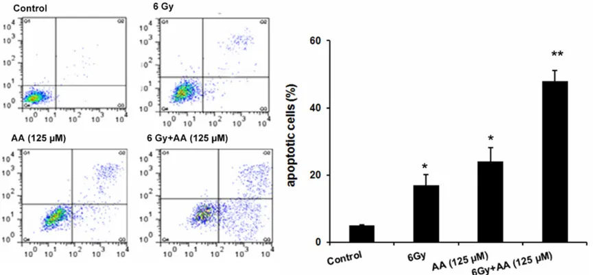
Figure 2. AA induced PC cell death via apoptosis. Apoptosis for all treatments of PC3 cells. Combination of radiation
and AA induced significant apoptosis in PC cell line. Bargraphs represent the total apoptosis of all conditions. The
[image:3.629.100.533.310.511.2]tumor effect of radiation, especially the patients who are insensitive to radiation.
AA sensitized PC cells to radiation by increas-ing apoptotic cell death
Apoptosis is a mode of cell death in response
[image:4.629.100.529.83.260.2]cytometry analysis was performed for PC3 cells treated with radiation (6 Gy) or AA (125 µM) alone or the combination. As shown in Figure 2, radiation alone only induced a small amount of cell apoptotic in PC3 cells (P=0.024) compared with control. Apoptosis rate for PC3 cells was approximately 26% when treated with AA alone.
Figure 3. AA sensitized PC cells to radiation by inhibiting cell invasion. Invasion for all treatments of PC3 cells.
Com-bination of radiation and AA induced significant invasion in PC cell line. Bargraphs represent the total apoptosis of
all conditions. Data were shown as mean ± SD of three experiments. *P<0.05 versus control or radiation alone; **P<0.05 versus control or other treatment alone.
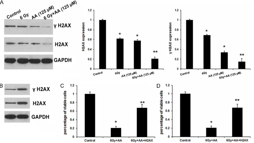
Figure 4. H2AX involved AA-induced radio sensitivity in PC cells. (A) The effects of AA and radiation on H2AX
expres-sion and γ-H2AX expresexpres-sion. Data were showed as mean ± SD of three experiments. *P<0.05 versus control or
[image:4.629.101.534.331.575.2]apoptosis rate increased to 48% for PC3, significantly higher rate compared with radia -tion or AA treatment alone (P<0.05). These results indicated that AA inhibited cell growth and enhanced radiation effect by inducing apoptosis.
AA sensitized PC cells to radiation by inhibiting cell invasion in PC cell lines
To further reveal the effects of AA on radio sensitivity of PC, we analyzed the invasion abil-ity of PC3 cells using transwell invasion assay. As shown in Figure 3, radiation alone or AA treatment alone inhibited cell invasion in PC3 cells compared with control. When AA was combined with radiation, the invasion ability greatly decreased, compared with radiation or AA treatment alone (P<0.05). These results indicated that AA inhibited cell invasion and enhanced radiation effect by repressing cell invasion.
H2AX involved AA-induced radio sensitivity in PC cells
High expression of phospho-H2AX predicts a poor prognosis in various types of cancers [12]. Here, we found that AA or radiation evi-dently repressed H2AX and p-H2AX expression in PC3 cells (Figure 4A).
To further confirm the function of H2AX in PC cell proliferation and invasion, we successfully engineered H2AX overexpression in PC3 cells (Figure 4B). Gamma-H2AX (γ-H2AX) is a phos -phorylated H2AX which exists nearby double-strand DNA breaks and is required for DNA damage repair [13]. As shown in Figure 4B, H2AX overexpression results in increased γ-H2AX level and restored the cell growth (Figure 4C) compared with AA and radiation group. Moreover, both cell invasion assays exhibited a similar effect as well (Figure 4D). In conclusion, these results indicated that H2AX involved AA-induced radio sensitivity in PC cells.
Discussion
Drugs from plants have become an important resource for discovery of anticancer drugs [10]. Although AA has already been reported to have significant anti-proliferative effect on a variety of cancer cells [7, 9, 14], its mechanism of action in prostate cancer is still unclear. In this
study, we report that AA inhibits PC3 cell prolif-eration in dose-and time-dependent manners. Based on these results, we investigated the molecular mechanism of action of AA on the suppression of tumor growth. Apoptosis is a form of programmed cell death that is charac-terized by a variety of morphological features, including cell shrinkage, chromatin condensa-tion, and chromosomal DNA fragmentation. In this study, we found that AA induces apoptosis in PC3 cells, as evident by Annexin V/PI assay with flow cytometry. Furthermore, transwell invasion assay revealed that AA inhibited cell invasion and enhanced radiation effect by repressing cell invasion.
DNA damage response (DDR) is an important cellular guard that protects genetic material from a constant barrage of genotoxic agents. Increased DNA damage is often associated with growth inhibition, which is also the case for cancer cells [15, 16]. Phosphorylation of the H2AX histone (p-H2AX) is an early indicator of DNA double-strand breaks and resulting DDR [17]. When DNA double-strand breaks occur, a PI3-like kinase and DNA-dependent protein kinases become activated and phosphorylate H2AX on acarboxylserine residue (Ser139) to generate γ-H2AX [17-19]. The overexpression of p-H2AX has been observed in multiple types of cancer, including, breast cancer, lung cancer, cervical cancer, renal cancer and bladder can-cer [12, 20, 21]. Recently, p-H2AX has been used as a biomarker for cancer, as abiodosim-eter for drug development and radiation expo-sure, and for clinical trials, for cancer chemo-and radiotherapy [22, 23]. Furthermore, emerg-ing uses for p-H2AX include detection of toxic environmental agents and chronic inflamma -tion [24]. In this study, we found that radia-tion and AA treatment decreased the H2AX and γ-H2AX expression levels in PC3 cells. Further-more, overexpressed H2AX reversed cell grow- th and invasion mediated by radiation, which suggests that AA-induced radio sensitivity by repressing H2AX protein expression.
In summary, we have demonstrated the antitu-mor activity of AA in vitro, which is at least par-tially associated with H2AX-mediated DDR. Acknowledgements
Hunan Province (Grant No. 2014SK3142) and a grant of Science Foundation of Healthy and Family Planning Commission of Hunan Province (Grant No. B2014-032).
Disclosure of conflict of interest
None.
Address correspondence to: Dr. Jing Tan, Depart- ment of Urology, The Third Hospital of Central Sou- th University, Changsha 410013, China. Tel: +86-731-88618578; Fax: +86-+86-731-88618578; E-mail: jt4485366@163.com
References
[1] Carlsson S, Vickers AJ, Roobol M, Eastham J,
Scardino P, Lilja H, Hugosson J. Prostate can-cer screening: facts, statistics, and interpreta-tion in response to the US Preventive Services Task Force Review. J Clin Oncol 2012; 30: 2581-2584.
[2] De Marzo AM, DeWeese TL, Platz EA, Meeker AK, Nakayama M, Epstein JI, Isaacs WB, Nelson WG. Pathological and molecular me- chanisms of prostate carcinogenesis: Impli- cations for diagnosis, detection, prevention, and treatment. J Cell Biochem 2004; 91: 459-477.
[3] Nupponen N, Visakorpi T. Molecular biology of
progression of prostate cancer. Eur Urol 1999; 35: 351-354.
[4] Kubo I, Kinst-Hori I, Yokokawa Y. Tyrosinase in-hibitors from anacardium occidentale fruits. J Nat Prod 1994; 57: 545-551.
[5] Toyomizu M, Okamoto K, Ishibashi T, Chen Z, Nakatsu T. Uncoupling effect of anacardic ac-ids from cashew nut shell oil on oxidative phos-phorylation of rat liver mitochondria. Life Sci 2000; 66: 229-234.
[6] Acevedo HR, Rojas MD, Arceo SD, Soto
Hernández M, Martínez Vázquez M, Terrazas T, del Toro GV. Effect of 6-nonadecyl salicylic acid
and its methyl ester on the induction of micro-nuclei in polychromatic erythrocytes in mouse peripheral blood. Mutat Res 2006; 609: 43-46.
[7] Xiu YL, Zhao Y, Gou WF, Chen S, Takano Y, Zheng HC. Anacardic acid enhances the prolif-eration of human ovarian cancer cells. PLoS One 2014; 9: e99361.
[8] Sung B, Pandey MK, Ahn KS, Yi T, Chaturvedi MM, Liu M, Aggarwal BB. Anacardic acid (6-nonadecylsalicylicacid), an inhibitor of his-toneacetyl transferase, suppresses expression of nuclear factor-kappab-regulated gene prod-ucts involved in cell survival, proliferation,
inva-sion, and inflammation through inhibition of
the inhibitory subunit of nuclear factor-kap-pabalphakinase, leading to potentiation of apoptosis. Blood 2008; 111: 4880-4891. [9] Tan J, Chen B, He L, Tang Y, Jiang Z, Yin G,
Wang J, Jiang X. Anacardic acid (6-pentadecyl-salicylicacid) induces apoptosis of prostate cancer cells through inhibition of androgen re-ceptor and activation of p53 signaling. Chin J Cancer Res 2012; 24: 275-283.
[10] Sun Y, Jiang X, Chen S, Price BD. Inhibition of histone acetyltransferase activity by anacardic acid sensitizes tumor cells to ionizing radia-tion. FEBS Lett 2006; 580: 4353-4356. [11] Sukumari-Ramesh S, Singh N, Jensen MA,
Dhandapani KM, Vender JR. Anacardic acid
induces caspase-independent apoptosis and radiosensitizes pituitary adenoma cells. J Neurosurg 2011; 114: 1681-1690.
[12] Lee YC, Yin TC, Chen YT, Chai CY, Wang JY, Liu MC, Lin YC, Kan JY. High expression of phos-pho-h2AX predicts a poor prognosis in colorec-tal cancer. Anticancer Res 2015; 35: 2447-2453.
[13] Furuta T, Takemura H, Liao ZY, Aune GJ, Redon C, Sedelnikova OA, Pilch DR, Rogakou EP, Celeste A, Chen HT, Nussenzweig A, Aladjem MI, Bonner WM, Pommier Y. Phosphorylation of histone h2AX and activation of mre11, rad50, and nbs1 in response to replication-dependent DNA double-strand breaks induced by mammalian DNA topoisomerase I cleavage complexes. J Biol Chem 2003; 278: 20303-20312.
[14] Seong YA, Shin PG, Yoon JS, Yadunandam AK, Kim GD. Induction of the endoplasmic reticu-lum stress and autophagy in human lung carcinoma A549 cells byanacardic acid. Cell Biochem Biophys 2014; 68: 369-377.
[15] Gangopadhyay NN, Luketich JD, Opest A, Landreneau R, Schuchert MJ. PARP inhibitor activates the intrinsic pathway of apoptosis in primary lung cancer cells. Cancer Invest 2014; 32: 339-348.
[16] Qu A, Wang H, Li J, Wang J, Liu J, Hou Y, Huang L, Zhao Y. Biological effects of (125)i seeds ra-diation on A549 lung cancer cells: G2/M ar-rest and enhanced cell death. Cancer Invest 2014; 32: 209-217.
[17] Redon C, Pilch D, Rogakou E, Sedelnikova O, Newrock K, Bonner W. Histone h2A variants h2AX and h2AZ. Curr Opin Genet Dev 2002; 12: 162-169.
[19] Kinner A, Wu W, Staudt C, Iliakis G. Gamma-h2AX in recognition and signaling of DNA dou-ble-strand breaks in the context of chromatin. Nucleic Acids Res 2008; 36: 5678-5694. [20] Zhao J, Wang Q, Li J, Si TB, Pei SY, Guo Z, Jiang
C. Comparative study of phosphorylated his-tone h2AX expressions in the cervical cancer patients of pre- and post-neoadjuvant chemo-therapy. Eur J Gynaecol Oncol 2015; 36: 318-322.
[21] Nagelkerke A, van Kuijk SJ, Martens JW, Sweep FC, Hoogerbrugge N, Bussink J, Span PN. Poor prognosis of constitutive gamma-h2AX expre- ssing triple-negative breast cancers is associ-ated with telomere length. Biomark Med 2015; 9: 383-390.
[22] Dickey JS, Redon CE, Nakamura AJ, Baird BJ, Sedelnikova OA, Bonner WM. H2AX: Functional roles and potential applications. Chromosoma 2009; 118: 683-692.
[23] Du LL, Nakamura TM, Russell P. Histone
modi-fication-dependent and -independent path -ways for recruitment of checkpoint protein Crb2 to double-strand breaks. Genes Dev 2006; 20: 1583-1596.