Original Article
ADAM12
expression predicts clinical outcome in
estrogen receptor-positive breast cancer
Bo Ma1,2, Qianqian Ma2, Chunhui Jin3, Xiaohong Wang1, Guolei Zhang1, Huiying Zhang1, Harald Seeger2, Alfred O Mueck2
1Affiliated Central Hospital of Huzhou Teachers College, Huzhou, Zhejiang 313000, China; 2University Hospital,
Tuebingen, Germany; 3Wuxi Hospital of Traditional Chinese Medicine, China
Received April 22, 2015; Accepted June 20, 2015; Epub October 1, 2015; Published October 15, 2015
Abstract: Objectives: Our study was aimed to make sure whether ADAM12 could serve as a prognostic biomarker of estrogen receptor (ER) -positive breast cancer. Methods: 127 patients with ER-positive breast cancer were in-cluded in the present study. The level of ADAM12 wasassayed through real-time quantitative PCR (RT-qPCR). Levels of ADAM12 in tumor tissues and adjacent normal tissues were compared with paired t-test. The association of
ADAM12 expression with clinical characteristics was analyzed via χ2 test. Kaplan-Meier survival curve was used to
evaluate the role of ADAM12 expression in overall survival (OS) of patients. Cox-regression analysis was performed to judge if ADAM12 could serve as a prognostic marker in breast cancer. Results: The level of ADAM12 was upregu-lated in tumor tissues of breast cancer compared to that of adjacent normal tissues (P < 0.05). The expression of
ADAM12 was closely related to the Ki-67 and HER2 status (P < 0.05 for both). The results of Kaplan-Meier survival curve showed that patients with higher level of ADAM12 exhibited shorter survival time compared to that of low level of ADAM12 (P < 0.001). Cox regression analysis showed that ADAM12 might be a biomarker in predicting prognosis of patients with ER-positive breast cancer (HR = 7.116, 95% CI = 3.329-15.212). Conclusion: ADAM12 appears to be a prognostic marker in ER-positive breast cancer.
Keywords: ADAM12, clinical outcome, estrogen receptor, breast cancer
Introduction
Breast cancer is a leading cause of cancer-related death in women [1], which is character-ized by diverse clinical behavior and outcomes [2]. The newly diagnosed patients are common-ly presented with earcommon-ly-stage breast cancer, however 20% of them will suffer recurrence in 10 years after diagnosis [3]. In recent years, despite the advances in early diagnosis and treatment therapy, there are still many patients died from cancer recurrence and metastasis. Until now, there has been many studies that proposed biomarkers for tumor behavior and clinical outcome in breast cancer [4-8], howev-er, the identification of new markers is still nec-essary [9]. Among the factors related with can-cer progression, abnormal genes expression has attracted a lot of attention, some of which have been demonstrated as independent bio-markers for breast cancer.
ADAMs, a group of membrane-associated metalloproteinases, plays an important role in regulating integrin-mediated cell adhesion, cell signal transduction and proteolytic release [10-12]. The family contains 20 members, 9 out of which are produced beyond the reproductive system: ADAM8, -9, -10, -12, -15, -17, -19, -28 and -33. The expression of ADAM12 is upregu-lated in various cancers [13-15]. Moreover,
ADAM12 has been demonstrated as a suscep-tibility gene of breast cancer [16]. Fröhlich et al. reported that overexpression of ADAM12 accel-erated the progression of breast cancer [17]. The related study also suggested that the level of ADAM12 was associated with disease status and pathologic stage in breast cancer [14, 17]. However, there are no studies investigating the effects of ADAM12 expression on the survival of patients with breast cancer.
Our study was aimed to ensure if ADAM12
breast cancer. The expression level of ADAM12
in serum and tissues (tumor tissues and adja-cent normal tissues) were tested with real-time quantitative PCR (RT-qPCR). Kaplan-Meier and Cox analyses were conducted to the role of
ADAM12 in breast cancer. Materials and methods
Participants
In this research, 205 women from Affiliated Central Hospital of Huzhou Teachers College with breast cancer were selected. Among them, 76 individuals were diagnosed as estrogen receptor (ER) negative and 129 were ER posi-tive. 129 women with ER positive breast cancer were included. They were aged 25-75 years old at the time of diagnosis. The patients who had metastatic disease, tumor history and rece- ived neoadjuvant chemotherapy were exclud-ed. After the selection, 127 patients were selected.
Pathological information was collected: primary tumor size, Ki-67 status and HER2 status. Before surgery, the blood samples of each indi-vidual were collected. Meanwhile, 127 tumor samples and adjacent normal samples were also collected for further analysis. The follow-ups were performed after the treatments. In the survey, survival time, date of death and date of last follow-up were recorded. The over-all survival time was defined from the time as
treatment began to death time or date of last follow-up.
All the participants signed the written consent before the study and the study was approved by The Ethical Committee of the hospital.
RT-qPCR assay
Total RNA was extracted from serum and tissue samples by Qiagen DNeasy Blood & Tissue Kit, followed by cDNA synthesis with First Strand cDNA Synthesis Kit (Thermo Scientific). GAPDH
was used as internal control. The expression of
ADAM12 was analyzed with Applied Biosy- stems 7500 real-time PCR system (Applied Biosystems, Foster City, CA, USA). The adopted calculation method was 2-ΔΔCT. All the samples were tested in triplicate.
Statistics
All the analysis was completed in SPSS 12.0. The differences in the level of ADAM12 between tumor tissues and adjacent normal tissues were compared with paired t-test. The relation-ship of expression of ADAM12 with clinical fea-tures was analyzed via χ2 test. Kaplan-Meier analysis was adopted to evaluate the effects of
ADAM12 expression on the overall survival (OS) of patients with breast cancer. Whether
ADAM12 could serve as a prognostic marker in breast cancer was estimated by Cox-regression analysis. The figures were finished in Graphpad prism 5. All the tests were two-tailed, and P < 0.05 indicated that there was significant difference.
Results
The expression level of ADAM12 in serum and tissues
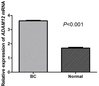
The level of ADAM12 in serum and tissues were tested with RT-qPCR technology. The relative content of ADAM12 in serum was 3.74. It was obvious that ADAM12 expression level was higher in tumor tissues compared to that of adjacent normal tissues (P < 0.05) (Figure 1).
Basic characteristic of patients with ER-positive breast cancer
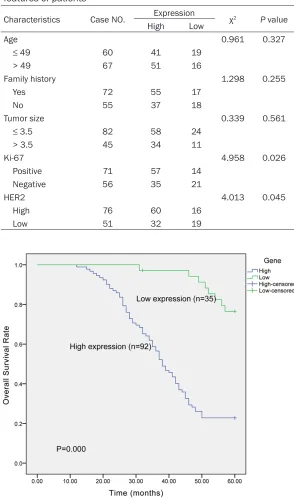
[image:2.612.89.290.71.245.2]The clinical characteristics of patients with ER-positive breast cancer were listed in Table 1. The patients were divided into two groups
according to the level of ADAM12. There were no obvious differences in age, family history and tumor size between two groups (P > 0.05 for all). While, it turned out that ADAM12 level was significantly related with Ki-67 and HER2 status (P < 0.05 for both), which indicates that
ADAM12 may be correlated with prognosis in breast cancer.
Kaplan-Meier survival curve and Cox regression analysis
Kaplan-Meier survival cur- ve was constructed to determine the differences in survival situation of two patients. The result illus-trated that high level of
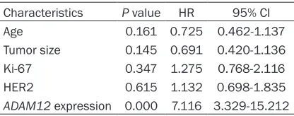
ADAM12 predicted worse prognosis of patients with ER-positive breast cancer (P < 0.001) (Figure 2). Further analysis of Cox regression provided evi-dence that ADAM12 was a promising prognostic biomarker in ER-positive breast cancer (HR = 7.116, 95% CI = 3.329-15.212) (Table 2).
Discussion
[image:3.612.89.383.91.590.2]ADAMs have specific extra-cellular domains, such as a prodomain, metalloprotein-ases domain, epidermal growth factor (EGF)-like repeat domain, disintegrin domain, cysteine-rich do- main, transmembrane do- main and cytoplasmic domain. ADAMs show many important biological activi-ties through these doma- ins. First, ADAMs can shed growth factors of TGF-α and HB-EGF, which results in promoted cell prolifera-tion. Second, ADAMs serve as adhesion molecules by binding to inregrins with Table 1. Relationship between ADAM12 expression and clinical
features of patients
Characteristics Case NO. Expression χ2 P value
High Low
Age 0.961 0.327
≤ 49 60 41 19
> 49 67 51 16
Family history 1.298 0.255
Yes 72 55 17
No 55 37 18
Tumor size 0.339 0.561
≤ 3.5 82 58 24
> 3.5 45 34 11
Ki-67 4.958 0.026
Positive 71 57 14
Negative 56 35 21
HER2 4.013 0.045
High 76 60 16
[image:3.612.92.384.104.371.2]Low 51 32 19
Figure 2. Kaplan-Meier survival curve. Increased expression of ADAM12 pre-dicted worse clinical outcome of patients with ER-positive breast cancer.
is that ADAMs contribute to the occurrence of many cancers [18].
ADAM12 is a member of ADAM family. It has certain crucial biological functions in tumors. Like other ADAMs, ADAM12 also could regulate proteolytic ectodomain and result in shedding of growth factors [11, 19, 20]. Besides, it is involved in nonproteolytic protein-protein inter-actions. The expression level of ADAM12 is always low. Nevertheless, its increased level was observed in liver cancer, bladder cancer, lung cancer and breast cancer [21-24]. ADAM12
is commonly expressed as two forms: trans-membrane form (ADAM12-L) and secreted form (ADAM12-S). The research of Roy et al. found that overexpression of ADAM12-L and ADAM- 12-S in breast cancer cells could promote the estrogen-independent proliferation [25]. In terms of the abnormality behavior of ADAM12
in cancers or diseases, there has been many studies investigating the specific role in single cancer or disease. Yang et al. concluded that
ADAM12 was a promising diagnostic marker for complete spontaneous abortion and ectopic pregnancy in symptomatic women [26]. A study on oral squamous cell carcinoma indicated that high level of ADAM12 could accelerate cell proliferation [27]. The positive activation loop between ADAM12 and HER2 may contribute to tumor cell migration and invasion in human head and neck cancer [28]. It also appears to be a prognostic marker in resected pethologi-cal stage I lung adenocarcinoma [23].
Based on the previous studies, our study test-ed the expression level of ADAM12 in serum and tissues. The results showed that the level of ADAM12 was upregulated in tumors serum and tissues. Moreover, its expression was associated with Ki-67 and HER2 status. The Kaplan-Meier survival curve illustrated that the patients with high level ADAM12 were more
likely to experience short survival time. Cox regression analysis indicated that ADAM12
was an independent marker for predicting clini-cal outcome in breast cancer.
Taken together, ADAM12 may serve as a prom-ising biomarker for predicting prognosis of patients with breast cancer. The present study only considered the effects of single gene on the survival situation, future researches should take more factors into account to get better understand on the pathological mechanism of breast cancer. ADAMs play its roles by many ways, so further studies to make sure the func-tion mechanism of ADAM12 in breast cancer are necessary for promoting the treatments of patients.
Disclosure of conflict of interest
None.
Address correspondence to: Dr. Xiaohong Wang, Affiliated Central Hospital of Huzhou Teachers College, Huzhou, Zhejiang 313000, China. E-mail: [email protected]; Dr. Harald Seeger, University Hospital, Tuebingen, Calwer Strasse 7, Tuebingan, Cermany 72076, Germany. E-mail: [email protected]
References
[1] Malvezzi M, Bertuccio P, Levi F, La Vecchia C and Negri E. European cancer mortality predic-tions for the year 2014. Ann Oncol 2014; 25: 1650-1656.
[2] Cadoo KA, Fornier MN and Morris PG. Biological subtypes of breast cancer: current concepts and implications for recurrence patterns. Q J Nucl Med Mol Imaging 2013; 57: 312-321. [3] Early Breast Cancer Trialists’ Collaborative
Group (EBCTCG), Effects of chemotherapy and hormonal therapy for early breast cancer on recurrence and 15-year survival: an overview of the randomised trials. Lancet 2005; 365: 1687-1717.
[4] Sorensen KP, Thomassen M, Tan Q, Bak M, Cold S, Burton M, Larsen MJ and Kruse TA. Long non-coding RNA expression profiles pre -dict metastasis in lymph node-negative breast cancer independently of traditional prognostic markers. Breast Cancer Res 2015; 17: 55. [5] Tanaka M, Ichikawa-Tomikawa N, Shishito N,
[image:4.612.90.298.95.177.2]Nishiura K, Miura T, Hozumi A, Chiba H, Yoshida S, Ohtake T and Sugino T. Co-expression of S100A14 and S100A16 corre-lates with a poor prognosis in human breast Table 2. Multivariate analysis of ADAM12
ex-pression in ER-positive breast cancer Characteristics P value HR 95% CI
Age 0.161 0.725 0.462-1.137
Tumor size 0.145 0.691 0.420-1.136
Ki-67 0.347 1.275 0.768-2.116
HER2 0.615 1.132 0.698-1.835
cancer and promotes cancer cell invasion. BMC Cancer 2015; 15: 53.
[6] Akter S, Choi TG, Nguyen MN, Matondo A, Kim JH, Jo YH, Jo A, Shahid M, Jun DY, Yoo JY, Ngo YN, Seo SW, Ali L, Lee JS, Yoon KS, Choe W, Kang I, Ha J, Kim J and Kim SS. Prognostic value of a 92-probe signature in breast cancer. Oncotarget 2015; 6: 15662-80.
[7] Hedau S, Batra M, Singh UR, Bharti AC, Ray A and Das BC. Expression of BRCA1 and BRCA2 proteins and their correlation with clinical stag-ing in breast cancer. J Cancer Res Ther 2015; 11: 158-163.
[8] Cheng CL, Thike AA, Tan SY, Chua PJ, Bay BH and Tan PH. Expression of FGFR1 is an inde-pendent prognostic factor in triple-negative breast cancer. Breast Cancer Res Treat 2015; 151: 99-111.
[9] Banin Hirata BK, Oda JM, Losi Guembarovski R, Ariza CB, de Oliveira CE and Watanabe MA. Molecular markers for breast cancer: predic-tion on tumor behavior. Dis Markers 2014; 2014: 513158.
[10] Blobel CP, Carpenter G and Freeman M. The role of protease activity in ErbB biology. Exp Cell Res 2009; 315: 671-682.
[11] Murphy G. The ADAMs: signalling scissors in the tumour microenvironment. Nat Rev Cancer 2008; 8: 929-941.
[12] Murphy G. Fell-Muir Lecture: Metalloprotei- nases: from demolition squad to master regu-lators. Int J Exp Pathol 2010; 91: 303-313. [13] Iba K, Albrechtsen R, Gilpin BJ, Loechel F and
Wewer UM. Cysteine-rich domain of human ADAM 12 (meltrin alpha) supports tumor cell adhesion. Am J Pathol 1999; 154: 1489-1501. [14] Kveiborg M, Frohlich C, Albrechtsen R, Tischler
V, Dietrich N, Holck P, Kronqvist P, Rank F, Mercurio AM and Wewer UM. A role for ADAM12 in breast tumor progression and stromal cell apoptosis. Cancer Res 2005; 65: 4754-4761. [15] Tian B, Wen J, Zhang M, Li Q and Xie D.
[Expression of ADAM12 (Meltrin-alpha) gene in giant cell tumor of bone]. Zhonghua Bing Li Xue Za Zhi 2001; 30: 350-352.
[16] Sjoblom T, Jones S, Wood LD, Parsons DW, Lin J, Barber TD, Mandelker D, Leary RJ, Ptak J, Silliman N, Szabo S, Buckhaults P, Farrell C, Meeh P, Markowitz SD, Willis J, Dawson D, Willson JK, Gazdar AF, Hartigan J, Wu L, Liu C, Parmigiani G, Park BH, Bachman KE, Papadopoulos N, Vogelstein B, Kinzler KW and Velculescu VE. The consensus coding se-quences of human breast and colorectal can-cers. Science 2006; 314: 268-274.
[17] Frohlich C, Nehammer C, Albrechtsen R, Kronqvist P, Kveiborg M, Sehara-Fujisawa A, Mercurio AM and Wewer UM. ADAM12 pro-duced by tumor cells rather than stromal cells accelerates breast tumor progression. Mol
[18] Duffy MJ, Mullooly M, O’Donovan N, Sukor S, Crown J, Pierce A and McGowan PM. The ADAMs family of proteases: new biomarkers and therapeutic targets for cancer? Clin Proteomics 2011; 8: 9.
[19] Kveiborg M, Albrechtsen R, Couchman JR and Wewer UM. Cellular roles of ADAM12 in health and disease. Int J Biochem Cell Biol 2008; 40: 1685-1702.
[20] Jacobsen J and Wewer UM. Targeting ADAM12 in human disease: head, body or tail? Curr Pharm Des 2009; 15: 2300-2310.
[21] Le Pabic H, Bonnier D, Wewer UM, Coutand A, Musso O, Baffet G, Clement B and Theret N. ADAM12 in human liver cancers: TGF-beta-regulated expression in stellate cells is associ-ated with matrix remodeling. Hepatology 2003; 37: 1056-1066.
[22] Frohlich C, Albrechtsen R, Dyrskjot L, Rudkjaer L, Orntoft TF and Wewer UM. Molecular profil -ing of ADAM12 in human bladder cancer. Clin Cancer Res 2006; 12: 7359-7368.
[23] Mino N, Miyahara R, Nakayama E, Takahashi T, Takahashi A, Iwakiri S, Sonobe M, Okubo K, Hirata T, Sehara A and Date H. A disintegrin and metalloprotease 12 (ADAM12) is a prog-nostic factor in resected pathological stage I lung adenocarcinoma. J Surg Oncol 2009; 100: 267-272.
[24] Lendeckel U, Kohl J, Arndt M, Carl-McGrath S, Donat H and Rocken C. Increased expression of ADAM family members in human breast cancer and breast cancer cell lines. J Cancer Res Clin Oncol 2005; 131: 41-48.
[25] Roy R and Moses MA. ADAM12 induces estro-gen-independence in breast cancer cells. Breast Cancer Res Treat 2012; 131: 731-741. [26] Yang J, Wu J, Guo F, Wang D, Chen K, Li J, Du L
and Yin A. Maternal serum disintegrin and me-talloprotease protein-12 in early pregnancy as a potential marker of adverse pregnancy out-comes. PLoS One 2014; 9: e97284.
[27] Uehara E, Shiiba M, Shinozuka K, Saito K, Kouzu Y, Koike H, Kasamatsu A, Sakamoto Y, Ogawara K, Uzawa K and Tanzawa H. Upregulated expression of ADAM12 is associ-ated with progression of oral squamous cell carcinoma. Int J Oncol 2012; 40: 1414-1422. [28] Rao VH, Kandel A, Lynch D, Pena Z, Marwaha