warwick.ac.uk/lib-publications
Original citation:Phillips, Daniel J., Harrison, James, Richards, Sarah-Jane, Mitchell, Daniel E., Tichauer, Esther, Hubbard, Alasdair T. M., Guy, Collette S., Hands-Portman, Ian, Fullam, Elizabeth and Gibson, Matthew I.. (2017) Evaluation of the antimicrobial activity of cationic polymers against mycobacteria : toward antitubercular macromolecules. Biomacromolecules, 18 (5). pp. 1592-1599.
Permanent WRAP URL:
http://wrap.warwick.ac.uk/88342
Copyright and reuse:
The Warwick Research Archive Portal (WRAP) makes this work of researchers of the University of Warwick available open access under the following conditions.
This article is made available under the Creative Commons Attribution 4.0 International license (CC BY 4.0) and may be reused according to the conditions of the license. For more details see: http://creativecommons.org/licenses/by/4.0/
A note on versions:
The version presented in WRAP is the published version, or, version of record, and may be cited as it appears here.
Evaluation of the Antimicrobial Activity of Cationic Polymers against
Mycobacteria: Toward Antitubercular Macromolecules
Daniel J. Phillips,
†James Harrison,
‡Sarah-Jane Richards,
†Daniel E. Mitchell,
†Esther Tichauer,
‡Alasdair T. M. Hubbard,
‡Collette Guy,
†,‡Ian Hands-Portman,
‡Elizabeth Fullam,
*
,‡and Matthew I. Gibson
*
,†,§†Department of Chemistry,‡School of Life Sciences, and§Warwick Medical School, University of Warwick, Coventry, CV4 7AL,
United Kingdom
ABSTRACT: Antimicrobial resistance is a global healthcare problem with a dwindling arsenal of usable drugs. Tuberculosis, caused by Mycobacterium tuberculosis, requires long-term combination therapy and multi- and totally drug resistant strains have emerged. This study reports the antibacterial activity of cationic polymers against mycobacteria, which are distinguished from other Gram-positive bacteria by their unique cell wall comprising a covalently linked mycolic acid−arabinogalactan−peptidoglycan complex (mAGP), interspersed with additional complex lipids which helps them persist in their host. The present study finds that poly(dimethylaminoethyl methacrylate) has particularly potent antimycobacterial activity and high selectivity over two Gram-negative strains. Removal of the backbone methyl group (poly(dimethylaminoethyl acrylate)) decreased antimycobacterial activity, and poly(aminoethyl methacrylate) also had no activity against mycobacteria. Hemolysis assays revealed poly(dimethylaminoethyl
methacry-late) did not disrupt red blood cell membranes. Interestingly, poly(dimethylaminoethyl methacrymethacry-late) was not found to permeabilize mycobacterial membranes, as judged by dye exclusion assays, suggesting the mode of action is not simple membrane disruption, supported by electron microscopy analysis. These results demonstrate that synthetic polycations, with the correctly tuned structure are useful tools against mycobacterial infections, for which new drugs are urgently required.
■
INTRODUCTIONThe emergence and spread of antimicrobial resistance (AMR) is a rapidly growing, global, healthcare threat. There have been no new classes of antibiotics since 1987 with the pipeline of new antibiotics being scarce.1Each year in the United States, at least 2 million people become infected with bacteria that are resistant to antibiotics and at least 23 000 people die each year as a direct result of these infections.2 Estimates of the cost of antibiotic-resistant infections in the United States alone are US $ 21 billion to US$ 34 billion3 needing a rethink of the approach to tackle this threat.4New and innovative treatments and diagnostics are urgently required.5−9A re-emerging threat is multi- and totally drug resistant Mycobacterium tuberculosis
(Mtb), the causative agent of tuberculosis (TB). It is estimated that there are up to 2 billion latent infections globally (with London known as the TB capital of Europe10), and it is now found in wealthy countries where it had previously been eradicated. In 2014 alone, there were about 480 000 new cases
of multidrug-resistant Mtb (MDR-TB). Extensively
drug-resistantMtb(XDR-TB) has been identified in 100 countries.11 The slow growth rate of Mtband complex lipid-rich cell wall contribute to its persistence inside host organisms and there are few new candidate drugs against it.12,13To counter the threat of AMR, new and innovative approaches to drug design are needed, which might not be limited to the “small
molecule-single target”paradigm or strict adherence to Lipinski’s rules of small hydrophobic drugs.14 An interesting class of antimicro-bials are cationic antimicrobial peptides (CAMPS).15−17 CAMPS are thought to function by interaction with the anionic bacterial cell membrane followed by cell wall permeabilization, or pore formation which ultimately leads to cell death, although the exact mechanisms are not fully understood and are under debate.18,19 Examples of CAMPS include the peptide Nisin which is widely used in the dairy industry as an antibiotic20 and Colistin a (polymyxin cyclic peptide with a hydrophobic tail) which is an antibiotic of last resort for infections such asPseudomonas aeruginosabut its use is limited by its high toxicity. The biocompatibility of CAMPS is a key issue as they can also disrupt the anionic membrane of eukaryotic (mammalian) cells, particularly red blood cells (RBCs). Reduction of the net cationic charge mitigates toxicity but reduces activity meaning a precise balance must be maintained.21
In an effort to create synthetic mimics of antimicrobial peptides (SMAMPS), cationic polymers have emerged as an easy-access class of antibacterial materials which have been
Received: February 10, 2017
Revised: March 28, 2017
Published: April 3, 2017
Article
pubs.acs.org/Biomac License, which permits unrestricted use, distribution and reproduction in any medium,
extensively reviewed.22−25 Synthetic polymers are particular appealing as their composition and architecture can be finely tuned using controlled (e.g., radical or ring-opening) polymer-ization methods and a huge range of monomers are available. These have been widely incorporated as biocidal coatings26or as mediators of bacteria aggregation and signaling which can effect quorum sensing as well as killing.27Fernandez-Trillo and co-workers recently demonstrated that polyionic complexes of an antibacterial polymer with an anionic peptide which upon exposure to elastase secreted by P. aeruginosa released the polymer leading to antibacterial activity, as a new route to targeted antimicrobials.28
The mechanism of the membrane disrupting polycations, is thought to make resistance less likely to develop as there is no single protein target. Cationic, second generation PAMAM (poly(amidoamine)) dendrimers have been reported to not induce resistance in penicillin resistant Escherichia coli and methicillin-resistantStaphylococcus aureus.29In contrast, CAMP resistance through neutralization of anionic surface charges or
changes in efflux pumps have been found, indicating that
resistance is possible and that the mechanism of action varies between polycations.30,31 Ikeda et al. found that molecular weight of the polycations was crucial. Using cationic polyacrylates, it was found that a molecular weight of 5−10 kDa was optimal with shorter or longer polymers leading to a decrease in antimicrobial activity againstS. aureus.32 Bacterial clustering triggered by polycations has recently been found to play a complex role in cell signaling beyond their cell lytic behavior also.33Generally, Gram-negative bacterial membranes are harder to disrupt than Gram-positive due to the presence of an inner and outer membrane structure, which limits the action of simple polycations. However, Tew et al. have developed a range of facially amphiphilic polymers, which reproduce the cationic/lipophilic character of natural CAMPs such as magainins which are broad spectrum antimicrobials.34
Considerable effect has been placed on the development of cationic polymers for Gram-negative bacteria due to their widespread role in human disease. Gram-positive bacteria and theMycobacteriumgenus, includingMtb, have been less studied in the context of polymer antimicrobials. The double versus single membrane cell wall structure between Gram-negative and Gram-positive (respectively) results in vastly different susceptibility to cationic polymers.35The cell wall components of Gram-positive and mycobacteria equally display vastly different components, with mycobacteria having a distinct mAGP cell wall complex, rich in complex lipids and carbohydrates. Synthetic CAMPS with just 10 amino acids
have been found to be active against Mtb at 10 μM
concentrations36 as have naturally occurring anti-TB peptides from several kingdoms of life.37 Synergistic anti-Mtb activity was seen with CAMPS in combination with the front line drug rifampicin, potentially due to the CAMPS disrupting the membrane to promote drug uptake.38 It has also been found that antimicrobial peptides attach to teichoic acids in cell wall of Gram-positives (which are not present in mycobacteria) and not the anionic cell membrane supporting the concept that the design rules to target mycobacteria will be significantly different for that of Gram-negative and Gram-positive organisms.39Tew and co-workers showed that facially amphiphilic SMAMPS were broad spectrum Gram-negative/positive active, but disruption of this amphiphilicity results in only maintaining activity against Gram-positives (S. aureus) again suggesting that
a simplicity driven approach toward antituberculars might be possible.40
Considering the above observations, it would seem that the development of new synthetic materials that can selectively target mycobacteria is possible, especially as other Gram-positive strains seem particularly susceptible. This study reports a preliminary investigation into the use of RAFT-derived (reversible addition−fragmentation chain transfer) synthetic cationic polymers against mycobacteria, which may enable them to be used as a new tool for treating such infections. Poly(dimethylaminoethyl methacrylate) in particular is shown to have potent, and selective activity against mycobacteria relative to Gram-negative strains.
■
EXPERIMENTAL SECTIONMaterials. 4-Cyano-4-(phenylcarbonothioylthio)pentanoic acid (>97.0%), 4,4′-azobis(4-cyanovaleric acid) (≥98.0%), mesitylene (analytical standard), 2-mercaptoethanol (≥99.0%), tribasic potassium phosphate (reagent grade,≥98.0%), carbon disulfide (≥99.0%), and benzyl bromide (98.0%) were purchased from Sigma-Aldrich. Monomers were purchased from Sigma-Aldrich and inhibitors removed by passing through a column of basic alumina prior to polymerization. RAFT agents were prepared as previously described.45 Physical and Analytical Methods.SEC analysis was performed using a Varian 390-LC MDS system equipped with a PL-AS RT/MT autosampler, a PL-gel 3μm (50×7.5 mm) guard column, two PL-gel 5 μm (300 × 7.5 mm) mixed-D columns using DMF with 5 mM NH4BF4at 50°C as eluent at aflow rate of 1.0 mL.min−1. The SEC system was equipped with ultraviolet (UV)/visible (set at 280 and 461 nm) and differential refractive index (DRI) detectors. Narrow molecular weight PMMA standard (200−1.0 × 10⁶ g mol−1) were used for calibration using a second order polynomial fit. NMR spectroscopy (1H,13C) was conducted on a Bruker DPX-300, Bruker DRX-500 or Bruker AV III 600 spectrometer using deuterated chloroform or deuterated methanol as solvent. All chemical shifts are reported in ppm (δ) relative to the solvent used. FTIR spectra were acquired using a Bruker Vector 22 FTIR spectrometer with a Golden Gate diamond attenuated total reflection cell. A total of 64 scans were collected on samples in their native state. Microscopy was performed using a Zeiss LSM 880 microscope. SYTO-9 dye was imaged by excitation at 488 nm and emission at 530 nm for greenfluorescence. Propidium iodide was imaged by excitation at 561 nm and emission at 646 nm for redfluorescence.
Synthetic Section. Polymerization of Dimethylaminoethyl Methacrylate. Dimethylaminoethyl methacrylate (2.00 g, 12.7 mmol), 2-cyano-2-propyl dodecyl trithiocarbonate (87.9 mg, 255 μmol), and azobis(isobutyronitrile) (4.18 mg, 25.5 μmol) were dissolved in dioxane (2 mL) in a glass vial containing a stir bar. The mixture was added to a dry ampule under a N2 atmosphere and degassed by three freeze−pump−thaw cycles. The reaction mixture was stirred and heated by an oil bath thermostated at 65°C. After 6 h, the reaction mixture was opened to air and quenched in liquid nitrogen. An aliquot was removed and conversion determined by1H NMR spectroscopic analysis. The product was purified by dialysis into deionized water (MWCO = 1000 g·mol−1). The solid was isolated by lyophilization to give a waxy, yellow solid. Conversion (NMR): 51.9%; Mn(theoretical): 4100 g·mol−1;Mn(SEC) 11 000 g·mol−1;Mw/Mn (SEC): 1.25.
Polymerization of Aminoethyl Methacrylate Hydrochloride. 2-Aminoethyl methacrylate hydrochloride (0.83 g, 5.01 mmol), 4-cyano-4-(phenylcarbonothioylthio)pentanoic acid (3.50 mg, 12.53 μmol) and 4,4′-azobis(4-cyanovaleric acid) (3.57 mg, 12.72 μmol) were dissolved in 0.6 mL of acetate buffer at pH 5.2 (produced using 0.27 mol·L−1 acetic acid and 0.73 mol·L−1 sodium acetate) in a 50 mL round bottomedflask, from a stock solution and subsequently diluted to 5 mL with the addition of further acetate buffer. The flask was purged with nitrogen for 45 min and placed in an oil bath at 70°C. After 180 min the reaction was quenched using liquid nitrogen.
Biomacromolecules Article
Dialysis using acetate buffer (24 h, 5 water changes) and lyophilization were then used to purify the product.1H NMR (D
2O):δ4.21 (br, 2H, −OCH2); δ 3.31 (br, 2H, −NH2CH2); δ 1.95 (br, 2H, backbone
CH2);δ0.83−1.36 (br, 3H, backbone CH3).
Polymerization of Dimethylaminoethyl Acrylate. Dimethylami-noethyl acrylate (1.00 g, 6.98 mmol), 2-(dodecylthiocarbonothioylth-io)-2-methylpropanoic acid (25.47 g, 69.80μmol) and 4,4′ -azobis(4-cyanovaleric acid) (3.92 mg, 14.00μmol) were dissolved in dioxane (4 mL) in a glass vial containing a stir bar. Mesitylene (200 μL) was added as an internal reference and the mixture stirred (5 min). An aliquot of this starting mixture was removed for 1H NMR spectroscopic analysis. The vial wasfitted with a rubber septum and degassed by bubbling with nitrogen gas (30 min). The vial was then placed in an oil bath thermostated at 70°C. After 6 h, the reaction mixture was opened to air and quenched in liquid nitrogen. An aliquot was removed and conversion determined by1H NMR spectroscopic analysis. The product was purified three times by precipitation from tetrahydrofuran into hexane, isolated by centrifugation and dried under vacuum overnight to give a waxy, yellow solid. Conversion (NMR): 77.6%;Mn (theoretical): 11 000 g·mol−1;Mn (SEC) 6900 g·mol−1; Mw/Mn(SEC): 1.43.
Microbiology Section. Bacterial Strains and Growth Con-ditions.Mycobacterium smegmatisMC2155 was grown in either Tryptic Soy broth with the addition 0.05% Tween 80 (TSBT) or Middlebrook 7H9 media supplemented with 0.2% glycerol and 0.05% Tween 80.
Escherichia coliTop10 andPseudomonas putidaKT4224 were grown in
Luria−Bertani (LB) media.
Determination of Antibacterial Activities of Compounds 1−6. Minimum inhibitory concentrations (MICs) of the compounds were determined againstM. smegmatis,P. putida, and E. coli. The bacteria were cultured to mid log phase and the inoculum standardized to 1× 10⁵CFU·mL−1before addition to a 96-well microtiter plate in which
P1−6 were serially diluted 2-fold across the plate. Control wells containing culture controls and a reference drug were undertaken. In the case ofM. smegmatisrifampicin was used, forE. coliampicillin was used and forP. putidatetracycline was used. The plates were incubated at 37°C for 72 h forM. smegmatis, at 37°C for 18 h forE. coliand at 30°C for 18 h forP. putida. Following this incubation period 25μL resazurin was added (one resazurin tablet (VWR) in 30 mL sterile PBS) and left for a further incubation period (24 h forM. smegmatis, 4
h forE. coliandP. putida. The MIC values were determined as the lowest concentration of drug concentration that prevented the color change of resazurin (blue, no bacterial growth) to resorufin (pink, bacterial growth). The MIC values were determined in triplicate.
Minimal Bactericidal Concentration (MBC) Determination. Minimal bactericidal concentrations (MBCs) against replicating M.
smegmatis were determined. M. smegmatis (inoculum of 105 CFU·
mL−1) was cultured in the presence ofP1−3in a 96-well microtiter plate for 24 h at 37°C. The highest concentrations of P1−3 were equivalent to afinal concentration of 10×MIC in thefirst wells and this was serially diluted 2-fold across the plate. Following incubation in the presence of compoundsP1−3, 100μL of the cells from each well were plated onto LB agar and incubated at 37°C for 72 h. After 72 h the colonies on each plate were counted and the MBC determined to be the lowest concentration of the compound that resulted in the observation of no growth of M. smegmatis. The MBC values were determined in triplicate.
Time Kill Assays.Cultures ofM. smegmatis(inoculum of 105CFU· mL−1) were incubated with 2× MBCP3 (62.5μg·mL−1) and 2 × MBC Rifampicin (6.25μg·mL−1) in 7H9 broth at 37°C. At defined intervals (0, 1, 2, 3, 4, 6, 24, and 48 h), 100μL of each culture was plated onto LB agar containing no antibiotics and incubated at 37°C for 2 days. Cell viability was assessed by determining CFU·mL−1 values. Each assay was performed in triplicate.
Confocal Microscopy.M. smegmatiswas grown to late log phase before being harvested by centrifugation at 3300gfor 15 min. The cell pellet was resuspended in 0.85% NaCl, then aliquoted and incubated with 0.5×MBC (15.625μg·mL−1) and 2×MBC (62.5μg·mL−1)P3, 0.5×MBC (62.5μg·mL−1) and 2×MBC (250μg·mL−1)P4for 15 min at room temperature. A single aliquot was used as a live cells control incubated with no drug and a further aliquot was used as a heat killed cells control, incubated at 80°C for 30 min. After incubation, all samples were washed with 0.85% NaCl twice, with the suspension volume halved the second time. A Live/Dead BacLight bacterial viability kit (Invitrogen) was used to check cell viability. Briefly, Dye A (Syto-9) and Dye B (propidium iodide) were used in a 1:1 ratio and added to all samples before incubation for 15 min in the dark. Samples were then analyzed using a Zeiss LSM 880 confocal microscope to check for green (Ex 488 nm Em 520 nm) and red (Ex 561 nm, Em 646 nm)fluorescence.
Scheme 1. RAFT Polymerization to Generate Cationic Polymersa
Hemolysis Assay. Samples containing 250 μL ovine red blood cells (RBCs) and 250 μL of polymer solution (at indicated concentration) were incubated at 37°C for 1 h. After centrifugation, 10μL of the supernatant was added to 90μL of PBS buffer in a 96 well plate. The absorbance was measured at 450 nm and compared against a PBS buffer and deionized water (to lyse cells) controls to determine the % hemolysis.
Transmission Electron Microscopy (TEM). Transmission electron microscopy (TEM) was performed as previously described.46
Briefly,M. smegmatiswas cultured in TBST for 72 h at 37°C, 200 rpm.
The culture was then diluted 1/100 in fresh TBST media andP1was added to the culture to give afinal concentration ofP1of 0.5×MIC99. The culture was incubated for 18 h at 37°C, 200 rpm. The cells were then centrifuged at 2400g for 10 min. The cell pellets were resuspended in 1×PBS and centrifuged and resuspended in 1×PBS twice more. The cell pellets were thenfixed and processed as described previously.46 Samples werefixed in 2% glutaraldehyde washed in 3× PBS, then fixed with 2% osmium tetroxide with ruthenium red, washed, dehydrated through an ethanol gradient, proplylene oxide then low viscocity epoxy resin. Polymerized at 60°C overnight and sectioned on a UltracutE ultramicrotome at 80 nm, stained with 2% uranyl acetate and Reynolds Lead Citrate before imaging. TEM imaging was carried out on a JEOL 2200FS at 200 kV using a Gatan K2 summit camera.
■
RESULTS AND DISCUSSIONTo evaluate if cationic polymers are potential lead candidates against mycobacteria, with a unique cell wall distinct from that of Gram-positive and Gram-negative organisms, a small panel of cationic polymers was selected, based on commercially available monomers. To this end, DMAEMA (dimethylami-noethyl methacrylate), DMAEA (dimethylami(dimethylami-noethyl acrylate), and AEAM (aminoethyl acrylate) were chosen based on previous reports on their antimicrobial activity and the commercial availability of the starting monomers.41To ensure control over polymer molecular weight and dispersity, RAFT polymerization was employed, Scheme 1, using the indicated chain transfer agents (CTAs).
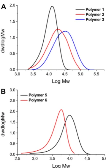
DMAEMA and DMAEA were polymerized in dioxane, and AEMA in acetate buffer (pH 5.2) to ensure protonation of the primary amine, which could otherwise undergo side reactions with the RAFT agent/ester group.42Following polymerization, the polymers were isolated by precipitation and dialyzed against Milli-Q water to remove excess monomer/solvent. The resulting polymers were characterized by SEC (size exclusion
chromatography) and NMR spectroscopy,Figure 1 andTable
1. SEC analysis revealed monomodal distributions but higher than expected dispersity values, which could be attributed to column interactions, as is common for amino-containing polymers. The amine side chains could also lead to some reduction in the fidelity of the RAFT agent also, leading to broadening. Nonetheless, these are accep for the purposes of this study (vide infra) where a library of different chain lengths was the target. The SEC molecular weights were also larger than expected from conversion and the feed ratio, supporting the assumption of some column interactions. PAEMA is challenging to characterize by SEC, as we have previously reported meaning only an estimatedMnbased on conversion and the [monomer]:[CTA] feed ratio.42
This focused library of polymers was then used to screen for antibacterial activity. Three representative strains of bacteria were chosen for this:Escherichia coli, a Gram-negative bacteria;
Pseudomonas putida, which is a Gram-negative bacteria closely related to pathogenic pseudomonas strains; andMycobacterium smegmatis, from the Mycobacterium genus, and an avirulent
model organism forMtb.12 The minimum inhibitory concen-trations (MIC) of the polymer library was determined against all the bacteria using a resazurin microtiter viability assay. In short, the polymers were serially diluted, added to the bacterial cultures, and the concentration where growth was inhibited reported as the MIC99 value. All assays were carried out in
triplicate and the results of this are shown inTable 2. Due to the presence of the inner and outer cell membranes, Gram-negative bacteria are challenging to kill using polycations, requiring careful installation of hydrophobic groups to promote insertion and disruption of the inner/outer membranes, or in the case of PDMAEMA a reduction in pH to increase the net positive charge (the aim here was to test at physiological pH hence it was maintained at 7.4). Our results were in agreement with this hypothesis, with all polymers having higher MIC99 values againstP. putidaandE. colithan againstM. smegmatis. In the case of P5 and P6, extremely high concentrations were needed (concentrations above 3125μg·mL−1,Table 2) despite the only structural difference compared to P1−3 being the
absence of the backbone methyl group (Scheme 1). These
subtle effects highlight the challenge of designing potent macromolecular antimicrobials as each individual strain has subtly different membrane components that govern the initial interactions, but also may offer a route to selectivity. Of particular interest was that P1−3 were very active against M. smegmatis, with the MIC99 value only 5×higher than that of
rifampicin (on a mass basis, more active on molar due to high MW of polymers), a front-line antibiotic used against mycobacteria. The observed potency is similar to what has been reported for other antimicrobial polymers against
nonmycobacteria, such as degradable polycarbonates25 or
[image:5.625.353.532.63.345.2]amino-terminated dendrimers with in vivo activity against E. coli infections in mice.29 The activity of these polymers is somewhat surprisingly considering the complex mAGP Figure 1.SEC analysis of polymers used in this study, as shown in
Table 1.
Biomacromolecules Article
complex that surrounds mycobacteria, which would be expected to reduce the electrostatic interactions between polycations and the anionic cell membrane components. AgainstM. smegmatis, there was no clear effect of molecular weight on activity, but against the Gram-negative organisms, the shortest (4500 g· mol−1) polymers (P1) showed increased activity (Table 2).
This is in line with observations by Ikeda et al., who saw that molecular weight effects on antimicrobial polycations were most pronounced below 10 kg mol−1.32
Based on their selectivity toward M. smegmatis the
PDMAEMAs (P1−3) were further tested to determine their
minimum bactericidal concentration (MBC). Briefly, the
bacteria were grown in the presence of serial dilutions of P1−3starting from 10×MIC99and after 24 h plated on solid agar media and counted. The minimum concentration required to kill all the bacteria was reported,Table 3. Pleasingly, these
values were found to closely correlate with those from the
resazurin MIC testing in liquid media, confirming the
bactericidal activity of the polymers. There was some evidence thatP1(lowestMn) had slightly higher activity than the longer P2andP3which agrees with the observations from the MIC99
results (fromTable 2, above).P3was also tested in a time-kill
assay at 2× MBC and compared to the front-line drug
rifampicin (also at 2× MBC), to determine the rate of
antibacterial action againstM. smegmatis,Figure 2. The polymer killed all the bacteria (down to the detection limit of the assay) within 6 h, but rifampicin took 48 h. This highlights the potent
nature of PDMAEMA against mycobacteria, which functions by a mechanism distinct (vide infra) from front-line drugs, which are slow to act, as they disrupt physiological pathways, which are themselves slow due to the slow-growing nature of mycobacteria.
[image:6.625.62.565.85.165.2]Previous reports of PDMAEMA against Gram-positive bacteria (not mycobacteria) suggested that the mode of action involved membrane binding, followed by permeabilization of the cell membrane, enabling leakage of cytoplasmic contents.41 This is also a potential route to cytotoxicity through lysis of mammalian cell membranes. In particular, this is an issue for red blood cells (RBCs) a key clinical side-effect in thefield of antimicrobial polymers/peptides. P2−4 were tested up to 5 mg·mL−1at 37°C against (ovine) RBCs (Figure 3).P2andP3, which were active against M. smegmatis, showed very little hemolysis, below 2% even at the highest concentration tested as would be expected for this polymer.43Higher concentrations of PDMAEMA (>100μg·mL−1) and molecular weights above 20 kDa than used here have been reported to lead to RBC agglutination, but was not seen here.43In contrast,P4 with a Table 1. Cationic Polymers Synthesized by RAFT Polymerization
polymer monomer [M]:[CTA] conversion (%)a M
n(th)(g mol‑1)b Mn(SEC)(g mol‑1) Mw/Mnc
P1 DMAEMA 50 51.9 4500 11 000 1.25
P2 DMAEMA 50 77.3 6100 13 900 1.61
P3 DMAEMA 100 71.4 11 200 21 800 1.64
P4 AEMA 100 71.4 11 200 N/A N/A
P5 DMAEA 100 77.6 11 000 6900 1.43
P6 DMAEA 25 89.6 3200 3300 1.47
[image:6.625.69.568.211.327.2]aDetermined by1H NMR against an internal mesitylene standard. bDetermined by the [M]:[CTA] ratio and conversion, assuming 100% CTA efficiency.cDetermined by SEC against PMMA standards.
Table 2. Minimum Inhibitory Concentrations of Polymers P1−6a
polymer M. smegmatis(Mycobacteria) (μg·mL−1) P. putida(Gram-negative) (μg·mL−1) E. coli(Gram-negative) (μg·mL−1)
P1 31.25 156.25 62.5
P2 31.25 >5000 250
P3 31.25 >5000 250
P4 500 625 1000
P5 >5000 >5000 >5000
P6 3125 >5000 >5000
tetracycline × 156.25 ×
rifampicin 6.25 × ×
ampicillin × × 3.125
aAll values from three repeats;×indicates the antibiotic was not used against that strain. Antibiotics chosen based on their front-line use against associated infections.
Table 3. Minimum Bactericidal Concentration of P1−3 againstM. smegmatis
polymer μg·mL−1
P1 31.25
P2 31.25−62.5
P3 31.25−62.5
Figure 2. Time-kill assay of P3 against M. smegmatis. Errors bars represent the standard deviation fromn= 3.
[image:6.625.349.549.353.497.2] [image:6.625.59.303.574.625.2]primary amine group showed significant hemolysis at all
concentrations. These results show that the identified
PDMAEMA are more selective toward mycobacteria and less hemolytic than the primary amine containing polymers. (It should be noted that this does not necessarily indicate a complete lack of cytotoxicity44).
The lack of activity against RBCs even at high concentrations raises questions about the mechanism of action against bacteria. Tew and co-workers have extensively studied the mechanism of facially amphiphilic cations against Gram-negative bacteria, revealing a complex process which appears to involve the formation of pores in the membrane enabling leaking of cellular components.19To probe the nature of the polymer−bacteria interaction the polymers were evaluated for their ability to
permeabilize membranes using fluorescence microscopy in
conjugation with membrane permeable/imperable dyes (“live/ dead”), Figure 4. Control experiments of live M. smegmatis
show green color due to the SYTO 9 dye associated with live
(membrane intact) bacteria. Conversely, heat killed M.
smegmatis (which does not lyse the bacteria) showed no green color but some were red, indicative of propidium iodide (dye) being able to cross damage cell membranes. Incubation of theM. smegmatiswithP3(PDMEAMA) for 30 min (as this
effect would be direct and rapid and avoiding secondary
interactions which could be mistaken for membrane damage and hence longer periods were not used, and guided by the kill-curve in Figure 2) both above (2× MIC) and below (0.5× MIC) MIC99 values resulted in green colored bacteria being
obtained in both cases. As the assay does not actually probe if they are alive, but rather if the membrane is damaged, this result suggested that PDMAEMA does not immediately damage the cell membrane, in contrast to the expected mode of action of a polycation.P4(PAEMA) with a primary amine side chain, resulted in red bacteria, due to extensive damage to the cell membrane, consistent with how other polycations function, and suggesting a unique mode of action between PDMAEMA and mycobacteria.
PDMAEMA (P1−3) was the most active polymer class
against mycobacteria, with high selectivity toward these compared to Gram-negative bacteria and erythrocytes while also having relatively low membrane lytic activity. These assays do not, however, prove the lack of interaction/impact between the polymers and the bacterial membranes, just that they are intact and resistant to permeation. To visualize the impact on the membrane, transmission electron microscopy (TEM) was conducted onM. smegmatis, which was grown with and without
the polymers at sublethal doses at 0.5×MIC99,Figure 5. After
incubation, the bacteria were fixed, stained and imaged. The
wild type M. smegmatis without the polymer showed the
expected cell morphology and membrane structure indicating it was intact when fixed. However, the M. smegmatis incubated withP1clearly showed signs of distress, with significant signs of puckering strongly suggesting that the cell wall has been stressed. A possible explanation for this observation would be electrostatic interactions between the polymer and the unique mycobacterial cell wall lipids and/or anionic cell membranes. Nonetheless, these images clearly support a mechanism based on cell wall surface interactions of the polymer.
■
CONCLUSIONSHere it is demonstrated that poly(dimethylaminoethyl methacrylate), PDMAEMA, has potent and selective anti-mycobacterial activity against a model strain forM. tuberculosis. Conversely, it was weakly active against two Gram-negative strains, indicating selectivity. Relatively low molecular weight PDMAEMA (11 kDa) was shown to have minimum inhibitory values as low as 30 μg·mL−1. An acrylate equivalent and polymers with primary amine side chains were found to be significantly less active. Mechanistic studies revealed that the PDMAEMA did not affect the integrity of the mycobacterial cell membrane, confirmed byfluorescent microscopy, indicat-Figure 3.Assessment of hemolytic activity of polymersP2−4after 1 h
[image:7.625.96.260.65.203.2]of exposure against ovine red blood cells. Error bars represent the standard deviationn= 3.
Figure 4.Fluorescence microscopy ofM. smegmatisupon exposure to varying concentrations of P3 and P4. Green channel is SYTO-9 (nucleic acid stain), and red is propidium iodide (damage membrane stain). Each image is 85×85μm2.
Biomacromolecules Article
[image:7.625.323.565.69.428.2]ing an intact membrane/cell wall, unlike primary amine-containing polymers, which despite having lower activity clearly disrupted the cell membranes. TEM analysis supported a mechanism where the PDMAEMA is binding to, and affecting the outer-cell components but without causing lysis. Finally, PDMAEMA was shown to be nonhemolytic to red blood cells, confirming that it has a unique mode of action in exerting its
effect on mycobacteria as well as demonstrating some
biocompatibility. The underlying reasons for the selectivity toward mycobacteria, compared to Gram-negative strains is not clear at this time but could be due to the unique mAGP complex layer surrounding mycobacteria, which are rich in anionic mycolic acids and interspersed with complex lipids that are not found in Gram-negative (or Gram-positive) organisms. Taken together, this shows that nontraditional antibiotics which target bacterial extracellular components, rather than traditional single drug/single protein drugs might offer a solution to the emergence of multidrug resistant mycobacteria and that (relatively) simple polycations could supplement the arsenal of antimycobacterial drugs, in particular againstM. tuberculosis
which is a devastating global disease.
■
AUTHOR INFORMATIONCorresponding Authors
*Fax: +44 247 652 4112. E-mail:[email protected].
*Fax: +44 247 652 2052. E-mail:[email protected]. ORCID
Matthew I. Gibson:0000-0002-8297-1278 Notes
The authors declare no competingfinancial interest.
■
ACKNOWLEDGMENTSEquipment used was supported by the Innovative Uses for Advanced Materials in the Modern World (AM2), with support from Advantage West Midlands (AWM) and part funded by the European Regional Development Fund (ERDF). The microscopy imaging facilities are funded by University of Warwick Advanced BioImaging Research Technology Platform
and BBSRC ALERT14 award BB/M01228X/1. The Royal Society provided funds for the plate reader (Grant number RG120405). M.I.G. acknowledges the ERC for a starter grant (CRYOMAT 638661). E.F. has a Sir Henry Dale Fellowship jointly funded by the Wellcome Trust and the Royal Society (Grant Number 104193/Z/14/Z). D.E.M. thanks the MOAC DTC for a Ph.D. studentship EP/F500378/1. D.P. and S.-J.R. thank the University of Warwick and Institute of Advanced Study for Early Career Fellowships. E.F. and M.I.G. acknowl-edge EPSRC (EP/M027503/1) for supporting JH with a Training Fellowship. E.F., M.I.G., and C.G. acknowledge the Antimicrobial Resistance Cross Council Initiative, support by BBSRC and MRC (MR/N006917/1).
■
REFERENCES(1) Wise, R.; Carrs, O.; Cassell, G.; Fishman, N.; Guidos, R.; Levy, S.; Powers, J.; Norrby, R.; Tillotson, G.; Davies, R.; Projan, S.; Dawson, M.; Monnet, D.; Keogh-Brown, M.; Hand, K.; Garner, S.; Findlay, D.; Morel, C.; Wise, R.; Bax, R.; Burke, F.; Chopra, I.; Czaplewski, L.; Finch, R.; Livermore, D.; Piddock, L. J. V.; White, T.J. Antimicrob.
Chemother.2011,66(9), 1939−1940.
(2) Centre for Disease Control; Antibiotic/Antimicrobial Resistance,
https://www.cdc.gov/drugresistance/.
(3) World Economic Forum; Global Risks, 2013; http://reports. weforum.org/global-risks-2013/.
(4) Nathan, C.Nature2004,431(7011), 899−902.
(5) O’Neil, J. The Review on Antimicrobial Resistance; O’Neil Report.
(6) Richards, S.-J.; Jones, M. W.; Hunaban, M.; Haddleton, D. M.; Gibson, M. I.Angew. Chem., Int. Ed.2012,51(31), 7812−7816.
(7) Otten, L.; Gibson, M. I.RSC Adv.2015,5(66), 53911−53914. (8) Otten, L.; Fullam, E.; Gibson, M. I.Mol. BioSyst.2016,12(2), 341−344.
(9) Andersson, D. I.; Hughes, D.Nat. Rev. Microbiol.2010,8 (4), 260.
(10)The Lancet; Elsevier, April 27, 2013; p 1431.
[image:8.625.131.494.66.295.2](11) World Health Organisation; AMR Factsheet 194, 2015. (12) Makarov, V.; Manina, G.; Mikusova, K.; Möllmann, U.; Ryabova, O.; Saint-Joanis, B.; Dhar, N.; Pasca, M. R.; Buroni, S.; Lucarelli, A. P.; Milano, A.; De Rossi, E.; Belanova, M.; Bobovska, A.; Dianiskova, P.; Kordulakova, J.; Sala, C.; Fullam, E.; Schneider, P.;
Figure 5.TEM analysis ofM. smegmatiswith and withoutP1.P1applied at 10μg·mL−1(0.3×MIC
99). Top three panels, scale bar = 1μm. Bottom three panels, scale bar = 100 nm.
McKinney, J. D.; Brodin, P.; Christophe, T.; Waddell, S.; Butcher, P.; Albrethsen, J.; Rosenkrands, I.; Brosch, R.; Nandi, V.; Bharath, S.; Gaonkar, S.; Shandil, R. K.; Balasubramanian, V.; Balganesh, T.; Tyagi, S.; Grosset, J.; Riccardi, G.; Cole, S. T.Science2009,324(5928), 801− 804.
(13) Neres, J.; Pojer, F.; Molteni, E.; Chiarelli, L. R.; Dhar, N.; Boy-Röttger, S.; Buroni, S.; Fullam, E.; Degiacomi, G.; Lucarelli, A. P.; Read, R. J.; Zanoni, G.; Edmondson, D. E.; De Rossi, E.; Pasca, M. R.; McKinney, J. D.; Dyson, P. J.; Riccardi, G.; Mattevi, A.; Cole, S. T.; Binda, C.Sci. Transl. Med.2012,4(150), 150ra121.
(14) Leeson, P.Nature2012,481(7382), 455−456. (15) Zasloff, M.Nature2002,415(6870), 389−395.
(16) Seo, M.-D.; Won, H.-S.; Kim, J.-H.; Mishig-Ochir, T.; Lee, B.-J.
Molecules2012,17(10), 12276−12286.
(17) Gordon, Y. J.; Romanowski, E. G.; McDermott, A. M.Curr. Eye Res.2005,30(7), 505−515.
(18) Brogden, K. A.Nat. Rev. Microbiol.2005,3(3), 238−250. (19) Yang, L.; Gordon, V. D.; Trinkle, D. R.; Schmidt, N. W.; Davis, M. A.; DeVries, C.; Som, A.; Cronan, J. E.; Tew, G. N.; Wong, G. C. L.
Proc. Natl. Acad. Sci. U. S. A.2008,105(52), 20595−20600.
(20) Thomas, L.; Clarkson, M.; Delves-Broughton, J. Natural food
antimicrobial systems; Naidu, A.., Ed.; CRC Press, 2000.
(21) Lopez, A. I.; Reins, R. Y.; McDermott, A. M.; Trautner, B. W.; Cai, C.Mol. BioSyst.2009,5(10), 1148−1156.
(22) Kenawy, E.-R.; Worley, S. D.; Broughton, R.Biomacromolecules
2007,8(5), 1359−1384.
(23) Jain, A.; Duvvuri, L. S.; Farah, S.; Beyth, N.; Domb, A. J.; Khan,
W.Adv. Healthcare Mater.2014,3(12), 1969−1985.
(24) Hasan, J.; Crawford, R. J.; Ivanova, E. P.Trends Biotechnol.2013, 31(5), 295−304.
(25) Chin, W.; Yang, C.; Ng, V. W. L.; Huang, Y.; Cheng, J.; Tong, Y. W.; Coady, D. J.; Fan, W.; Hedrick, J. L.; Yang, Y. Y.Macromolecules
2013,46, 8797−8807.
(26) Siedenbiedel, F.; Tiller, J. C.Polymers (Basel, Switz.)2012,4(4), 46−71.
(27) Lui, L. T.; Xue, X.; Sui, C.; Brown, A.; Pritchard, D. I.; Halliday, N.; Winzer, K.; Howdle, S. M.; Fernandez-Trillo, F.; Krasnogor, N.; Alexander, C.Nat. Chem.2013,5(12), 1058−1065.
(28) Insua, I.; Liamas, E.; Zhang, Z.; Peacock, A. F. A.; Krachler, A. M.; Fernandez-Trillo, F.Polym. Chem.2016,7(15), 2684−2690.
(29) Xue, X.; Chen, X.; Mao, X.; Hou, Z.; Zhou, Y.; Bai, H.; Meng, J.; Da, F.; Sang, G.; Wang, Y.; Luo, X.AAPS J.2013,15(1), 132−142. (30) Li, M.; Cha, D. J.; Lai, Y.; Villaruz, A. E.; Sturdevant, D. E.; Otto,
M.Mol. Microbiol.2007,66(5), 1136−1147.
(31) Tzeng, Y.-L.; Ambrose, K. D.; Zughaier, S.; Zhou, X.; Miller, Y. K.; Shafer, W. M.; Stephens, D. S.J. Bacteriol.2005,187(15), 5387− 5396.
(32) Ikeda, T.; Yamaguchi, H.; Tazuke, S. Antimicrob. Agents
Chemother.1984,26(2), 139−144.
(33) Louzao, I.; Sui, C.; Winzer, K.; Fernandez-Trillo, F.; Alexander,
C.Eur. J. Pharm. Biopharm.2015,95(Pt A), 47−62.
(34) Tew, G. N.; Liu, D.; Chen, B.; Doerksen, R. J.; Kaplan, J.; Carroll, P. J.; Klein, M. L.; DeGrado, W. F.Proc. Natl. Acad. Sci. U. S. A.2002,99(8), 5110−5114.
(35) Lienkamp, K.; Kumar, K.-N.; Som, A.; Nüsslein, K.; Tew, G. N.
Chem. - Eur. J.2009,15(43), 11710−11714.
(36) Ramón-García, S.; Mikut, R.; Ng, C.; Ruden, S.; Volkmer, R.; Reischl, M.; Hilpert, K.; Thompson, C. J. Antimicrob. Agents
Chemother.2013,57(5), 2295−2303.
(37) Abedinzadeh, M.; Gaeini, M.; Sardari, S. J. Antimicrob.
Chemother.2015,70(5), 1285−1289.
(38) Khara, J. S.; Wang, Y.; Ke, X.-Y.; Liu, S.; Newton, S. M.; Langford, P. R.; Yang, Y. Y.; Ee, P. L. R.Biomaterials2014,35(6), 2032−2038.
(39) Scott, M. G.; Gold, M. R.; Hancock, R. E.Infect. Immun.1999, 67(12), 6445−6453.
(40) Thaker, H. D.; Cankaya, A.; Scott, R. W.; Tew, G. N.ACS Med.
Chem. Lett.2013,4(5), 481−485.
(41) Rawlinson, L.-A. B.; Ryan, S. M.; Mantovani, G.; Syrett, J. A.; Haddleton, D. M.; Brayden, D. J. Biomacromolecules 2010, 11 (2), 443−453.
(42) Mitchell, D. E.; Lilliman, M.; Spain, S. G.; Gibson, M. I.
Biomater. Sci.2014,2(12), 1787−1795.
(43) Cerda-Cristerna, B. I.; Flores, H.; Pozos-Guillén, A.; Pérez, E.; Sevrin, C.; Grandfils, C.J. Controlled Release2011,153(3), 269−277. (44) Rawlinson, L.-A. B.; O’Brien, P. J.; Brayden, D. J.J. Controlled
Release2010,146(1), 84−92.
(45) Phillips, D. J.; Wilde, M.; Greco, F.; Gibson, M. I.
Biomacromolecules2015,16(10), 3256−3264.
(46) Etienne, G.; Villeneuve, C.; Billman-Jacobe, H.; Astarie-Dequeker, C.; Dupont, M. A.; Daffe, M.́ Microbiology 2002, 148 (10), 3089−3100.
Biomacromolecules Article