ORIGINAL RESEARCH
Reduced Cortical Thickness in Children with
New-Onset Seizures
E. Widjaja S. Zarei Mahmoodabadi C. Go C. Raybaud S. Chuang O.C. Snead M.L. Smith
BACKGROUND AND PURPOSE: Children with new-onset seizures may have antecedent neurobiologic alterations that predispose them to developing seizures. Our aim was to evaluate hippocampal and thalamic volumes and lobar cortical thickness of children with new-onset seizures.
MATERIALS AND METHODS: Twenty-nine children with new-onset seizures and normal MR imaging findings were recruited. Ten patients had generalized seizures, 19 had partial seizures, and 15 were on antiepileptic medications. Twenty-three age-matched healthy controls were also recruited. Hippocam-pal and thalamic volumes and lobar cortical thickness, including frontal, medial temporal, lateral temporal, parietal, cingulate, and occipital cortical thickness, were assessed by using volumetric T1-weighted imaging and were compared between patients and controls.
RESULTS: There were no significant differences in hippocampal and thalamic volumes of patients with new-onset seizures, including the subgroups with generalized and partial seizures and those on and off antiepileptic medications, compared with controls (P⬎.01). There was significant reduction in cortical thickness in right cingulate (P⫽.004), right medial temporal (P⫽.006), and left frontal (P⫽.007) cortices in patients with new-onset seizures. Patients with generalized seizures did not demonstrate a significant reduction in cortical thickness (P⬎.01). Patients with partial seizures demonstrated a significant reduction in cortical thickness in the right frontal (P⫽.008), right parietal (P⫽.003), and left frontal (P⫽.007) cortices. There were no significant differences in cortical thickness among patients on or off antiepileptic medications (P⬎.01).
CONCLUSIONS:We found reduced cortical thickness in children with new-onset seizures. Further studies are necessary to elucidate the neurobiologic relevance of these structural changes.
ABBREVIATIONS:EEG⫽electroencephalography; PD⫽proton density
P
atients with chronic localization-related epilepsy, includ-ing adults and children, have demonstrated structural ab-normalities, including cortical thickness and volume reduc-tion and reduced hippocampal volume.1-7 Volumetricabnormalities have been reported in neuronal regions in-volved in the generation and propagation of seizures, includ-ing the hippocampus,8-11 amygdala,12 entorhinal cortex,13
thalamus,14 and also extratemporal regions15,16 in patients
with chronic temporal lobe epilepsy. Children with idiopathic generalized epilepsies have also shown distributed patterns of abnormality affecting the thalamus and frontal lobe.17-21It is
not known whether the structural alterations developed dur-ing a vulnerable phase of cerebral development or are the cu-mulative effects of recurrent seizures22or are a combination of these factors acting synergistically.11To determine whether
antecedent neurobiologic alterations are present, assessment could be performed in a population of patients with new-onset seizures, which would exclude the confounding effects of recurrent seizures and long-term antiepileptic medications on the brain.
Children with new-onset seizures or new-onset epilepsy are known to have neuropsychological deficits and academic
underachievement,23-26 even when they are intellectually
healthy. It is possible that there is an antecedent neurobiologic alteration that predisposes to both seizures and neuropsycho-logical deficits. Volumetric imaging of the brain has been used to quantify total and lobar gray and white matter volumes in children with new-onset epilepsy but did not identify overall differences in total cerebral or lobar gray and white matter volumes.24It is possible that voxel-based morphometry of
to-tal and lobar volumes is not sufficiently sensitive to assess sub-tle changes in brain structures. Alternatively, other structures such as the hippocampi or thalami could be more susceptible to the neurobiologic alterations and are, therefore, more likely to manifest structural changes. In this study, we evaluated the hippocampal and thalamic volumes and lobar cortical thick-ness of the cerebral hemispheres of children with new-onset seizures. Our hypothesis was that antecedent structural alter-ations, including changes in cortical thickness and other gray matter structures, were present in children with new-onset seizures, which predisposed the patients to seizures.
Materials and Methods
This study had the approval of the research ethics board. We retro-spectively recruited children between 5 and 18 years of age with new-onset seizures who had undergone an MR imaging from 2008 to 2010. The exclusion criteria were children with febrile seizures and those with a mass or focal or diffuse abnormality on MR imaging as assessed by the pediatric neuroradiologist. Twenty-three consecutive children with unprovoked seizures and normal MR imaging findings were recruited. The type of seizures, either focal or generalized seizures, was obtained from the MR imaging requisitions and patient charts and was available in the EEG records. We also noted whether the
Received March 17, 2011; accepted after revision June 25.
From the Department of Diagnostic Imaging (E.W., S.Z.M., C.R., S.C.), Division of Neurology (C.G., O.C.S.), and Department of Psychology (M.L.S.), Hospital for Sick Children, Toronto, Ontario, Canada.
Please address correspondence to E. Widjaja, MD, 555 University Ave, Toronto, Ontario M5G 1X8, Canada; e-mail: Elysa.Widjaja@sickkids.ca
Indicates article with supplemental on-line table. http://dx.doi.org/10.3174/ajnr.A2982
PEDIATRICS
ORIGINAL
patients were on antiepileptic medications. Twenty-three healthy age-matched controls with no neurologic or psychiatric disorders who participated in another epilepsy study were also included in this study. All 23 controls had normal MR imaging findings.
MR Imaging and Image Processing
MR imaging was performed on 3T scanner (Achieva, Philips Medical Systems, Best, the Netherlands) by using an 8-channel phased-array head coil in patients and controls. The imaging in patients consisted of axial and coronal FLAIR (TR/TE⫽10,000/140 ms, section thick-ness⫽3 mm, FOV⫽22 cm, matrix⫽316⫻290), T2 and PD (TR/TE⫽4200/80/40 ms, section thickness⫽3 mm, FOV⫽22 cm, matrix⫽400⫻272), and volumetric 3D T1 (TR/TE⫽4.9/2.3 ms, section thickness⫽1 mm, FOV⫽22 cm, matrix⫽220⫻220). The imaging in controls included axial FLAIR (TR/TE⫽10,000/140 ms, section thickness⫽3 mm, FOV⫽22 cm, matrix⫽316⫻290), T2 and PD (TR/TE⫽4200/80/40 ms, section thickness⫽3 mm, FOV⫽ 22 cm, matrix⫽400⫻272), and volumetric 3D T1 (TR/TE⫽4.9/2.3 ms, section thickness⫽1 mm, FOV⫽22 cm, matrix⫽220⫻220). FreeSurfer (version 3.05, https://surfer.nmr.mgh.harvard.edu) was used for hippocampal and thalamic volume assessment, as well as cortical surface reconstruction and cortical thickness estimation of the patients and controls.27-34Nonuniform intensity-correction was
performed. Nonuniform nonparametric intensity normalization was applied. The data were skull-stripped and linear and nonlinear nor-malized to the Montreal Neurological Institute 305 atlas within Free-Surfer. Segmentation of the white matter was obtained by using a connected-components algorithm. Subcortical structures, including the basal ganglia, thalamus, amygdala, hippocampus, and the ventri-cles were labeled by using a probabilistic atlas and Bayesian classifica-tion rule for label assignment. The hippocampal and thalamic vol-umes were obtained.
To assess the cortical thickness, we divided the brain into 2 hemi-spheres and filled the white matter region and ventricles to obtain a single white matter volume for each hemisphere, which was then cov-ered with a polygonal tessellation and smoothed to reduce metric distortions. The obtained surface was inflated and topologic defects were automatically corrected. Subsequently, the gray-white matter boundary was reconstructed by segmenting all white matter voxels in the MR imaging, and the resulting white matter surface was refined to obtain submillimeter accuracy in delineating the gray-white matter surface. This surface was then outward deformed to identify the gray-CSF boundary. The cortical thickness at each vertex across the cortical mantle was defined by calculating the average of the following: 1) the shortest distance between the gray-white boundary and the gray-CSF boundary, and 2) the shortest distance between the gray-CSF bound-ary and the gray-white boundbound-ary at each vertex on the tessellated
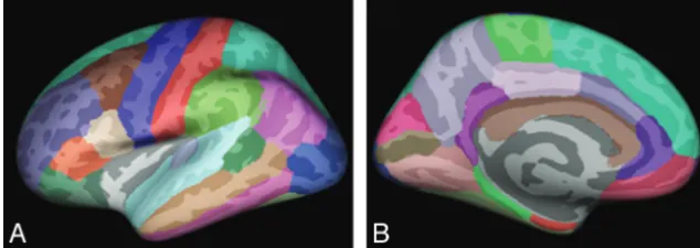
surface. Thickness measures were then mapped to the inflated surface of each brain reconstruction, allowing optimal visualization in both sulcal and gyral regions across the entire neocortex without being obscured by cortical folding. Sulcal and gyral features across individ-ual subjects were aligned by morphing each subject’s brain to an av-erage spheric representation by using a nonrigid high-resolution sur-face based an averaging method that allowed accurate matching of cortical locations among subjects while minimizing metric distortion. The data were then smoothed on the tessellated surface by using a 10-mm full width half maximum Gaussian kernel to improve the signal intensity–to-noise ratio. An automated parcellation technique was used to subdivide each hemisphere into 34 gyral labels (11 frontal, 4 medial temporal, 5 lateral temporal, 5 parietal, 4 occipital, and 5 cingulate) (Fig 1).35Six lobar regions of interest were subsequently
defined in each hemisphere: frontal, medial temporal, lateral tempo-ral, parietal, cingulate, and occipital.36The mean thickness of each
lobe was obtained by using the weighted average of the thickness within each gyral-based region of interest—that is, mean regional thickness multiplied by the number of vertices for that region and divided by the total number of vertices.
Statistical Analysis
Statistical analysis was performed by using the Statistical Package for the Social Sciences, Version 15 (SPSS, Chicago, Illinois). The hip-pocampal volume, thalamic volume, and lobar cortical thickness of patients with new-onset seizures and controls were compared by us-ing the Mann-WhitneyUtest. The hippocampal volume, thalamic volume, and lobar cortical thickness of subgroups of patients with generalized and partial seizures and those who were on or off antiepi-leptic medications were compared with those in controls by using analysis of variance and subsequently the Mann-WhitneyUtest. To minimize the likelihood of type I error, aPvalue of⬍.01 was consid-ered statistically significant.
Results
Subjects
Twenty-nine patients with new-onset seizures and normal MR imaging findings (17 males and 12 females; mean age, 9.2⫾ 2.2 years) were included. Ten patients had generalized sei-zures, including tonic clonic, absence, and myoclonic seisei-zures, and 19 had partial seizures. Fifteen patients were on antiepi-leptic medications at the time of the MR imaging, 8 patients were on carbamazepine, 6 patients were on valproic acid, and 1 was on oxcarbazepine. Thirteen patients were not on anti-epileptic medications at the time of MR imaging; in 1 patient, the medication status could not be obtained. The time interval
[image:2.594.134.451.45.157.2]between seizure onset and MR imaging varied from 9 days to 1.1 years, with a mean of 5.8 months. Twenty-three healthy controls with normal MR imaging findings (14 males and 9 females; mean age, 10.0⫾2.7 years) were included. There was no significant difference in the age of patients and controls.
Hippocampal and Thalamic Volumes
There were no significant differences in hippocampal and tha-lamic volumes of patients with new-onset seizures and con-trols (P⬎.01 for all) (On-line Table).
Subgroup analysis of patients with generalized or partial seizures also did not show any significant differences in hip-pocampal and thalamic volumes of both patient groups com-pared with controls (P⬎.01 for all) or between patients with generalized and those with partial seizures (P⬎.01 for all).
Subgroup analysis among patients on or off antiepileptic medications did not show any significant differences in hip-pocampal and thalamic volumes of both patient groups com-pared with controls (P⬎.01 for all) or between the 2 groups of patients on and off antiepileptic medications (P⬎.01 for all).
Lobar Cortical Thickness
Patients with new-onset seizures demonstrated a significant reduction in cortical thickness in the right cingulate (P ⫽ .004), medial temporal (P⫽.006), and left frontal (P⫽.007) cortices and a tendency to reduced cortical thickness in the right frontal (P⫽.015), parietal (P⫽.018), and left cingulate (P⫽.054) cortices (On-line Table).
Subgroup analysis of patients with generalized seizures demonstrated a tendency toward reduced cortical thickness in the right cingulate (P⫽.028) and medial temporal (P⫽.042) cortices compared with controls. Subgroup analysis of pa-tients with partial seizures demonstrated significant reduction in cortical thickness in the right frontal (P⫽.008), right pari-etal (P⫽ .003), and left frontal (P⫽.007) cortices, and a tendency toward reduced cortical thickness in the right cingu-late (P⫽.010) and medial temporal (P⫽.013) cortices. There were no significant differences in lobar cortical thicknesses of patients with generalized seizures and those with partial sei-zures (P⬎.01 for all).
Subgroup analysis of patient who were on antiepileptic medications showed a tendency toward reduced cortical thickness in the right cingulate (P⫽.035), right medial tem-poral (P⫽.041), right parietal (P⫽.044), and left frontal (P⫽ .030) cortices compared with controls. Patients who were not on antiepileptic medications showed a tendency toward re-duced cortical thickness in the right frontal (P⫽.031), right cingulate (P⫽.012), right medial temporal (P⫽.020), and left frontal (P⫽.034) cortices. There were no significant dif-ferences in the cortical thickness of patients on antiepileptic medications and those not on antiepileptic medications (P⬎ .01 for all).
Discussion
We found that children with new-onset seizures and normal routine MR imaging findings demonstrated significant reduc-tion in cortical thickness in the right cingulate, medial tempo-ral, and left frontal cortices. However, we did not detect a significant alteration in thalamic and hippocampal volumes. The significance of the reduction in cortical thickness in the
cingulate, medial temporal, and frontal cortices remains to be elucidated. A previous study in children with new-onset epi-lepsy found impairment in several cognitive domains, includ-ing intelligence, executive function, psychomotor speed, and language.24The reduction in cortical thickness in the frontal
lobe could be related to impairment in executive function and psychomotor speed, and the reduced cortical thickness in the cingulate may reflect impairment in executive function.
Few studies have examined the structural consequences of predominantly adults with new-onset seizures.37,38Liu et al37
evaluated the hippocampal, cerebellar, total brain, gray and white matter, and intracranial volumes and hippocampal T2 relaxation times in patients with new-onset seizures, and they found no significant change in brain volumes and hippocam-pal T2 relaxation times in patients compared with controls at baseline. The observed lack of alteration in total gray and white matter volumes and cerebral volume may be due to a variety of reasons. Volume loss may occur within specific brain regions rather than diffusely in the whole brain. Measurements using total brain and cerebral gray and white matter volumes may not be sufficiently sensitive to identify volumetric changes within specific regions of the brain. In a subsequent study by the same group, the authors found a reduction in hippocam-pal volume in a subgroup of patients with new-onset temporal lobe epilepsy.38 We also found no significant differences in
hippocampal volume in children with new-onset seizures compared with controls, similar to findings in the earlier study by Liu et al.37Although subgroup analysis was performed in patients with generalized and partial seizures and did not dem-onstrate a difference in hippocampal or thalamic volume, we did not distinguish the different subtypes of partial seizures due to the relatively small sample size. Liu et al included pa-tients with normal structural imaging findings as well as those with lesions such as focal cortical dysplasia, cavernoma, and dysembryoplastic neuroepithelial tumor. We excluded pa-tients with lesions due to potential for these lesions to affect cortical thickness measurements.
Hermann et al24evaluated children with new-onset
epi-lepsy and found no significant change in total cerebral gray and white matter volumes and also no significant change in frontal, parietal, temporal, and occipital volumes within 1 year of the diagnosis of epilepsy. The authors also found no signif-icant differences in total gray and white matter and lobar vol-umes in patients with partial and generalized epilepsy. How-ever, the subgroup with academic underachievement revealed significantly lower gray matter volumes in the parietal and occipital regions compared with controls and patients without academic problems. We have not distinguished patients with or without academic problems because such data were not available in this retrospective study. The reduction in cortical thickness in our patients may be related to differences in pop-ulation characteristics or methodologic assessment of the cor-tex, in that thickness measures by using surface-based mor-phometry are more sensitive than volume measures by using voxel-based morphometry. Pulsipher et al39also assessed
epilepsy compared with controls. We have not found a reduc-tion in frontal cortical thickness in patients with generalized seizures. Failure to identify such a reduction in may be related to the sample size.
One of the potential confounding factors of our study was that approximately half of the patients were on antiepileptic medications at the time of MR imaging. However, subgroup analysis results of those who were on antiepileptic medication were not significantly different compared with those who were not on antiepileptic medications. Another potential con-founding factor was the time interval between seizure onset and MR imaging. This is partly related to wait times for a pediatric neurologist consult and also for MR imaging, in par-ticular if the MR imaging had to be performed with the patient under general anesthesia. In the prospective studies by Her-mann et al24and Pulsipher et al,39the time interval between the diagnosis of epilepsy and imaging was also within 1 year of diagnosis, similar to that in our study. Further study that eval-uates structural changes in the brain within a shorter time interval between the diagnosis of new seizures or epilepsy and neuroimaging is needed to exclude any potential effects of these ongoing changes from as-yet-unknown predisposing factors.
We excluded children with a mass or focal or diffuse ab-normalities on MR imaging as assessed by the pediatric neu-roradiologist. However, it is possible that some patients with subtle focal cortical dysplasia were not identified and that some cases with focal cortical dysplasia may have had de-creased cortical thickness, which confounded the results. We have minimized the likelihood of missing a lesion by perform-ing the MR imagperform-ing on a 3T system with a high-resolution epilepsy protocol in all patients. We have analyzed children with generalized and partial seizures as a group due to the size of the study cohort, similar to the analysis in the study by Hermann et al24and Tosun et al.40Subsequently, subgroup
analysis was performed in patients with generalized and par-tial seizures. Details of the specific seizure localization were not available in all patients with partial seizures because short EEG recordings were obtained in patients with new-onset sei-zures and none had prolonged video-EEG recordings. Also the EEG recordings were not available for review in some patients who were referred by external neurologists.
A longitudinal study of children with new-onset epilepsy has found differences in baseline gray and white matter vol-umes of patients compared with controls, suggesting that an-tecedent anomalies in brain development were present.40In
addition to changes in baseline brain volumes, the authors also found slowed white matter expansion and changes in gray matter volume in patients at 2-year follow-up. The image pro-cessing was done by using deformation-based morphometry, which is a more sensitive tool for detecting changes in brain volumes because it avoids the need for image segmentation. The authors identified changes in brain volumes in children with new-onset epilepsy40 that were not revealed by using
voxel-based morphometry in an earlier study.24
We have assessed cortical thickness rather than cortical vol-ume and have also identified changes in the cortex in children with new-onset seizures, similar to the assessment in the study by Tosun et al,40 again providing support that antecedent
anomalies were present in these patients. Janssen et al41
eval-uated cortical thickness, surface area, and cortical volume in adolescents with first-episode early-onset psychosis. They found more widespread areas of cortical thinning relative to volume reduction, attributed to changes in surface area, which counteracted volume changes. We have used surface-based morphometry to evaluate cortical thickness because this methodology is less influenced by individual gyral variations than traditional voxel-based morphometry. The FreeSurfer software allows assessment of not only the cortical thickness but also volumetric measurement of other gray matter struc-tures, including the hippocampus and thalamus. However, one of the disadvantages of our method of analysis is that it does not include white matter measurements.
Conclusions
We found reduced cortical thickness in the right medial temporal, cingulate, and left frontal cortices but no significant differences in hippocampal and thalamic volumes in children with new-onset seizures. Structural changes in the gray matter suggest that antecedent developmental anomalies are present and may predispose the patients to seizures. Our findings in-dicate that cortical thickness is a sensitive measure of gray matter integrity and is compromised in children with new-onset seizures. Further studies are needed to verify the relation between gray matter integrity and neurocognitive function as well as to assess these changes longitudinally.
References
1. Lawson JA, Cook MJ, Bleasel AF, et al.Quantitative MRI in outpatient child-hood epilepsy.Epilepsia1997;38:1289 –93
2. Lawson JA, Nguyen W, Bleasel AF, et al.ILAE-defined epilepsy syndromes in children: correlation with quantitative MRI.Epilepsia1998;39:1345– 49 3. Lawson JA, Vogrin S, Bleasel AF, et al.Predictors of hippocampal, cerebral, and
cerebellar volume reduction in childhood epilepsy.Epilepsia2000;41:1540 – 45 4. Lawson JA, Vogrin S, Bleasel AF, et al.Cerebral and cerebellar volume
reduc-tion in children with intractable epilepsy.Epilepsia2000;41:1456 – 62 5. Lawson JA, Cook MJ, Vogrin S, et al.Clinical, EEG, and quantitative MRI
differences in pediatric frontal and temporal lobe epilepsy. Neurology
2002;58:723–29
6. Mueller SG, Laxer KD, Barakos J, et al.Widespread neocortical abnormalities in temporal lobe epilepsy with and without mesial sclerosis.Neuroimage
2009;46:353–59
7. McDonald CR, Ahmadi ME, Hagler DJ, et al.Diffusion tensor imaging corre-lates of memory and language impairments in temporal lobe epilepsy. Neu-rology2008;71:1869 –76
8. Jack CR Jr, Sharbrough FW, Cascino GD, et al.Magnetic resonance image-based hippocampal volumetry: correlation with outcome after temporal lo-bectomy.Ann Neurol1992;31:138 – 46
9. Quigg M, Bertram EH, Jackson T, et al.Volumetric magnetic resonance imag-ing evidence of bilateral hippocampal atrophy in mesial temporal lobe epi-lepsy.Epilepsia1997;38:588 –94
10. Woermann FG, Barker GJ, Birnie KD, et al.Regional changes in hippocampal T2 relaxation and volume: a quantitative magnetic resonance imaging study of hippocampal sclerosis.J Neurol Neurosurg Psychiatry1998;65:656 – 64 11. Tasch E, Cendes F, Li LM, et al.Neuroimaging evidence of progressive
neuro-nal loss and dysfunction in temporal lobe epilepsy.Ann Neurol1999;45: 568 –76
12. Kalviainen R, Salmenpera T, Partanen K, et al.MRI volumetry and T2 relaxom-etry of the amygdala in newly diagnosed and chronic temporal lobe epilepsy.
Epilepsy Res1997;28:39 –50
13. Bernasconi N, Bernasconi A, Andermann F, et al.Entorhinal cortex in temporal lobe epilepsy: a quantitative MRI study.Neurology1999;52:1870 –76 14. DeCarli C, Hatta J, Fazilat S, et al.Extratemporal atrophy in patients with
complex partial seizures of left temporal origin.Ann Neurol1998;43:41– 45 15. Marsh L, Morrell MJ, Shear PK, et al.Cortical and hippocampal volume
defi-cits in temporal lobe epilepsy.Epilepsia1997;38:576 – 87
16. Sandok EK, O’Brien TJ, Jack CR, et al.Significance of cerebellar atrophy in intractable temporal lobe epilepsy: a quantitative MRI study.Epilepsia
17. Betting LE, Mory SB, Li LM, et al.Voxel-based morphometry in patients with idiopathic generalized epilepsies.Neuroimage2006;32:498 –502
18. Betting LE, Mory SB, Lopes-Cendes I, et al.MRI volumetry shows increased anterior thalamic volumes in patients with absence seizures.Epilepsy Behav
2006;8:575– 80
19. Betting LE, Mory SB, Lopes-Cendes I, et al.MRI reveals structural abnormal-ities in patients with idiopathic generalized epilepsy.Neurology2006;67: 848 –52
20. Pardoe H, Pell GS, Abbott DF, et al.Multi-site voxel-based morphometry: methods and a feasibility demonstration with childhood absence epilepsy.
Neuroimage2008;42:611–16
21. Pulsipher DT, Seidenberg M, Guidotti L, et al.Thalamofrontal circuitry and executive dysfunction in recent-onset juvenile myoclonic epilepsy.Epilepsia
2009;50:1210 –19
22. Pringle CE, Blume WT, Munoz DG, et al.Pathogenesis of mesial temporal sclerosis.Can J Neurol Sci1993;20:184 –93
23. Fastenau PS, Johnson CS, Perkins SM, et al.Neuropsychological status at sei-zure onset in children: risk factors for early cognitive deficits.Neurology
2009;73:526 –34
24. Hermann B, Jones J, Sheth R, et al.Children with new-onset epilepsy: neuro-psychological status and brain structure.Brain2006;129:2609 –19 25. Oostrom KJ, Smeets-Schouten A, Kruitwagen CL, et al.Not only a matter of
epilepsy: early problems of cognition and behavior in children with “epilepsy only”—a prospective, longitudinal, controlled study starting at diagnosis. Pe-diatrics2003;112:1338 – 44
26. Berg AT, Smith SN, Frobish D, et al.Special education needs of children with newly diagnosed epilepsy.Dev Med Child Neurol2005;47:749 –53
27. Fischl B, Sereno MI, Tootell RB, et al.High-resolution intersubject averaging and a coordinate system for the cortical surface. Hum Brain Mapp
1999;8:272– 84
28. Fischl B, Sereno MI, Dale AM.Cortical surface-based analysis. II. Inflation, flattening, and a surface-based coordinate system.Neuroimage1999;9:195– 207
29. Fischl B, Dale AM.Measuring the thickness of the human cerebral cortex from magnetic resonance images.Proc Natl Acad Sci U S A2000;97:11050 –55 30. Fischl B, Liu A, Dale AM.Automated manifold surgery: constructing
geomet-rically accurate and topologically correct models of the human cerebral cor-tex.IEEE Trans Med Imaging2001;20:70 – 80
31. Fischl B, Salat DH, Busa E, et al.Whole brain segmentation: automated label-ing of neuroanatomical structures in the human brain.Neuron2002;33: 341–55
32. Fischl B, van der Kouwe A, Destrieux C, et al.Automatically parcellating the human cerebral cortex.Cereb Cortex2004;14:11–22
33. Dale AM, Fischl B, Sereno MI.Cortical surface-based analysis. I. Segmentation and surface reconstruction.Neuroimage1999;9:179 –94
34. Segonne F, Grimson E, Fischl B.A genetic algorithm for the topology correc-tion of cortical surfaces.Inf Process Med Imaging2005;19:393– 405 35. Desikan RS, Segonne F, Fischl B, et al.An automated labeling system for
sub-dividing the human cerebral cortex on MRI scans into gyral based regions of interest.Neuroimage2006;31:968 – 80
36. McDonald CR, Hagler DJ Jr, Ahmadi ME, et al.Regional neocortical thinning in mesial temporal lobe epilepsy.Epilepsia2008;49:794 – 803
37. Liu RS, Lemieux L, Sander JW, et al.Seizure-associated hippocampal volume loss: a longitudinal magnetic resonance study of temporal lobe epilepsy.Ann Neurol2002;52:861, author reply 862
38. Liu RS, Lemieux L, Bell GS, et al.Cerebral damage in epilepsy: a population-based longitudinal quantitative MRI study.Epilepsia2005;46:1482–94 39. Pulsipher DT, Dabbs K, Tuchsherer V, et al.Thalamofrontal
neurodevelop-ment in new-onset pediatric idiopathic generalized epilepsy.Neurology
2011;76:28 –33
40. Tosun D, Dabbs K, Caplan R, et al.Deformation-based morphometry of pro-spective neurodevelopmental changes in new onset paediatric epilepsy.Brain
2011;134(Pt 4):1003–14. Epub 2011 Mar 11