ORIGINAL RESEARCH
ADULT BRAIN
Mapping the Orientation of White Matter Fiber Bundles: A
Comparative Study of Diffusion Tensor Imaging, Diffusional
Kurtosis Imaging, and Diffusion Spectrum Imaging
XG.R. Glenn, XL.-W. Kuo, X Y.-P. Chao,X C.-Y. Lee, XJ.A. Helpern, and XJ.H. Jensen
ABSTRACT
BACKGROUND AND PURPOSE: White matter fiber tractography relies on fiber bundle orientation estimates from diffusion MR imaging. However, clinically feasible techniques such as DTI and diffusional kurtosis imaging use assumptions, which may introduce error into in vivo orientation estimates. In this study, fiber bundle orientations from DTI and diffusional kurtosis imaging are compared with diffusion spectrum imaging as a criterion standard to assess the performance of each technique.
MATERIALS AND METHODS:For each subject, full DTI, diffusional kurtosis imaging, and diffusion spectrum imaging datasets were acquired during 2 independent sessions, and fiber bundle orientations were estimated by using the specific theoretic assumptions of each technique. Angular variability and angular error measures were assessed by comparing the orientation estimates. Tractography generated with each of the 3 reconstructions was also examined and contrasted.
RESULTS:Orientation estimates from all 3 techniques had comparable angular reproducibility, but diffusional kurtosis imaging decreased angular error throughout the white matter compared with DTI. Diffusion spectrum imaging and diffusional kurtosis imaging enabled the detection of crossing-fiber bundles, which had pronounced effects on tractography relative to DTI. Diffusion spectrum imaging had the highest sensitivity for detecting crossing fibers; however, the diffusion spectrum imaging and diffusional kurtosis imaging tracts were qualitatively similar.
CONCLUSIONS: Fiber bundle orientation estimates from diffusional kurtosis imaging have less systematic error than those from DTI, which can noticeably affect tractography. Moreover, tractography obtained with diffusional kurtosis imaging is qualitatively comparable with that of diffusion spectrum imaging. Because diffusional kurtosis imaging has a shorter typical scan time than diffusion spectrum imaging, diffusional kurtosis imaging is potentially more suitable for a variety of clinical and research applications.
ABBREVIATIONS:b0⫽image in DWI dataset with no diffusion weighting; DKI⫽diffusional kurtosis imaging; dPDF⫽diffusion displacement probability distribution function; dODF⫽diffusion orientation distribution function; DSI⫽diffusion spectrum imaging; FA⫽fractional anisotropy
W
hite matter fiber tractography is used clinically to visualize functionally important WM tracts and aid neurosurgeons during presurgical planning.1,2Tractography is also an important research tool for studying structural connectivity because tractog-raphy is currently the only noninvasive technique for in vivo map-ping of anatomic neural connections in the human brain.3 How-ever, tractography relies on fiber bundle orientation estimatesderived from particular DWI techniques, which may have inher-ent methodologic limitations, potinher-entially resulting in clinically misleading information.4,5
Of the several proposed DWI methods for estimating the ori-entation of WM fiber bundles, a common approach uses the dif-fusion orientation distribution function (dODF), which quanti-fies the relative degree of diffusion mobility along a given
Received September 2, 2015; accepted after revision December 30.
From the Center for Biomedical Imaging (G.R.G., C.-Y.L., J.A.H., J.H.J.), Department of Neurosciences (G.R.G., J.A.H.), and Department of Radiology and Radiological Science (G.R.G., C.-Y.L., J.A.H., J.H.J.), Medical University of South Carolina, Charles-ton, South Carolina; Institute of Biomedical Engineering and Nanomedicine (L.-W.K.), National Health Research Institutes, Miaoli County, Taiwan; and Graduate Institute of Medical Mechatronics (Y.-P.C.), Chang Gung University, Taoyuan, Taiwan.
This work was supported by the National Institutes of Health research grant T32GM008716 (to P. Halushka), the Litwin Foundation (to J.A.H.), and grants NHRI-BN-104-PP-06 and MOST-103-2221-E-400-001 (to L.-W.K).
Please address correspondence to J.H. Jensen, PhD, Center for Biomedical Imaging, Department of Radiology and Radiological Science, Medical University of South Carolina, 96 Jonathan Lucas, MSC 323, Charleston, SC 29425-0323; e-mail: jense@ musc.edu; L.-W. Kuo, PhD, Institute of Biomedical Engineering and Nanomedicine, National Health Research Institutes, 35 Keyan Rd, Zhunan Town, Miaoli County, Taiwan 35053; e-mail: [email protected]
Indicates open access to non-subscribers at www.ajnr.org Indicates article with supplemental on-line table and appendix.
Indicates article with supplemental on-line photos. Indicates article with supplemental on-line video.
direction from physical properties of water diffusion.6-9Diffusion of water is assumed to be least restricted parallel to the orientation of WM fiber bundles, resulting in local maxima of the dODF. The dODF may be defined by
⌿共n兲⫽1 Z
冕
0
⬁
r␣P共rn,t兲dr,
wherenis a normalized orientation vector,ris a radial displace-ment magnitude,P(rn,t) is the diffusion displacement probabil-ity distribution function (dPDF) for diffusion displacementrn over a diffusion timet,␣is a constant radial weighting power, and
Zis a normalization constant.
Several distinct techniques exist for reconstructing the dODF from DWI data, which differ in their theoretic assumptions and optimal experimental implementation. These include DTI, which assumes that the diffusion of water can be completely described by Gaussian (normal) diffusion10-12; diffusional kurtosis imaging (DKI), which extends the DTI model to account for non-Gauss-ian diffusion effects13-16; Q-ball imaging, which applies the Funk transform to DWI data from high-angular-resolution diffusion-weighted imaging6,7; and diffusion spectrum imaging (DSI).8,9
In contrast to other methods, DSI quantifies the dODF by using an exact (in the narrow gradient pulse limit) Fourier trans-form relationship between the DWI signal and the dPDF. To ac-complish this requires a dense sampling ofq-space with relatively high maximum b-values. Thus, DSI effectively characterizes com-plex intravoxel microarchitecture without the need for intricate tissue models or ancillary approximations, though it tends to have more demanding data-acquisition requirements than alternative methods. Due to its rigorous mathematic formulation and com-prehensive description of intravoxel diffusion dynamics, DSI may be considered a reference standard for validating other dODF techniques for in vivo experiments.17Nonetheless, even the exact dODF may not give the precise orientation of WM fiber bundles, reflecting the complex and subtle relationship between diffusion and microstructure.
The DTI dODF has the same information as the diffusion ten-sor ellipsoid, and the global maximum of the DTI dODF gives the direction identical to the principal eigenvector of the diffusion tensor.7,16Although efficient in terms of image-acquisition time, DTI is not capable of directly resolving intravoxel fiber cross-ings,10-12which can lead to increased errors in orientation esti-mates from regions with complex tissue architecture.5,18
The motivation for considering the kurtosis dODF is 2-fold. First, there have been a considerable number of prior studies us-ing DKI to investigate neuropathology, includus-ing stroke,19-23 Alz-heimer disease,24-28cancer,29-31and numerous others.32 There-fore, a tractography method that is compatible with DKI can be of value. Second, DKI shares some of the practical advantages of DTI that make it particularly attractive for clinical settings, such as small maximum b-values and protocol options with relatively short scan times.14,21,33For example, in clinical settings, a whole-brain DKI dataset with good image quality may be acquired in approximately 7 minutes,21 and respectable whole-brain DKI tractography has been demonstrated with acquisition times as short as 5.3 minutes.33Moreover, DKI inherently provides
mea-sures of the diffusion and kurtosis tensors as well as all the corre-sponding tensor-derived quantitative measures (eg, mean diffu-sivity and mean kurtosis), which are of interest for characterizing tissue microstructure.34
In this study, dODFs derived from DSI, DKI, and DTI by using in vivo human measurements are directly compared, particularly with regard to their estimates of fiber bundle orientation. The errors intrinsic to the dODF orientations from DTI and DKI are calculated using the DSI orientations as benchmarks. In addition, the intrasubject variabilities of dODF orientation estimates are calculated across independent sessions for all 3 methods. A pri-mary goal of this study is to assess the degree to which the DKI dODF approximates the DSI dODF and improves the DTI dODF. Tractography results are also compared qualitatively for the 3 dODF reconstruction techniques.
MATERIALS AND METHODS
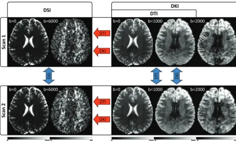
The study was approved by the institutional review board at the National Health Research Institutes (Taiwan), and informed con-sent was obtained from all participants before enrollment in the study. Experiments were performed on 3 healthy volunteers on a 3T MR imaging system (Tim Trio; Siemens, Erlangen, Germany); and for each participant, 2 full DSI and DKI datasets were ob-tained, with the DTI dataset being taken as a subset of the DKI dataset. Angular variabilities in the orientation estimates were quantified as the absolute, voxelwise angular difference for each reconstruction between repeat scans, and for DKI and DTI, an-gular errors were quantified as the absolute, voxelwise anan-gular differences from the corresponding DSI scan. For each subject, T1-weighted magnetization-prepared rapid acquisition of gradi-ent echo images were also acquired for anatomic reference. The experimental design is illustrated inFig 1, and the angular vari-ability and error measures are illustrated inFig 2. A detailed de-scription of our image-acquisition protocol and image-analysis steps is given in the On-line Appendix.
The angular error estimates, as quantified in this study, in-clude contributions from both random and systematic errors. Random error may result from thermal noise, incompleteq-space sampling distributions, and physiologic effects such as pulsatile flow and bulk subject motion, while systematic errors arise from the approximations inherent to the DTI and DKI dODFs. Al-though it is difficult to rigorously isolate the random and system-atic components of the angular error, a rough index of systemsystem-atic error is given by the difference between the angular error and angular variability for a given reconstruction because the angular variability is a measure of random error. We used this heuristic approach as a practical means of comparing systematic errors for the DTI and DKI dODFs.
trajec-tories, whereas differing colors indicate tracts following different overall trajectories.
RESULTS
Summary statistics for each subject and ROI are given in the On-line Table. DTI has the lowest angular variability in both the in-clusive and conservative WM ROIs as well as the single fiber
bun-dle ROI, while DSI has the lowest angular variability in both the 2 andⱖ3 crossing-fibers ROIs. Conversely, DKI has the highest angular variability in all ROIs, with the exception of theⱖ3 cross-ing-fibers ROI, where DTI has the highest angular variability. However, the angular variabilities for all reconstructions are com-parable within each of the ROIs, differing by, at most, 2.1° in the single fiber bundle ROI (On-line Table, “Single-fiber ROI”). On FIG 1. Experimental design illustrated with sample images from a single subject. For each subject, 2 separate scans are obtained, which include independent DSI and DKI acquisitions optimized for the respective reconstructions. The DTI reconstruction is calculated from a subset of the DKI acquisition and is fully independent of the DSI scan but not the DKI scan. Angular variability is calculated between scans (blue arrows), and angular error is calculated for DKI and DTI in reference to the corresponding DSI scan (red arrows). Units for the b-value are second/square millimeter, and the signal intensity ranges for each image are given by the corresponding color bar (in arbitrary units). DWIs from the highest b-value for each acquisition are given to illustrate the range of diffusion-weighting applied.
[image:3.594.55.533.45.331.2] [image:3.594.54.531.403.561.2]the other hand, DKI consistently improves angular error com-pared with DTI in all ROIs. Moreover, the DKI systematic errors are all substantially smaller than the DTI systematic errors, con-sistent with a higher degree of accuracy for the DKI dODFs.
For the ROIs tested, dODF performance measures are influ-enced by the fractional anisotropy (FA) value, with the smaller angular variability and angular error for regions with higher FA. Conversely, the occurrence of crossing fibers increased angular variability and angular error in dODF-derived orientation esti-mates. However, the accuracy of the DKI dODF is less affected than the DTI-derived dODF in crossing-fiber regions. Properties of the dODF reconstructions are explored further in On-line Figs 1 and 2.
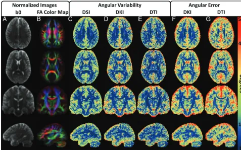
Mean normalized parameter maps are given inFig 3to illus-trate the group-wise performance of the dODF reconstructions. All 3 of the reconstruction techniques demonstrate similar angu-lar variability throughout the WM, but DTI shows improvement in angular variability in regions with high FA (eg, note the corpus callosum and corticospinal tracts in rows 2 and 3, which show high FA contrast). The DKI angular error estimates are relatively consistent throughout the WM, whereas the DTI angular error estimates show distinct WM regions where the angular error de-teriorates. When one compares these regions with the normalized FA color maps, it is likely that these regions represent voxels with more complex fiber bundle geometries owing to influences from
multiple fiber bundle orientations within a voxel (eg, note the intersecting regions between the corpus callosum and corona ra-diata, which are apparent in rows 1 and 3).
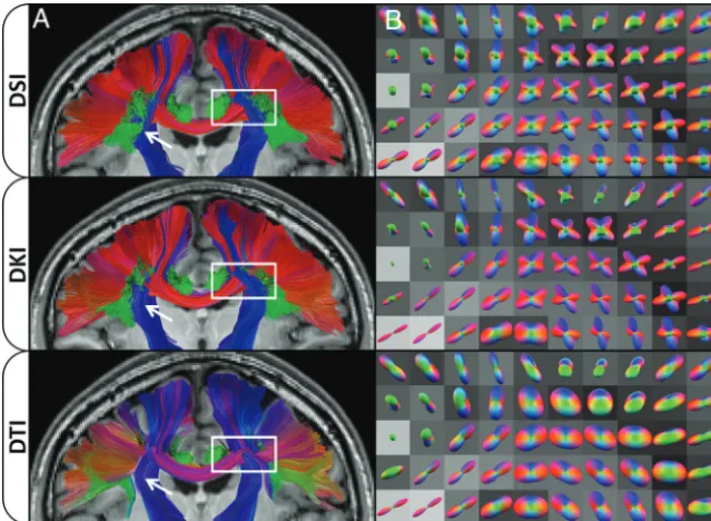
[image:4.594.53.531.47.345.2]shown inFig 4. Full-brain tractography results are compared in the On-line Video.
DISCUSSION
In this study, we have used DSI as a reference standard to assess the angular error in orientation estimates from DKI and DTI and quantified the intrasubject angular variability of WM fiber bundle orientation estimates from DTI, DKI, and DSI. We have focused primarily on comparing the estimated fiber orientations that the dODFs identify, because these are the inputs needed for con-structing tractography. However, these are only approximations for the true fiber orientations, which are, in general, not known, even if the dODF is measured exactly.
A primary motivation for this study is to help assess the po-tential of DKI tractography for data obtained with clinical MR imaging scanners. By estimating both the diffusion and kurtosis tensors, DKI more fully characterizes diffusion in complex neural tissue than conventional DTI; this feature, theoretically, should improve tractography. Our experimental results support this proposition because both the angular and systematic errors are markedly lower for DKI (On-line Table andFig 3). Moreover, tractography generated with DKI is qualitatively much more sim-ilar to that obtained with DSI than is DTI tractography (Fig 4and On-line Video). Given that DKI, in comparison with DTI, also
provides several additional diffusion measures (eg, mean kurtosis) that are sensitive to neuropathologic changes as-sociated with a variety of diseases,19-32 there are potentially compelling advan-tages to DKI vis-a`-vis DTI.
Overall, the angular variability esti-mates are comparable for all 3 recon-structions in all ROIs, differing by, at most, 2.1° in the single-fiber ROI (On-line Table, “Single-fiber ROI”). How-ever, DKI tends to have increased angu-lar variability compared with both DTI and DSI in all ROIs except for the ROI with ⱖ3 crossing-fiber bundles. Al-though the precise origin of the in-creased angular variability of DKI is un-clear, this could result from a trade-off between estimation error from incom-pleteq-space sampling distributions and subject motion. DTI, for example, re-quires the shortest acquisition time, which may result in the lowest contri-butions of subject motion to angular variability. DSI, on the other hand, uses a large number of diffusion-en-coding vectors to characterize diffu-sion dynamics, which could have lower angular variability from the dODF reconstruction but an increased likelihood of subject motion. DKI is also known to be sensitive to recon-struction artifacts resulting from Gibbs ringing35,36 and noise bias,37 though these are also expected to affect DSI.
To acquire high-quality, whole-brain DSI and DKI datasets for evaluation, we optimized our protocol for high SNR rather than a short acquisition time. Consequently, the total scan time used in this study was relatively long compared with typical clinical pro-tocols. To improve scan efficiency, one or more of several differ-ent strategies may be used. For example, there has been a success-ful effort to reduce theq-space sampling burden of DSI, including decreasing the q-space sampling density by sampling fewer points,38,39sampling only one-half of theq-space by assuming symmetry of theq-space data,40,41or sampling only a quarter of theq-space by using compressed sensing.42The acquisition time can also be reduced with simultaneous multisection EPI,43-47 while stronger diffusion-encoding gradients can be used to reduce the TE to improve the SNR.47Although DSI may show the largest improvement in acquisition time, these considerations are gener-ally applicable to DKI as well. There may be an increase in the angular error and variability if SNR is reduced, as may occur with accelerated acquisition schemes,45or if sparseq-space sampling schemes are used.40Nevertheless, DKI may be presumed to have shorter typical scan times than DSI because DKI requires only the second and fourth cumulants of the dPDF,48while DSI uses the full dPDF with the inherent greater data-acquisition burden. A FIG 4. Effects of dODF reconstructions on WM tractography. ColumnAshows a coronal
[image:5.594.55.375.47.281.2]valuable follow-up study would be to quantitatively investigate the differences in the orientation estimates by using protocols with acquisition times that are more suitable for routine clinical scanning.
A variety of alternative techniques can resolve the orientations of crossing-fiber bundles for tractography. Compared with sev-eral other dODF reconstructions, the kurtosis dODF has been shown to have a comparable or improved resolving power49; however, numeric simulations indicate that the kurtosis dODF may sometimes have a greater angular error than other dODFs for larger fiber-crossing angles.16,49 Fiber bundle orientations can also be estimated from directional diffusional kurtosis estimates provided by DKI without estimating the dODF directly,50or the white matter fiber bundles may be modeled mathematically and used to estimate a model-dependent fiber orientation distribu-tion funcdistribu-tion—for example, by using fiber ball imaging51or con-strained spherical deconvolution.52,53Because none of these tech-niques are directly analogous to the dODF, they were not included in the present study. In addition, model-based approaches make detailed assumptions about the relationship between WM and the DWI signal that have yet to be fully validated. Nevertheless, the directional diffusional kurtosis approach has been shown to in-crease fiber detection through the corpus callosum,50and con-strained spherical deconvolution can be highly sensitive to cross-ing fibers.18,54
To summarize, in this study we acquired, from 3 healthy vol-unteers, a unique dataset with 6 full DSI and DKI acquisitions, to quantify dODF performance measures from DTI, DKI, and DSI. In general, DKI substantially decreases the error of dODF orien-tation estimates relative to DTI. Moreover, DKI enables the de-tection of crossing fibers, which results in pronounced improve-ment relative to DTI for tractography throughout regions with complex fiber bundle geometries.15,16,33,36Indeed, our results in-dicate that the tractography obtained with DKI is qualitatively quite comparable with that for DSI, despite DKI sampling a much smaller portion ofq-space. With enhanced tractography relative to DTI and shorter typical scan times than DSI, DKI-based trac-tography is potentially advantageous, particularly in clinical set-tings where time considerations are crucial. However, further study will be needed to more fully investigate the comparative utility of DKI-based tractography.
CONCLUSIONS
The higher order information provided by the kurtosis tensor enables DKI to directly resolve crossing fibers and improves the accuracy of DKI relative to DTI for tractography. Both DKI and DTI are capable of mapping the single predominant fiber bundle orientation, but the angular error of DTI deteriorates in regions with complex fiber orientations due to its theoretic limitation under the assumption of Gaussian diffusion. DSI, DKI, and DTI all have comparable angular variabilities; however, DKI has de-creased angular error in the dODF fiber orientation estimates relative to DTI. Unlike DTI, DKI is thus able to generate white matter fiber tractography comparable with that of DSI, and due to its shorter typical scan time than DSI, DKI is potentially more suitable for a variety of clinical and research applications.
Disclosures: Li-Wei Kuo—RELATED:Grant: NHRI-BN-104-PP-06*; MOST-103-2221-E-400-001.* Joseph A. Helpern—RELATED:Grant: Litwin Foundation*;UNRELATED:
Grants/Grants Pending: National Institutes of Health.* Jens H. Jensen—RELATED:
Grant: Litwin Foundation,*Comments: I have partial salary support from this grant;
UNRELATED:Patents (planned, pending or issued) and Royalties: US Patent 8811706,
Comments: I am a coinventor on a patent that covers one of the imaging methods investigated in this article (DKI). The patent is owned by my former employer (New York University), but I could be entitled to royalties at some point. To date, I have not received royalties from this patent. *Money paid to the institution.
REFERENCES
1. Romano A, D’Andrea G, Minniti G, et al.Pre-surgical planning and MR-tractography utility in brain tumour resection.Eur Radiol 2009;19:2798 – 808CrossRef Medline
2. Mormina E, Longo M, Arrigo A, et al.MRI tractography of cortico-spinal tract and arcuate fasciculus in high-grade gliomas per-formed by constrained spherical deconvolution: qualitative and quantitative analysis. AJNR Am J Neuroradiol 2015;36:1853–58
CrossRef Medline
3. Lazar M.Mapping brain anatomical connectivity using white mat-ter tractography.NMR Biomed2010;23:821–35CrossRef Medline
4. Tournier JD, Mori S, Leemans A.Diffusion tensor imaging and be-yond.Magn Reson Med2011;65:1532–56CrossRef Medline
5. Farquharson S, Tournier JD, Calamante F, et al.White matter fiber tractography: why we need to move beyond DTI.J Neurosurg2013; 118:1367–77CrossRef Medline
6. Tuch DS, Reese TG, Wiegell MR, et al.Diffusion MRI of complex neural architecture.Neuron2003;40:885–95CrossRef Medline
7. Tuch DS. Q-ball imaging. Magn Reson Med 2004;52:1358 –72
CrossRef Medline
8. Wedeen VJ, Hagmann P, Tseng WY, et al.Mapping complex tissue architecture with diffusion spectrum magnetic resonance imaging. Magn Reson Med2005;54:1377– 86CrossRef Medline
9. Wedeen VJ, Wang RP, Schmahmann JD, et al.Diffusion spectrum magnetic resonance imaging (DSI) tractography of crossing fibers. Neuroimage2008;41:1267–77CrossRef Medline
10. Basser PJ, Mattiello J, LeBihan D.Estimation of the effective self-diffusion tensor from the NMR spin echo.J Magn Reson1994;103: 247–54CrossRef Medline
11. Basser PJ, Pierpaoli C.Microstructural and physiological features of tissues elucidated by quantitative-diffusion-tensor-MRI.J Magn Reson B1996;111:209 –19CrossRef Medline
12. Basser PJ, Pajevic S, Pierpaoli C, et al.In vivo fiber tractography using DT-MRI data.Magn Reson Med2000;44:625–32Medline
13. Jensen JH, Helpern JA, Ramani A, et al. Diffusional kurtosis imaging: the quantification of non-Gaussian water diffusion by means of magnetic resonance imaging.Magn Reson Med2005;53: 1432– 40CrossRef Medline
14. Jensen JH, Helpern JA.MRI quantification of non-Gaussian water diffusion by kurtosis analysis. NMR Biomed. 2010;23:698 –710
CrossRef Medline
15. Lazar M, Jensen JH, Xuan L, et al.Estimation of the orientation distribution function from diffusional kurtosis imaging. Magn Reson Med2008;60:774 – 81CrossRef Medline
16. Jensen JH, Helpern JA, Tabesh A.Leading non-Gaussian corrections for diffusion orientation distribution function.NMR Biomed2014; 27:202–11CrossRef Medline
17. Hagmann P, Jonasson L, Maeder P, et al.Understanding diffusion MR imaging techniques: from scalar diffusion-weighted imaging to diffusion tensor imaging and beyond.Radiographics2006;26(suppl 1):S205–23CrossRef Medline
18. Jeurissen B, Leemans A, Tournier JD, et al.Investigating the preva-lence of complex fiber configurations in white matter tissue with diffusion magnetic resonance imaging.Hum Brain Mapp2013;34: 2747– 66CrossRef Medline
19. Jensen JH, Falangola MF, Hu C, et al.Preliminary observations of increased diffusional kurtosis in human brain following recent ce-rebral infarction.NMR Biomed2011;24:452–57CrossRef Medline
in ischemic stroke.Neuroimaging Clin N Am2011;21:345–77, xi
CrossRef Medline
21. Hui ES, Fieremans E, Jensen JH, et al.Stroke assessment with diffu-sional kurtosis imaging.Stroke2012;43:2968 –73CrossRef Medline
22. Grinberg F, Ciobanu L, Farrher E, et al.Diffusion kurtosis imaging and log-normal distribution function imaging enhance the visuali-sation of lesions in animal stroke models.NMR Biomed2012;25: 1295–304CrossRef Medline
23. Umesh Rudrapatna S, Wieloch T, Beirup K, et al.Can diffusion kur-tosis imaging improve the sensitivity and specificity of detecting microstructural alterations in brain tissue chronically after exper-imental stroke? Comparisons with diffusion tensor imaging and histology.Neuroimage2014;97:363–73CrossRef Medline
24. Falangola MF, Jensen JH, Tabesh A, et al.Non-Gaussian diffusion MRI assessment of brain microstructure in mild cognitive impair-ment and Alzheimer’s disease.Magn Reson Imaging2013;31:840 – 46
CrossRef Medline
25. Benitez A, Fieremans E, Jensen JH, et al.White matter tract integrity metrics reflect the vulnerability of late-myelinating tracts in Alz-heimer’s disease.Neuroimage Clin2013;9:64 –71CrossRef Medline
26. Fieremans E, Benitez A, Jensen JH, et al.Novel white matter tract integrity metrics sensitive to Alzheimer disease progression.AJNR Am J Neuroradiol2013;34:2105–12CrossRef Medline
27. Vanhoutte G, Pereson S, Delgado Y Palacios R, et al.Diffusion kur-tosis imaging to detect amyloidosis in an APP/PS1 mouse model for Alzheimer’s disease.Magn Reson Med2013;69:1115–21CrossRef Medline
28. Gong NJ, Wong CS, Chan CC, et al.Correlations between micro-structural alterations and severity of cognitive deficiency in Alzhei-mer’s disease and mild cognitive impairment: a diffusional kurtosis imaging study. Magn Reson Imaging 2013;31:688 –94 CrossRef Medline
29. Raab P, Hattingen E, Franz K, et al.Cerebral gliomas: diffusional kurtosis imaging analysis of microstructural differences.Radiology 2010;254:876 – 81CrossRef Medline
30. Van Cauter S, Veraart J, Sijbers J, et al.Gliomas: diffusion kurtosis MR imaging in grading. Radiology 2012;263:492–501 CrossRef Medline
31. Rosenkrantz AB, Sigmund EE, Johnson G, et al.Prostate cancer: fea-sibility and preliminary experience of a diffusional kurtosis model for detection and assessment of aggressiveness of peripheral zone cancer.Radiology2012;264:126 –35CrossRef Medline
32. Steven AJ, Zhuo J, Melhem ER.Diffusion kurtosis imaging: an emerging technique for evaluating the microstructural environ-ment of the brain.AJR Am J Roentgenol2014;202:W26 –33CrossRef Medline
33. Glenn GR, Helpern JA, Tabesh A, et al.Optimization of white matter fiber tractography with diffusional kurtosis imaging.NMR Biomed 2015;28:1245–56CrossRef Medline
34. Tabesh A, Jensen JH, Ardekani BA, et al.Estimation of tensors and tensor-derived measures in diffusional kurtosis imaging.Magn Reson Med2011;65:823–36CrossRef Medline
35. Veraart J, Fieremans E, Jelescu IO, et al.Gibbs ringing in diffusion MRI.Magn Reson Med2015 Aug 10. [Epub ahead of print]CrossRef Medline
36. Perrone D, Aelterman J, Pizˇurica A, et al.The effect of Gibbs ringing artifacts on measures derived from diffusion MRI.Neuroimage 2015;120:441–55CrossRef Medline
37. Glenn GR, Tabesh A, Jensen JH.A simple noise correction scheme
for diffusional kurtosis imaging. Magn Reson Imaging 2015;33: 124 –33CrossRef Medline
38. Kuo LW, Chen JH, Wedeen VJ, et al.Optimization of diffusion spec-trum imaging and q-ball imaging on clinical MRI system. Neuroim-age2008;41:7–18CrossRef Medline
39. Tefera GB, Zhou Y, Juneja V, et al.Evaluation of fiber tracking from subsampled q-space data in diffusion spectrum imaging. Magn Reson Imaging2013;31:820 –26CrossRef Medline
40. Kuo LW, Chiang WY, Yeh FC, et al.Diffusion spectrum MRI using body-centered-cubic and half-sphere sampling schemes.J Neurosci Methods2013;212:143–55CrossRef Medline
41. Yeh CH, Cho KH, Lin HC, et al.Reduced encoding diffusion spec-trum imaging implemented with a bi-Gaussian model.IEEE Trans Med Imaging2008;27:1415–24CrossRef Medline
42. Menzel MI, Tan ET, Khare K, et al.Accelerated diffusion spectrum imaging in the human brain using compressed sensing.Magn Reson Med2011;66:1226 –33CrossRef Medline
43. Feinberg DA, Setsompop K.Ultra-fast MRI of the human brain with simultaneous multi-slice imaging.J Magn Reson2013;229:90 –100
CrossRef Medline
44. Larkman DJ, Hajnal JV, Herlihy AH, et al.Use of multicoil arrays for separation of signal from multiple slices simultaneously excited. J Magn Reson Imaging2001;13:313–17Medline
45. Setsompop K, Cohen-Adad J, Gagoski BA, et al.Improving diffusion MRI using simultaneous multi-slice echo planar imaging. Neuroim-age2012;63:569 – 80CrossRef Medline
46. Reese TG, Benner T, Wang R, et al.Halving imaging time of whole brain diffusion spectrum imaging and diffusion tractography using simultaneous image refocusing in EPI.J Magn Reson Imaging2009; 29:517–22CrossRef Medline
47. Setsompop K, Kimmlingen R, Eberlein E, et al.Pushing the limits of in vivo diffusion MRI for the Human Connectome Project. Neuro-image2013;80:220 –33CrossRef Medline
48. Kiselev VG.The cumulant expansion: an overarching mathematical framework for understanding diffusion NMR.In: Jones DK, ed. Dif-fusion MRI: Theory, Methods and Applications. New York: Oxford University Press; 2011:152– 68
49. Jensen JH, Helpern JA.Resolving power for the diffusion orienta-tion distribuorienta-tion funcorienta-tion.Magn Reson Med2015 Oct 7. [Epub ahead of print]CrossRef Medline
50. Neto Henriques R, Correia MM, Nunes RG, et al.Exploring the 3D geometry of the diffusion kurtosis tensor: impact on the develop-ment of robust tractography procedures and novel biomarkers. Neuroimage2015;111:85–99CrossRef Medline
51. Jensen JH, Russell Glenn G, Helpern JA.Fiber ball imaging. Neuro-image2016;124:824 –33CrossRef Medline
52. Tournier JD, Calamante F, Connelly A.Robust determination of the fibre orientation distribution in diffusion MRI: non-negativity constrained super-resolved spherical deconvolution.Neuroimage 2007;35:1459 –72CrossRef Medline
53. Tournier JD, Yeh CH, Calamante F, et al.Resolving crossing fibres using constrained spherical deconvolution: validation using diffu-sion-weighted imaging phantom data.Neuroimage2008;15:617–25
CrossRef Medline