ORIGINAL RESEARCH
The Efficacy of Endovascular Stenting in the
Treatment of Supraclinoid Internal Carotid Artery
Blister Aneurysms Using a Stent-in-Stent
Technique
J.R. Gaughen, Jr D. Hasan A.S. Dumont M.E. Jensen J. Mckenzie A.J. Evans
BACKGROUND AND PURPOSE: Blister aneurysms of the supraclinoid ICA represent a rare but well-documented cause of subarachnoid hemorrhage. These aneurysms are difficult to detect, and their surgical treatment is challenging, with high morbidity and mortality rates. The reports currently in the literature that describe the surgical and endovascular treatment of these aneurysms offer no clear consensus on the optimal treatment. We describe a staged endovascular treatment entailing stenting using a stent-in-stent technique, as well as planned but delayed embolization as the aneurysm increases in size to allow the introduction of coils.
MATERIALS AND METHODS: We performed a retrospective review of all cerebral angiograms per-formed at our institution over an 8-month period for evaluation of subarachnoid hemorrhage, identi-fying 6 ICA blister aneurysms.
RESULTS: All 6 blister aneurysms were located in the supraclinoid ICA. The stent-in-stent technique was used for the initial treatment of all patients. Three patients had no residual or recurrent aneurysm following initial treatment. Three patients required retreatment with coils after continued growth of the aneurysm, identified on follow-up angiography. Five patients had good recovery (average mRS score of 1), and 1 patient had poor neurologic recovery (mRS score of 3) due to a large hemorrhagic infarction.
CONCLUSIONS:Our case series suggests that staged endovascular treatment entailing the use of a stent-in-stent technique, augmented with subsequent coil embolization as necessary for progressive disease, is a viable endovascular option for treating ruptured supraclinoid blister aneurysms, allowing for parent artery preservation.
ABBREVIATIONS:CTA⫽CT angiography; DSA⫽digital subtraction angiography; ICA⫽internal carotid artery; mRS⫽modified Rankin Scale; SAH⫽subarachnoid hemorrhage
F
irst described by Sundt and Murphey,1the term “blister aneurysm” (also known as blister-type or dorsal wall aneu-rysm) has been used to describe aneurysms arising from non-branching sites of the wall of the supraclinoid ICA. As a result of their anatomy and histopathology, which appear to differ from saccular aneurysms, these rare lesions offer challenges in both diagnosis and treatment.As the literature supports, routine microsurgical and endo-vascular treatment approaches carry a relatively high morbid-ity and mortalmorbid-ity,2 leading to attempts at numerous novel treatment techniques. Endovascular techniques usually em-ploy some combination of coil embolization and/or stent placement, with definitive endovascular treatment limited to parent artery occlusion, with or without surgical bypass.3-7
We report our experience with a series of 6 patients pre-senting with blister aneurysms of the supraclinoid ICA treated with a staged endovascular treatment entailing stent place-ment by using a stent-in-stent technique, and planned but
delayed embolization as the aneurysm increased in size to al-low the introduction of coils.
Materials and Methods
Patients and Techniques
Following internal review board approval, we performed a retrospec-tive review of all catheter cerebral angiograms performed at our insti-tution from July 2007 through March 2008 for evaluation of sub-arachnoid hemorrhage, identifying 6 cases diagnosed as ruptured ICA blister aneurysms. We reviewed all relevant imaging studies and records pertaining to the diagnosis and treatment of these aneurysms, including admission and discharge summaries, progress notes, initial diagnostic imaging studies, procedural images and reports, and fol-low-up imaging studies. The review was performed by 2 interven-tional neuroradiologists (M.E.J., A.J.E.) and 2 neurosurgeons (D.H., A.S.D.).
In each of these patients, the blister aneurysm was initially too small and shallow to allow for coils to be introduced on the first day of treatment. Consequently, the treatment for this cohort included a staged endovascular protocol entailing stent placement by using a stent-in-stent technique, with a planned coiling procedure to follow in the case of expected aneurysm enlargement. In 3 patients the an-eurysm enlarged as expected. Two of these anan-eurysms were treated successfully with coiling, one with coiling and then subsequent parent vessel occlusion.
Received June 2, 2009; accepted after revision November 20.
From the Departments of Radiology (J.R.G., M.E.J., A.J.E.) and Neurological Surgery (A.S.D.), University of Virginia Health System, Charlottesville, Virginia; Department of Neurosurgery (D.H.), University of Iowa Hospitals and Clinics, Iowa City, Iowa; and private practice of Drs. Mori, Bean, and Brooks, P.A. (J.M.), Jacksonville, Florida
Please address correspondence to John R. Gaughen, Jr, MD, Department of Radiology, Box 800170, Charlottesville, VA 22908; e-mail: jrg4@virginia.edu
Endovascular stent placement has been reported to be a poten-tially effective treatment of uncoilable intradural pseudoaneurysms.8 The aneurysms were deemed at the time of initial treatment to be too small to safely coil, even after the placement of the endovascular stents. Therefore, the patients were initially treated with endovascular stent placement by using a stent-in-stent technique, and all patients subsequently underwent short-term (7–13 days) follow-up conven-tional angiography. Given our knowledge of the malignant natural history of blister aneurysms, the expectation at follow-up angiogra-phy was to perform adjunct coil embolization or parent vessel occlu-sion, though 3 patients showed improvement in the aneurysms at follow-up and were therefore observed.
The stent-in-stent technique entails the deployment of 2 endovas-cular stents, a longer stent and a shorter stent. The longer stent is deployed first, spanning a large portion of the supraclinoid ICA seg-ment and covering the aneurysm neck. A second, shorter stent is then deployed within the lumen of the longer stent, also spanning the an-eurysm neck. Enterprise stents (Cordis Endovascular, Miami Lakes, Florida) were used in 5 cases, and Neuroform stents (Boston Scien-tific, Natick, Massachusetts) were used in 1 case. The stent selection was based solely on operator preference. All patients were initiated on dual antiplatelet therapy following the procedure, consisting of aspirin (325 mg) and clopidogrel (75 mg) daily.
Results
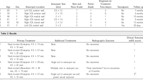
Demographics are summarized in Table 1.
All 6 patients presented with subarachnoid hemorrhage, diagnosed by unenhanced head CT. Each patient underwent CTA followed by DSA in the diagnostic evaluation of sub-arachnoid hemorrhage. Of the 6 blister aneurysms, 4 (67%) were identified prospectively on the CTA, and all were identi-fied with DSA. The average interval between the diagnostic and initial treatment procedures was 1.5 days (range, 0 – 4
days). Importantly, 4 of the aneurysms showed interval growth and change in the size and configuration of the dome between the diagnostic DSA and initial treatment procedure. All 6 patients were treated with endovascular stent place-ment, by using the above-described stent-in-stent technique. Short-term (7–13 days) follow-up DSA was performed during the inpatient hospital stay in all 6 cases to evaluate for residual disease and/or vasospasm. Additional short-term follow-up was performed as deemed necessary to monitor the disease status. This subsequent follow-up ranged from 7 days to 3 months. Subsequent intermediate follow-up (routinely per-formed at 6 months) DSA imaging has been perper-formed on all 6 patients, with an average follow-up interval of 6.3 months (range, 20 days to 12 months).
Results are summarized in Table 2. Contrary to our expec-tations, 3 of the aneurysms (50%) did not enlarge to the point that coils could be placed. In fact in these 3 patients, no resid-ual or recurrent aneurysms were detected at early or interme-diate follow-up (Fig 1). On the other hand, 3 of the aneurysms did enlarge and subsequent interventions were performed as detailed below. Five of the 6 patients (83%) currently have no aneurysm recurrence at intermediate follow-up, with the sixth patient having undergone definitive treatment with aneurysm trapping and parent vessel occlusion. Five patients had good recovery (average mRS score of 1), and the patient who re-bled had poor neurologic recovery (mRS score of 3), due to a large hemorrhagic infarction. All patients (with the exception of the patient with the parent vessel occlusion) were placed on long-term dual antiplatelet therapy, with none demonstrating any significant degree of in-stent stenosis at intermediate follow-up.
[image:2.594.54.532.61.329.2]One of the patients with progressive disease showed slight enlargement of the aneurysm on follow-up DSA performed Table 1: Demographics
Age Sex Aneurysm Location
Aneurysm Size (mm) Hunt and Hess Grade Fisher Grade Diagnosis-to-Treatment
Time (days) Vasospasm Follow-up
1 69 F Left ICA: anterior wall 2.9⫻1.2 1 1 2 No 7 months
2 39 F Right ICA: anterior wall 2⫻1.5 3 4 0 No 4 months
3 46 F Right ICA: medial wall 2⫻1 2 2 1 No 10 months
4 61 F Right ICA: lateral wall 2.9⫻1.4 2 4 1 No 4 months
5 42 M Right ICA: lateral wall 2⫻1.2 2 4 4 No 12 months
[image:2.594.51.538.67.325.2]6 41 F Left ICA: medial wall 3⫻2 3 4 1 Yes 20 days
Table 2: Results
Primary Treatment Additional Treatments Radiographic Outcome
Clinical Outcome (mRS score)
1 Stent-in-stent (Enterprise: 4.5⫻14 mm, 4.5⫻14 mm)
None No recurrence 1
2 Stent-in-stent (Enterprise: 4.5⫻37 mm, 4.5⫻22 mm)
None No recurrence 1
3 Stent-in-stent (Enterprise: 4.5⫻37 mm, 4.5⫻14 mm)
None No recurrence 1
4 Stent-in-stent (Enterprise: 4.5⫻28 mm, 4.5⫻22 mm)
Single coil in aneurysm sac No recurrence 1
5 Stent-in-stent (Neuroform: 4.5⫻20 mm, 4.5⫻15 mm)
Multiple coils in aneurysm sac Three recurrencesabut no recurrence
at 9 months
1
6 Stent-in-stent (Enterprise: 4.5⫻37 mm, 4.5⫻14 mm)
Single coil in aneurysm sac and parent vessel occlusion
No recurrence 3b
a
At 1, 3, and 6 months.
bExpressive aphasia, moderate right facial palsy, antigravity (3/5) strength in upper and lower extremities, ambulates with assistance.
INTERVENTIONAL
ORIGINAL
7 days following stent placement. A single HydroSoft 2 mm⫻ 1 cm coil (MicroVention Terumo, Aliso Viejo, California) was then placed into the aneurysm sac, with sluggish filling of the aneurysm on the immediate postprocedural runs. Subsequent follow-up imaging at 2 weeks, 3 months, and 6 months showed no residual or recurrent aneurysm (Fig 2).
Another of the patients with progressive disease showed mild sequential enlargement and change in the configuration of the aneurysm on follow-up DSA performed 7 days, 14 days, and 30 days following stent placement. This residual aneu-rysm, measuring 3.4 mm, was then coil embolized by using a single HyperSoft 2 mm⫻3 cm coil (MicroVention Terumo), with minimal residual filling of the aneurysm neck on the im-mediate postprocedural runs. However, follow-up DSA at 3 months demonstrated aneurysm recurrence (3.8 mm), which was again coil embolized with 7 coils (including a Micrus Spherical 3 mm⫻5.4 cm coil [Micrus Endovascular, San Jose, California], a Compass 3.5 mm⫻4.5 mm coil [MicroVention
Terumo], 3 Complex 2 mm ⫻ 4 cm coils [MicroVention
Terumo], and 2 HyperSoft-10 2 mm⫻4 cm coils), with a small (1.7 mm) residual neck remnant. Follow-up imaging at 6 months again demonstrated aneurysm regrowth (3.3 mm), which was again coil embolized with 4 coils (including a
Com-pass 2 mm⫻4 cm coil, a HydroSoft 2 mm⫻2 cm coil, and 2 HyperSoft 2 mm⫻1 cm coils), with no residual aneurysm on the immediate postprocedural runs. MR angiography per-formed at 8 months (to evaluate for headaches) showed no evidence of aneurysm recurrence, and DSA follow-up at 9 and 12 months showed minimal filling at the base of the aneurysm with otherwise no evidence of recurrence (Fig 3).
The final patient with progressive disease demonstrated a complicated course (Fig 4), beginning with the stent place-ment procedure. Overlapping Enterprise stents were deployed across the aneurysm neck, but the second stent did not deploy properly. Close examination revealed that the distal tines of the second stent were entrapped in 1 of the cells of the first stent, thus preventing the second stent from opening properly and resulting in poor apposition of the second stent to the wall of the vessel. Ten days later, the patient sustained aneurysmal rebleeding, with associated deterioration of her neurologic ex-amination. A cerebral angiogram revealed significant enlarge-ment of the previously treated left ICA blister aneurysm. A temporary balloon occlusion test was performed, which the patient passed. Under proximal balloon occlusion of the ICA, a single coil (GDC 360 2 mm⫻4 cm; Target Therapeutics, Fremont, California) was detached in the dome of the
[image:3.594.54.537.36.412.2]rysm. This was followed by complete occlusion of the left ICA spanning from supraclinoid ICA (just proximal to the origin of the fetal posterior cerebral artery) to the cavernous ICA (just distal to the persistent trigeminal artery, which filled the basilar trunk). Postprocedurally, the left hemisphere was fill-ing through collateral flow from the anterior communicatfill-ing artery and fetal left posterior cerebral artery. A week following the ICA occlusion, the patient developed severe vasospasm, treated with medical management and multiple intra-arterial verapamil infusions, but eventually developed a large, hemor-rhagic left middle cerebral artery infarction. The patient re-covered during the course of the next several weeks and was discharged to an inpatient rehabilitation facility. The stroke resulted in severe, persistent neurologic deficit, including ex-pressive aphasia, right facial palsy, and right hemiparesis.
Discussion
Ruptured blister aneurysms of the supraclinoid ICA present with SAH and are estimated to represent 0.9%– 6.5% of rup-tured intracranial aneurysms.9Compared with saccular aneu-rysms, these lesions tend to have a more precipitous course, enlarging rapidly and rebleeding frequently. Morphologically, they appear as wide-necked, shallow outpouchings of non-branching sites of the supraclinoid ICA. At surgery, they often contain a fragile, thin wall and a broad communication with the parent vessel, without an identifiable neck. Histologically, these lesions have been shown to represent focal wall defects covered with thin, fibrous tissue and adventitia, lacking the usual collagen layer, findings indicative of pseudoaneurysm.10 As such, these lesions have shown an association with arterial dissections.11
Because of their broad-based, shallow profiles, these aneu-rysms often represent a diagnostic challenge, both with CTA and DSA. The imaging appearance of supraclinoid ICA blister aneurysms has been described in detail elsewhere.12In addi-tion to an understanding of the morphology and distribuaddi-tion of these lesions, meticulous angiographic technique and a high index of suspicion are often both necessary to make this diag-nosis. Any patient with SAH in an aneurysmal pattern and no detectable berry aneurysm should be carefully scrutinized for a blister aneurysm.
The 6 patients included in this study presented with acute SAH on unenhanced head CT and were diagnosed by CTA and/or DSA with blister aneurysms. In these patients, coil em-bolization was not performed at the initial treatment session because the operators deemed that the morphology of the aneurysms did not allow for safe coil deposition, even after stent placement. Instead, these patients were treated with a staged protocol: immediate treatment by using a stent-in-stent technique in an effort to temporize, preserve the artery, and redirect flow to prevent rehemorrhage. The second stage consisted of careful and rapid follow-up with coil emboliza-tion of the aneurysms that enlarged. Interestingly, in 3 pa-tients, the aneurysms did not enlarge, and the stent-in-stent technique was curative.
Importantly, our patients also fell into 2 groups in terms of their initial imaging findings. In 1 group, we deemed the CTA and DSA to show definitive evidence of a blister aneurysm as the cause of the SAH. These patients were treated per our protocol as described. The second group of patients presented with suspicious but not definitive findings of blister aneu-rysms and were not treated immediately, but rather they were
[image:4.594.53.534.43.321.2]reevaluated with short-term follow-up DSA (within a few days). The reasoning for this decision stems from the concept that blister aneurysms tend to enlarge and change configura-tion rather rapidly. Three of our patients (50%) fell into the “suspicious but not definitive” category, all showing increase
in the size and change in the configuration of the aneurysm on follow-up DSA. It should be noted that during DSA evaluation for endovascular treatment of blister aneurysms, consider-ation should be given to evaluating collateral flow to the af-fected circulation (cross-compression views, Alcock test) and
Fig 3.Right supraclinoid ICA blister aneurysm. Preprocedural DSA (A) shows a shallow outpouching (white arrow) arising from a nonbranching site along the lateral wall of the supraclinoid segment of the right ICA, consistent with a blister aneurysm. One-month follow-up DSA (B) following endovascular stent placement again shows the blister aneurysm (white arrow), now increased in size and more round in shape. The patient subsequently underwent adjunct coil embolization with a single coil. Subsequent 3-month follow-up DSA (C) again shows the blister aneurysm, again increased in size and more round in shape. The patient subsequently underwent adjunct coil embolization with multiple coils. Subsequent 6-month follow-up DSA (D) again shows the blister aneurysm (white arrow), with recurrence at the aneurysm base. The patient subsequently underwent adjunct coil embolization with multiple coils. Twelve-month follow-up DSA (E) shows wide patency of the stents and minimal residual filling of the aneurysm sac (white arrow), with no filling of the coil interstices or aneurysm dome.
[image:5.594.53.535.41.379.2] [image:5.594.55.535.448.623.2]possibly temporary balloon occlusion, as parent vessel occlu-sion remains the definitive endovascular treatment of ICA blister aneurysms, though the complications of parent vessel occlusion in the setting of acute subarachnoid hemorrhage are well documented.13-16
Given the fragile nature of these aneurysms, they are as-sociated with high surgical morbidity and mortality due to intraoperative rupture, regrowth, and rehemorrhage, even after the adequate aneurysm clip application.2 Thus, the optimum treatment of these lesions remains uncertain, lead-ing to attempts at numerous novel surgical17-20 and endo-vascular2-7,9,12,17treatment techniques.
Given the histopathology of these lesions (resembling pseudoaneurysms with an incomplete wall or fibrous capsule) and the hemodynamic stress these lesions experience, it is not surprising that these aneurysms react unfavorably after coil embolization or clipping or even after stent-assisted coil embolization. The small size and shallow, wide-necked mor-phology of these aneurysms makes safe deposition of coil material unlikely, due to the risk of further aneurysm rupture or coil migration. Additionally, the lack of a true aneurysm wall allows for frequent aneurysm regrowth.
Our treatment strategy in the acute setting, that is, endo-vascular treatment with a stent-in-stent approach, attempts to diminish the flow into and hemodynamic stresses placed upon these lesions through flow diversion. Theoretically, the overlapping stents at the aneurysm neck would create better flow diversion than a single stent. In fact, in our experience, overlapping stents markedly decrease the flow into these an-eurysms, at least to visual inspection on the angiogram. Post-procedural DSA following overlapping stent placement dem-onstrated uniformly decreased flow within the aneurysm sacs of our cohort. Indeed, this treatment alone proved effective in 3 of our patients (50%), who have not needed further treat-ment or shown aneurysm regrowth at intermediate follow-up. Note that the stent-in-stent protocol should always include a plan for immediate follow-up and coiling of aneurysms that enlarge.
In our small cohort, 3 of 6 aneurysms enlarged (as ex-pected) and underwent subsequent coil embolization as part of our planned staged procedure. In 1 patient, this goal was achieved with 1 coiling session, by using a single coil. In an-other patient, this entailed 3 coiling sessions and several coils. This treatment has proved effective in both of these patients, who have not shown aneurysm regrowth at intermediate follow-up.
Our final patient and only complication warrants some discussion. This patient had rebleeding from the blister aneu-rysm following overlapping stent deployment, necessitating eventual parent vessel occlusion. The treatment was compli-cated by initial maldeployment of 1 of the stents (the second stent in our stent-in-stent procedure). In retrospect, we think that this was an avoidable technical complication. As the sec-ond stent was being placed, the operator, in an effort to posi-tion it properly, maintained significant forward pressure on the stent, inadvertently forcing it forward in such a way that as it deployed, the distal end of the stent became trapped in 1 of the cells of the previously placed stent, preventing the second stent from opening properly, and resulting in poor apposition of the second stent to the wall of the vessel. We hypothesize
that the poor treatment response at least partially related to abnormal flow alterations in the ICA and blister aneurysm. The second stent was at least ineffective at remodeling the artery and directing flow away from the aneurysm, and it may even have directed flow into the neck of the aneurysm, unfa-vorably altering the hemodynamic stresses on the wall of the aneurysm.
Park et al7published a case series comparing coil emboli-zation (with or without stent assistance or balloon remodel-ing) with aneurysm trapping and parent vessel occlusion, re-porting much better clinical outcomes in patients undergoing parent vessel occlusion. The results of our study resemble those of their aneurysm trapping group. Given the similarity in clinical outcomes, we think that these treatment strategies should be considered as salvage procedures when initial treat-ments with endovascular stent placement with or without subsequent coiling have failed. This approach allows for par-ent vessel preservation, with concomitant reduction in the risk of subsequent stroke development. Proximal occlusion of the ICA as a definitive treatment for ruptured blister aneurysm, despite good collateral flow, has a potential of exaggerated short- and long-term cerebral ischemia, particularly in the event that these patients experience cerebral vasospasm. This point is illustrated in our patient treated with proximal ICA occlusion, who subsequently developed severe cerebral vaso-spasm, which severely diminished the flow to her occluded territory. This resulted in a large hemorrhagic middle cerebral artery infarction, despite aggressive medical management and intra-arterial infusion of verapamil.
The limitations of our study should be noted, including its retrospective evaluation, small study size, and lack of a com-parative control group. Although our case series suggests a beneficial treatment outcome from the treatment algorithm studied, further investigation with larger patient populations and prospective evaluation (if possible) is clearly warranted to test the validity of these results.
Conclusions
Our case series suggests that staged endovascular treatment entailing the use of a stent-in-stent technique, augmented with subsequent coil embolization as necessary for progressive disease, is a viable endovascular option for treating ruptured supraclinoid blister aneurysms, allowing for parent artery preservation. These patients must be followed carefully to detect persistent filling or regrowth of the aneurysm so that additional treatment can be performed as necessary.
References
1. Sundt TM Jr, Murphey F.Clip grafts for aneurysm and small vessel surgery. 3. Clinical experience in intracranial internal carotid artery aneurysms.
J Neurosurg1969;31:59 –71
2. Kawashima A, Okada Y, Kawamata T, et al.Successful treatment of a blood blister-like aneurysm of the internal carotid artery by trapping with a high-flow bypass.J Clin Neurosci2008;15:797– 800
3. McNeely PD, Clarke DB, Mendez I, et al.Endovascular treatment of a “blister” aneurysm of the internal carotid artery.Can J Neurol Sci2000;27:247–50 4. Tanoue S, Kiyosue H, Matsumoto S, et al.Ruptured “blister like” aneurysm
with a pseudoaneurysm formation requiring delayed intervention with endo-vascular coil embolization: case report.J Neurosurg2004;101:159 – 62 5. Sim A, Shin Y, Cho KG, et al.Blood blister aneurysms at nonbranching sites of
6. Kim B, Chung E, Park S, et al.Treatment of blood blister-like aneurysm of the internal carotid artery with stent-assisted coil emboilization followed by stent-within-a-stent technique.J Neurosurg2007;107:1211–13
7. Park JH, Sung PI, Han D, et al.Endovascular treatment of blood blister-like aneurysms of the internal carotid artery.J Neurosurg2007;106:812–19 8. Fiorella D, Albuquerque FC, Deshmukh VR, et al.Endovascular reconstruction
with the Neuroform stent as monotherapy for the treatment of uncoilable intradural pseudoaneurysms.Neurosurgery2006;59:291–300
9. Ogawa A, Susuki M, Ogasawara K.Aneurysms at no branching sites in the supraclinoid portion of the internal carotid artery: internal carotid artery trunk aneurysms.Neurosurgery2000;47:578 – 86
10. Ishkawa T, Nakamura N, Houkin K, et al.Pathological consideration of a “blis-ter” aneurysm at the superior wall of the internal carotid artery: case report.
Neurosurgery1997;40:403– 06
11. da Silva JC, Faquini IV, Kitamura MAP, et al.Internal carotid artery blood blister-like aneurysm.Arq Neuropsiquiatr2008;66:563– 65
12. Gaughen JR, Raghavan P, Hasan D, et al.Utility of CT angiography in the identification and characterization of supraclinoid internal carotid artery blister aneurysms.AJNR Am J Neuroradiol2010;31:640 – 44
13. Matsuda M, Shiino A, Handa I.Rupture of previously unruptured giant
ca-rotid aneurysm after superficial temporal-middle cerebral artery bypass and internal carotid occlusion.Neurosurgery1985;16:177– 84
14. Anson JA, Stone JL, Crowell RM.Rupture of a giant carotid aneurysm after extracranial-to-intracranial bypass surgery.Neurosurgery1991;28:142– 47 15. Larson JJ, Tew JM Jr, Tomsick TA, et al.Treatment of aneurysm of the internal
carotid artery by intravascular balloon occlusion: long-term follow-up of 58 patients.Neurosurgery1995;36:23–30
16. Gurian JH, Vinuela F, Guglielmi G, et al.Aneurysm rupture after parent vessel sacrifice: treatment with Guglielmi detachable coil embolization via retro-grade catheterization: case report.Neurosurgery1995;37:1216 –21
17. Kubo Y, Ogasawara K, Tomitsuka N, et al.Wrap-clipping with polytetra-fluoroethylene for ruptured blisterlike aneurysms of the internal carotid artery.J Neurosurg2006;105:785– 87
18. Joo S, Kim T, Moon K, et al.Arterial suturing followed by clip reinforcement with circumferential wrapping for blister-like aneurysms of the internal ca-rotid artery.Surg Neurol2006;66:424 –29
19. Sekula R, Cohen D, Quigley M, et al.Primary treatment of a blister-like aneu-rysm with an encircling clip graft: technical case report.Neurosurgery2006; 59(suppl 1):ONS 168