(6
S
*)-6-[(1
S
*,2
R
*)-1,2-Dihydroxypentyl]-4-methoxy-5,6-dihydro-2
H
-pyran-2-one
Domenic J. Valenti,aAtta M. Arif,bGary A. Strobelcand James K. Harpera*
aDepartment of Chemistry, University of Central Florida, 4104 Libra Drive, Orlando,
FL 32816, USA,bUniversity of Utah, Department of Chemistry, 315 S. 1400 E. Rm.
2020, Salt Lake City, UT 84112, USA, andcDepartment of Plant Sciences and Plant
Pathology, Montana State University, 206 Plant Bioscience Building, Bozeman, MT 59717, USA
Correspondence e-mail: James.Harper@ucf.edu
Received 11 September 2013; accepted 1 October 2013
Key indicators: single-crystal X-ray study;T= 200 K; mean(C–C) = 0.005 A˚; disorder in main residue;Rfactor = 0.052;wRfactor = 0.121; data-to-parameter ratio = 8.6.
The title compound, C11H18O5, was isolated from a liquid
culture ofPestalotiopsis sp. In the molecule, the pyran-2-one ring assumes a half-chair conformation. The two terminal C atoms of the pentyl group were refined as disordered over two sets of sites, with refined occupancies of 0.881 (10) and 0.119 (10). In the crystal, molecules are linkedviaO—H O hydrogen bonds forming a three-dimensional network.
Related literature
For the first isolation of the title compound, see: McGahrenet al. (1973). For the natural and unnatural stereospecific synthesis, see: Kirihata et al. (1990, 1992a,b); Masaki et al.
(1994). For closely related products from other fungi, see: Kimuraet al.(1986); Kirihata et al.(1996); Leeet al.(1995); Davies-Coleman & Rivett (1989). For biological activity, see: Venkatasubbaiah & Van Dyke (1991). For crystal structures of related compounds, see: Yoshino & Nowacki (1972); Engel & Nowacki (1972a,b).
Experimental
Crystal data
C11H18O5
Mr= 230.25
Orthorhombic,P212121
a= 5.0375 (3) A˚ b= 11.4515 (13) A˚ c= 20.802 (2) A˚
V= 1200.02 (19) A˚3
Z= 4
MoKradiation
= 0.10 mm1
T= 200 K
0.280.180.08 mm
Data collection
Nonius KappaCCD diffractometer Absorption correction: multi-scan
(DENZO-SMN; Otwinowski & Minor, 1997)
Tmin= 0.973,Tmax= 0.992
2711 measured reflections 1616 independent reflections 1153 reflections withI> 2(I) Rint= 0.043
Refinement
R[F2> 2(F2)] = 0.052
wR(F2) = 0.121
S= 1.06 1616 reflections 188 parameters
H atoms treated by a mixture of independent and constrained refinement
max= 0.16 e A˚
3 min=0.18 e A˚
[image:1.610.80.261.608.705.2]3
Table 1
Hydrogen-bond geometry (A˚ ,).
D—H A D—H H A D A D—H A
O10—H10O O2i
0.85 (3) 1.93 (3) 2.778 (3) 177 (3) O20
—H20O O20ii
0.80 (4) 2.05 (4) 2.8178 (18) 163 (4)
Symmetry codes: (i)xþ1;y1 2;zþ
3 2; (ii)x
1 2;yþ
1 2;zþ2.
Data collection: COLLECT (Nonius, 1998); cell refinement:
DENZO-SMN (Otwinowski & Minor, 1997); data reduction:
DENZO-SMN; program(s) used to solve structure:SIR97(Altomare
et al., 1999); program(s) used to refine structure:SHELXL97 (Shel-drick, 2008); molecular graphics:WinGX(Farrugia, 2012),ORTEP-3 for Windows(Farrugia, 2012) andPLATON(Spek, 2009); software used to prepare material for publication:SHELXL97.
Supplementary data and figures for this paper are available from the IUCr electronic archives (Reference: LH5653).
References
Altomare, A., Burla, M. C., Camalli, M., Cascarano, G. L., Giacovazzo, C., Guagliardi, A., Moliterni, A. G. G., Polidori, G. & Spagna, R. (1999).J. Appl. Cryst.32, 115–119.
Davies-Coleman, M. T. & Rivett, D. E. A. (1989).Progress in the Chemistry of Organic Natural Products, Vol. 55, edited by W. Herz, H. Grise, G. W. Kirby & Ch. Tamm, pp. 1–35. Berlin: Springer-Verlag.
Engel, P. & Nowacki, W. (1972a).Z. Kristallogr.136, 437–452. Engel, P. & Nowacki, W. (1972b).Z. Kristallogr.136, 453–467. Farrugia, L. J. (2012).J. Appl. Cryst.45, 849–854.
Kimura, Y., Hamasaki, T. & Nakajima, H. (1986).Agric. Biol. Chem.50, 1649– 1650.
Kirihata, M., Kamihisa, Y., Ichimoto, I. & Ueda, H. (1992a).Chem. Express,7, 837–840.
Kirihata, M., Ohe, M., Ichimoto, I. & Kinura, Y. (1996).Biosci. Biotechnol. Biochem.60, 677–679.
Kirihata, M., Ohe, M., Ichimoto, I. & Ueda, H. (1992b).Biosci. Biotechnol. Biochem.56, 1825–1828.
Kirihata, M., Ohta, K., Ichimoto, I. & Ueda, H. (1990).Agric. Biol. Chem.54, 2401–2405.
Lee, J. C., Yang, X., Schwartz, M., Strobel, G. & Clardy, J. (1995).Chem. Biol. 2, 721–727.
Masaki, Y., Imaeda, T. & Kawai, M. (1994).Chem. Pharm. Bull.42, 179–181.
organic compounds
Acta Cryst.(2013). E69, o1657–o1658 doi:10.1107/S1600536813027025 Valentiet al.
o1657
Acta Crystallographica Section E
Structure Reports Online
McGahren, W. J., Ellestad, G. A., Morton, G. O., Kunstmann, M. P. & Mullen, P. (1973).J. Org. Chem.38, 3542–3544.
Nonius (1998).COLLECT. Nonius BV, Delft, The Netherlands.
Otwinowski, Z. & Minor, W. (1997). Methods in Enzymology, Vol. 276, Macromolecular Crystallography, Part A, edited by C. W. Carter Jr & R. M. Sweet, pp. 307–326. New York: Academic Press.
Sheldrick, G. M. (2008).Acta Cryst.A64, 112–122. Spek, A. L. (2009).Acta Cryst.D65, 148–155.
supporting information
sup-1
Acta Cryst. (2013). E69, o1657–o1658
supporting information
Acta Cryst. (2013). E69, o1657–o1658 [doi:10.1107/S1600536813027025]
(6
S
*)-6-[(1
S
*,2
R
*)-1,2-Dihydroxypentyl]-4-methoxy-5,6-dihydro-2
H
-pyran-2-one
Domenic J. Valenti, Atta M. Arif, Gary A. Strobel and James K. Harper
S1. Comment
The title compound was first isolated from an unidentified Penicillium sp. (McGahren et al., 1973). Multiple routes have been reported for the total synthesis (Kirihata et al., 1990; Kirihata et al., 1992a; Masaki et al., 1994) including syntheses creating unnatral stereoisomers (Kirihata et al., 1992b). Closely related products have been reported from other fungi (Kirihata et al., 1996; Lee et al., 1995). The 6-substituted 5,6-dihydropyran-2-one moiety present in the title compound is also found in natural products from several species of plants and fungi (Davies-Coleman & Rivett, 1989). Although many
structures with this moiety exhibit bioactivity, it appears that the title compound displays none of the reported activities.
Notably, the gibberellin synergistic activity of the very closely related compound pestalotin is not found (Kimura et al., 1986; Venkatasubbaiah & Van Dyke, 1991). In our lab we observed moderate antifungal activity. Crystal structures for
the related natural products, kavain, dihydrokavain and methysticin have been reported (Yoshino & Nowacki, 1972;
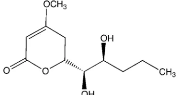
Engel & Nowacki, 1972a; Engel & Nowacki, 1972b). In the title compound, the atoms of the pyran-2-one assume a half-chair conformation. The conformations of the methoxy and dihydroxypentyl groups are shown in Fig. 1. Carbons 4′ and
5′ are disordered over two sites with occupancies of 0.881 (10):0.119 (10). In the crystal, molecules are linked via O—
H···O hydrogen bonds forming a three-dimensional network (see Table 1 and Fig. 2).
S2. Experimental
The title compound was obtained by liquid-liquid extraction (CH2Cl2/H2O) of a culture of an endophytic Pestalotiopsis
sp. The CH2Cl2 fraction was evaporated under reduced pressure then purified by chromatography on a silica column with
CHCl3/CH3OH (8/2) as eluent. After pooling common fractions, a crystal was grown by slow evaporation of a MeOH
solution.
S3. Refinement
The molecule exhibits orientational disorder at atoms C4 and C5. Hydrogen atoms were located and refined isotropically
except those on C4 and C5 which were placed in calculated positions of C—H = 0.98 and 0.99Å and assigned isotropic
displacement parameters of Uiso(H) = 1.2Ueq(C) or 1.5Ueq(Cmethyl), and their coordinates were allowed to ride on their
Figure 1
Molecular structure of the title compound. Displacement ellipsoids are shown at the 50% probability level on
non-hydrogen atoms. The disorder is not shown.
Figure 2
[image:4.610.120.486.373.608.2]supporting information
sup-3
Acta Cryst. (2013). E69, o1657–o1658
(6S*)-6-[(1S*,2R*)-1,2-Dihydroxypentyl]-4-methoxy-5,6-dihydro-2H-pyran-2-one
Crystal data
C11H18O5
Mr = 230.25
Orthorhombic, P212121
Hall symbol: P 2ac 2ab
a = 5.0375 (3) Å
b = 11.4515 (13) Å
c = 20.802 (2) Å
V = 1200.02 (19) Å3
Z = 4
F(000) = 496
Dx = 1.274 Mg m−3
Mo Kα radiation, λ = 0.71073 Å Cell parameters from 17230 reflections
θ = 1.0–27.5°
µ = 0.10 mm−1
T = 200 K Plate, colorless 0.28 × 0.18 × 0.08 mm
Data collection
Nonius KappaCCD diffractometer
Radiation source: fine-focus sealed tube Graphite monochromator
φ plus ω scans
Absorption correction: multi-scan
(DENZO-SMN; Otwinowski & Minor, 1997)
Tmin = 0.973, Tmax = 0.992
2711 measured reflections 1616 independent reflections 1153 reflections with I > 2σ(I)
Rint = 0.043
θmax = 27.5°, θmin = 3.4°
h = −6→6
k = −14→14
l = −26→26
Refinement
Refinement on F2
Least-squares matrix: full
R[F2 > 2σ(F2)] = 0.052
wR(F2) = 0.121
S = 1.06 1616 reflections 188 parameters 0 restraints
Primary atom site location: structure-invariant direct methods
Secondary atom site location: difference Fourier map
Hydrogen site location: inferred from neighbouring sites
H atoms treated by a mixture of independent and constrained refinement
w = 1/[σ2(F
o2) + (0.0544P)2 + 0.1236P]
where P = (Fo2 + 2Fc2)/3
(Δ/σ)max < 0.001
Δρmax = 0.16 e Å−3
Δρmin = −0.18 e Å−3
Extinction correction: SHELXL97 (Sheldrick, 2008), Fc*=kFc[1+0.001xFc2λ3/sin(2θ)]-1/4
Extinction coefficient: 0.033 (6)
Special details
Experimental. The program DENZO-SMN (Otwinowski & Minor, 1997) uses a scaling algorithm that effectively corrects for absorption effects. High redundancy data were used in the scaling program thus the 'multi-scan′ code word was used. No transmission coefficients are available from the program (only scale factors for each frame). The scale factors in the experimental table are calculated from the 'size′ command in the SHELXL97<i/> input file.
Geometry. All e.s.d.'s (except the e.s.d. in the dihedral angle between two l.s. planes) are estimated using the full covariance matrix. The cell e.s.d.'s are taken into account individually in the estimation of e.s.d.'s in distances, angles and torsion angles; correlations between e.s.d.'s in cell parameters are only used when they are defined by crystal symmetry. An approximate (isotropic) treatment of cell e.s.d.'s is used for estimating e.s.d.'s involving l.s. planes.
Refinement. Refinement of F2 against ALL reflections. The weighted R-factor wR and goodness of fit S are based on F2,
conventional R-factors R are based on F, with F set to zero for negative F2. The threshold expression of F2 >σ(F2) is used
only for calculating R-factors(gt) etc and is not relevant to the choice of reflections for refinement. R factors based on F2
Fractional atomic coordinates and isotropic or equivalent isotropic displacement parameters (Å2)
x y z Uiso*/Ueq Occ. (<1)
O1 0.4082 (4) 0.31378 (17) 0.78556 (9) 0.0429 (5) O2 0.5372 (5) 0.4250 (2) 0.70604 (12) 0.0678 (7) O4 −0.0742 (6) 0.5734 (2) 0.85714 (11) 0.0685 (8) O1′ 0.5361 (4) 0.1294 (2) 0.86362 (10) 0.0451 (6) H1′O 0.516 (7) 0.066 (3) 0.8434 (15) 0.050 (10)* O2′ 0.2491 (5) 0.21272 (19) 0.97762 (9) 0.0443 (5) H2′O 0.125 (8) 0.238 (3) 0.9970 (17) 0.051 (10)* C2 0.4146 (7) 0.4194 (3) 0.75666 (15) 0.0481 (8) C3 0.2702 (8) 0.5156 (3) 0.78511 (15) 0.0541 (8) H3 0.302 (8) 0.590 (3) 0.7637 (17) 0.070 (11)* C4 0.0890 (7) 0.4946 (3) 0.83017 (13) 0.0520 (8) C5 0.0460 (6) 0.3748 (3) 0.85552 (15) 0.0454 (7) H5A −0.004 (7) 0.381 (2) 0.9013 (15) 0.048 (8)* H5B −0.094 (8) 0.335 (3) 0.8314 (15) 0.056 (9)* C6 0.3007 (6) 0.3053 (3) 0.85045 (13) 0.0395 (7) H6 0.433 (6) 0.340 (2) 0.8789 (12) 0.028 (6)* C1′ 0.2742 (5) 0.1766 (3) 0.86336 (14) 0.0376 (6) H1′ 0.168 (5) 0.146 (2) 0.8299 (12) 0.026 (7)* C2′ 0.1275 (6) 0.1490 (3) 0.92562 (14) 0.0421 (7) H2′ −0.057 (6) 0.174 (3) 0.9211 (14) 0.044 (8)*
C3′A 0.1175 (9) 0.0209 (3) 0.94034 (14) 0.0598 (9) 0.881 (10) H3′A 0.3000 −0.0079 0.9481 0.072* 0.881 (10) H3′B 0.0454 −0.0213 0.9027 0.072* 0.881 (10) C4′A −0.0549 (13) −0.0058 (5) 0.99940 (18) 0.0624 (14) 0.881 (10) H4′A 0.0249 0.0315 1.0377 0.075* 0.881 (10) H4′B −0.2335 0.0283 0.9930 0.075* 0.881 (10) C5′A −0.0814 (18) −0.1325 (4) 1.0111 (3) 0.118 (3) 0.881 (10) H5′A −0.1905 −0.1454 1.0495 0.177* 0.881 (10) H5′B 0.0949 −0.1666 1.0178 0.177* 0.881 (10) H5′C −0.1660 −0.1695 0.9739 0.177* 0.881 (10) C3′B 0.1175 (9) 0.0209 (3) 0.94034 (14) 0.0598 (9) 0.119 (10) H3′C 0.2915 −0.0094 0.9258 0.072* 0.119 (10) H3′D −0.0147 −0.0109 0.9098 0.072* 0.119 (10) C4′B 0.064 (7) −0.043 (3) 1.0025 (13) 0.036 (7)* 0.119 (10) H4′C 0.1133 0.0035 1.0409 0.043* 0.119 (10) H4′D 0.1484 −0.1207 1.0041 0.043* 0.119 (10) C5′B −0.244 (5) −0.049 (2) 0.9920 (11) 0.038 (8)* 0.119 (10) H5′D −0.3267 −0.0887 1.0287 0.057* 0.119 (10) H5′E −0.2819 −0.0932 0.9526 0.057* 0.119 (10) H5′F −0.3161 0.0299 0.9881 0.057* 0.119 (10) C7 −0.0519 (12) 0.6930 (3) 0.83475 (18) 0.0924 (17)
supporting information
sup-5
Acta Cryst. (2013). E69, o1657–o1658
Atomic displacement parameters (Å2)
U11 U22 U33 U12 U13 U23
O1 0.0439 (11) 0.0449 (12) 0.0398 (10) −0.0023 (9) 0.0045 (9) 0.0010 (9) O2 0.0802 (18) 0.0588 (15) 0.0645 (14) 0.0010 (13) 0.0250 (14) 0.0148 (12) O4 0.0831 (18) 0.0673 (15) 0.0549 (13) 0.0342 (14) −0.0025 (13) −0.0027 (11) O1′ 0.0373 (11) 0.0457 (13) 0.0525 (12) 0.0014 (9) 0.0025 (9) −0.0052 (11) O2′ 0.0365 (11) 0.0575 (13) 0.0388 (10) −0.0012 (11) −0.0009 (9) −0.0102 (9) C2 0.0505 (18) 0.0443 (17) 0.0497 (17) −0.0040 (15) −0.0004 (15) 0.0035 (15) C3 0.071 (2) 0.0453 (19) 0.0460 (17) 0.0060 (18) −0.0063 (16) 0.0010 (16) C4 0.057 (2) 0.058 (2) 0.0406 (15) 0.0203 (18) −0.0105 (15) −0.0035 (15) C5 0.0386 (16) 0.0567 (19) 0.0408 (16) 0.0048 (14) 0.0013 (13) −0.0036 (14) C6 0.0356 (14) 0.0472 (17) 0.0356 (14) −0.0013 (12) −0.0022 (11) −0.0066 (12) C1′ 0.0315 (13) 0.0438 (16) 0.0375 (14) −0.0017 (12) −0.0029 (12) −0.0064 (13) C2′ 0.0328 (15) 0.0548 (19) 0.0388 (15) −0.0060 (13) −0.0019 (12) −0.0054 (14) C3′A 0.078 (2) 0.053 (2) 0.0477 (17) −0.0177 (19) 0.0093 (16) −0.0044 (16) C4′A 0.077 (4) 0.058 (3) 0.053 (2) −0.004 (3) 0.016 (2) 0.002 (2) C5′A 0.197 (8) 0.061 (3) 0.097 (4) −0.029 (4) 0.070 (5) 0.002 (3) C7 0.152 (5) 0.067 (3) 0.059 (2) 0.053 (3) −0.006 (3) 0.0042 (19)
Geometric parameters (Å, º)
O1—C2 1.351 (3) C2′—C3′A 1.499 (5) O1—C6 1.458 (3) C2′—H2′ 0.98 (3) O2—C2 1.222 (4) C3′A—C4′A 1.536 (5) O4—C4 1.343 (4) C3′A—H3′A 0.9900 O4—C7 1.451 (4) C3′A—H3′B 0.9900 O1′—C1′ 1.426 (3) C4′A—C5′A 1.477 (7) O1′—H1′O 0.85 (3) C4′A—H4′A 0.9900 O2′—C2′ 1.442 (3) C4′A—H4′B 0.9900 O2′—H2′O 0.80 (4) C5′A—H5′A 0.9800 C2—C3 1.447 (5) C5′A—H5′B 0.9800 C3—C4 1.330 (5) C5′A—H5′C 0.9800 C3—H3 0.98 (4) C4′B—C5′B 1.57 (4) C4—C5 1.486 (5) C4′B—H4′C 0.9900 C5—C6 1.513 (4) C4′B—H4′D 0.9900 C5—H5A 0.99 (3) C5′B—H5′D 0.9800 C5—H5B 0.97 (4) C5′B—H5′E 0.9800 C6—C1′ 1.504 (4) C5′B—H5′F 0.9800 C6—H6 0.98 (3) C7—H7A 0.9800 C1′—C2′ 1.524 (4) C7—H7B 0.9800 C1′—H1′ 0.94 (3) C7—H7C 0.9800
O2—C2—C3 124.5 (3) C2′—C3′A—H3′B 109.2 O1—C2—C3 119.2 (3) C4′A—C3′A—H3′B 109.2 C4—C3—C2 119.7 (3) H3′A—C3′A—H3′B 107.9 C4—C3—H3 126 (2) C5′A—C4′A—C3′A 112.3 (4) C2—C3—H3 113 (2) C5′A—C4′A—H4′A 109.1 C3—C4—O4 126.4 (3) C3′A—C4′A—H4′A 109.1 C3—C4—C5 121.1 (3) C5′A—C4′A—H4′B 109.1 O4—C4—C5 112.5 (3) C3′A—C4′A—H4′B 109.1 C4—C5—C6 109.7 (3) H4′A—C4′A—H4′B 107.9 C4—C5—H5A 108.2 (17) C4′A—C5′A—H5′A 109.5 C6—C5—H5A 108.9 (19) C4′A—C5′A—H5′B 109.5 C4—C5—H5B 110.5 (19) H5′A—C5′A—H5′B 109.5 C6—C5—H5B 109 (2) C4′A—C5′A—H5′C 109.5 H5A—C5—H5B 110 (3) H5′A—C5′A—H5′C 109.5 O1—C6—C1′ 105.3 (2) H5′B—C5′A—H5′C 109.5 O1—C6—C5 110.2 (2) C5′B—C4′B—H4′C 112.8 C1′—C6—C5 115.3 (2) C5′B—C4′B—H4′D 112.8 O1—C6—H6 106.3 (15) H4′C—C4′B—H4′D 110.3 C1′—C6—H6 110.7 (15) C4′B—C5′B—H5′D 109.5 C5—C6—H6 108.7 (15) C4′B—C5′B—H5′E 109.5 O1′—C1′—C6 106.9 (2) H5′D—C5′B—H5′E 109.5 O1′—C1′—C2′ 111.5 (2) C4′B—C5′B—H5′F 109.5 C6—C1′—C2′ 113.4 (2) H5′D—C5′B—H5′F 109.5 O1′—C1′—H1′ 112.6 (16) H5′E—C5′B—H5′F 109.5 C6—C1′—H1′ 106.5 (15) O4—C7—H7A 109.5 C2′—C1′—H1′ 106.0 (16) O4—C7—H7B 109.5 O2′—C2′—C3′A 110.9 (3) H7A—C7—H7B 109.5 O2′—C2′—C1′ 109.1 (2) O4—C7—H7C 109.5 C3′A—C2′—C1′ 113.1 (3) H7A—C7—H7C 109.5 O2′—C2′—H2′ 109.1 (18) H7B—C7—H7C 109.5
supporting information
sup-7
Acta Cryst. (2013). E69, o1657–o1658
Hydrogen-bond geometry (Å, º)
D—H···A D—H H···A D···A D—H···A
O1′—H1′O···O2i 0.85 (3) 1.93 (3) 2.778 (3) 177 (3)
O2′—H2′O···O2′ii 0.80 (4) 2.05 (4) 2.8178 (18) 163 (4)