Received 6 March 2018 Accepted 13 April 2018
Edited by L. Fabian, University of East Anglia, England
Keywords:crystal structure; lamotrigine; ethanolate.
CCDC reference:1826282
Supporting information:this article has supporting information at journals.iucr.org/e
Lamotrigine ethanol monosolvate
Charlie L. Hall, Jason Potticary, Hazel A. Sparkes, Natalie E. Pridmore and Simon R. Hall*
School of Chemistry, University of Bristol, Cantock’s Close, Bristol, England BS8 1TS, England. *Correspondence e-mail: [email protected]
Lamotrigine is an active pharmaceutical ingredient used as a treatment for epilepsy and psychiatric disorders. Single crystals of an ethanolate solvate, C9H7Cl2N5C2H5OH, were produced by slow evaporation of a saturated solution
from anhydrous ethanol. Within the crystal structure, the lamotrigine molecules form dimers through N—H N hydrogen bonds involving the amine N atoms in the ortho position of the triazine group. These dimers are linked into a tape motif through hydrogen bonds involving the amine N atoms in theparaposition. The ethanol and lamotrigine are present in a 1:1 ratio in the lattice with the ethyl group of the ethanol molecule exhibiting disorder with an occupancy ratio of 0.516 (14):0.484 (14).
1. Chemical context
Anticonvulsants are a group of drugs used principally in the treatment of epilepsy, which have also been shown to aid in the treatment of psychiatric conditions such as bipolar disorder. Although the drugs are effective when inside the body, many suffer from having low solubility and bioavail-ability. Prime examples of such drugs are carbamazepine (Uzunovic´ et al., 2010), phenytoin (Widanapathirana et al., 2015) and lamotrigine (Vaithianathanet al., 2015), which are all categorised as BCS (biopharmaceutical classification system) class II (low solubility, high permeability).
In an attempt to increase the solubility of BCS class II drugs, extensive studies have been undertaken to produce crystal structures including the active pharmaceutical ingre-dients (APIs) with lower crystal lattice energies. In the case of lamotrigine, Cheneyet al.(2010) investigated the solubility of 10 novel forms, including salts, co-crystals and solvates, showing the possibility of creating many stable lamotrigine compounds. The structures of lamotrigine co-crystals and solvates are stabilized due to the large number of hydrogen bonds that can form with the 1,2,4-triazine-3,5-diamine group.
In this work, the structure for the ethanolate (I), previously only obtained as a powder pattern (Garti et al., 2008), is defined. This new structure determination affords a deeper
insight into the different hydrogen-bonding networks that can form in the lamotrigine crystal.
2. Structural commentary
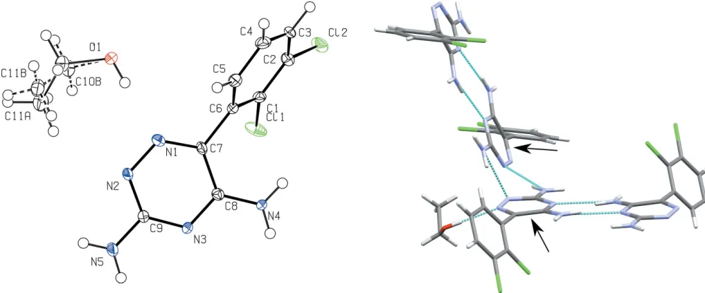
A displacement ellipsoid plot for lamotrigine ethanolate is shown in Fig. 1. The central dihedral, C1—C6—C7—C8, sits at an angle of 63.5 (9), the flexibility of which allows for the inclusion of solvent molecules to form hydrogen-bonding networks. Central dihedral angles for lamotrigine solvates are included in Table 1. Fig. 2 shows the unit cell for (I), which consists of eight lamotrigine molecules and eight ethanol molecules. The main motif within the structure is a lamotrigine dimer stabilized by two ethanol molecules. Here the lamo-trigine dimer forms using the amine N atoms in the ortho
position of the triazine group.
3. Supramolecular features
In the crystal, adjacent in-plane lamotrigine dimers are linked
viahydrogen bonding of the amines in theparaposition of the triazine group (Table 2). Each dimer sits at an angle of 67.2 (5) to the next closest dimer, measured with respect to the in-plane triazine rings, highlighted in Fig. 3.
4. Database survey
A database survey of the Cambridge Structural Database (CSD, version 5.38, last update May 2017; Groomet al., 2016) showed a list of 35 existing co-crystal/solvate structures for lamotrigine, including 6 structures incorporating alcohols, but no ethanol solvate. The most similar structure compositionally to (I) is the ethanol solvate monohydrate (Cheneyet al., 2010); however, the arrangement contrasts quite dramatically, with
research communications
Acta Cryst.(2018). E74, 678–681 Hallet al. C
[image:2.610.45.566.93.170.2]9H7Cl2N5C2H6O
679
Table 1Chosen parameters for the comparison of lamotrigine alcohol solvates.
Structure Central dihedral angle (
) Dimerization motif Density (g cm1)
Methanol disolvate 63.7 (2) para 1.50
Ethanol monohydrate 67.6 (0) para 1.49
Methanol monosolvate 80.1 (5) ortho 1.45
Ethanol solvate (I) 63.5 (9) ortho 1.42
2-Propanol solvate 69.6 (8) ortho 1.36
[image:2.610.315.565.321.469.2]Butan-1-ol solvate monohydrate 71.2 (1) para 1.34
Figure 2
[image:2.610.45.554.510.722.2]The crystal packing of (I), viewed along thecaxis.
Figure 1
A displacement ellipsoid plot of (I), showing the atom-labelling scheme. Displacement ellipsoids are drawn at the 50% probability level.
Figure 3
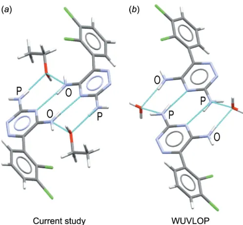
the dimer formation of the lamotrigine molecules using the amine N atoms in the para position, shown in Fig. 4. This change in dimerization motif leads to a reduction in density of the lamotrigine ethanolate over the lamotrigine ethanol monohydrate by 5%.
Analysis of the previously published lamotrigine alcohol solvates shows a trend between the alcohol chain length and whether the lamotrigine dimers form on the ortho or para
group of the triazine. The two densest structures are the methanol disolvate (Hanna et al., 2009) and the ethanol solvate monohydrate, where lamotrigine dimers are connected
viathe amines in theparaposition of the triazine. Conversely, the methanol monosolvate (Janes et al., 1989), isopropanol solvate (Qian et al., 2009) and title compound form dimers from the amine on the ortho positions. The least dense structure is the butan-1-ol solvate monohydrate (Sridhar & Ravikumar, 2011), which has similar arrangement to the dense structures, with the dimers held apart by the large butanol solvent molecules. The densities of the lamotrigine structures
are highlighted in Table 1. 5. Synthesis and crystallization
Lamotrigine (>98%, Acros Organics) was saturated in a solution of pure anhydrous ethanol (>99.5%, Sigma Aldrich) over several weeks. Crystals of lamotrigine ethanolate were producedvia slow evaporation of 1 ml of the solution over 72 h.
6. Refinement
Crystal data, data collection and structure refinement details are summarized in Table 3. All of the hydrogen atoms were located geometrically (aromatic C—H = 0.95 A˚ , methyl C—H = 0.98 A˚ , ethyl C—H = 0.99 A˚, O—H = 0.84 A˚ N—H= 0.88 A˚) and refined using a riding model [aromatic, ethyl and amine
Uiso(H) = 1.2 times parent atom Ueq, methyl and alcohol
Uiso(H) = 1.5 times parent atomUeq]. The ethanol solvent in
the lattice is disordered over two positions; the occupancies of the two positions were refined with the sum set to equal 1, refining to give relative occupancies of 52:48. Restraints (SIMU 0.01 0.02) were applied to maintain sensible thermal displacement parameters for the carbon atoms.
Funding information
[image:3.610.312.557.88.373.2]SRH, CLH and JP would like to thank MagnaPharm, a collaborative research project funded by the European Union’s Horizon 2020 Research and Innovation programme Table 2
Hydrogen-bond geometry (A˚ ,).
D—H A D—H H A D A D—H A
O1—H1A N1 0.84 2.01 2.848 (7) 179
N4—H4A N3i 0.88 2.10 2.972 (7) 172
N4—H4B O1ii 0.88 2.14 2.841 (7) 137
N5—H5A O1iii 0.88 2.16 3.014 (7) 163
N5—H5B N2iv 0.88 2.14 2.987 (8) 161
Symmetry codes: (i)xþ1 2;yþ
3
2;z; (ii)xþ 1 2;yþ
1 2;zþ
1
2; (iii)x;yþ1;z 1 2;
[image:3.610.45.294.460.691.2](iv)xþ1;y;zþ1 2.
Figure 4
(a) The dimerization motif in (I), held together with the amines in the
orthoposition of the triazine group. The amine in theorthoandpara
positions are labelled with O and P, respectively. (b) The dimerization motif in the ethanolate hydrate structure, held together with the amines in theparaposition of the triazine group.
Table 3
Experimental details.
Crystal data
Chemical formula C9H7Cl2N5C2H6O
Mr 302.16
Crystal system, space group Monoclinic,C2/c
Temperature (K) 100
a,b,c(A˚ ) 21.2458 (15), 10.2320 (8), 14.8428 (11)
(
) 118.808 (4)
V(A˚3) 2827.3 (4)
Z 8
Radiation type MoK
(mm1) 0.46
Crystal size (mm) 0.390.250.13
Data collection
Diffractometer Bruker APEXII CCD
Absorption correction Multi-scan (SADABS; Bruker, 2015)
Tmin,Tmax 0.602, 0.745
No. of measured, independent and observed [I> 2(I)] reflections
21376, 2925, 2634
Rint 0.053
(sin/)max(A˚
1
) 0.629
Refinement
R[F2> 2(F2)],wR(F2),S 0.098, 0.234, 1.41
No. of reflections 2925
No. of parameters 193
No. of restraints 48
H-atom treatment H-atom parameters constrained
max,min(e A˚
3) 0.62,0.87
Computer programs:APEX2andSAINT(Bruker, 2015),SHELXT(Sheldrick, 2015a),
(grant No. 736899), the Bristol Centre for Functional Nano-materials (EP/G036780/1) and the Centre for Doctoral Training in Condensed Matter Physics (EP/L015544/1).
References
Bruker (2015). APEX2, SAINT and SADABS. Bruker AXS Inc.,
Madison, Wisconsin USA.
Cheney, M. L., Shan, N., Healey, E. R., Hanna, M., Wojtas, L., Zaworotko, M. J., Sava, V., Song, S. & Sanchez-Ramos, J. R. (2010).
Cryst. Growth Des.10, 394–405.
Dolomanov, O. V., Bourhis, L. J., Gildea, R. J., Howard, J. A. K. &
Puschmann, H. (2009).J. Appl. Cryst.42, 339–341.
Garti, N., Berkovich, Y., Dolitzky, B. Z., Aronhime, J., Singer, C., Liebermann, A. & Gershon, N. (2008). US Patent Number. 7390807B2.
Groom, C. R., Bruno, I. J., Lightfoot, M. P. & Ward, S. C. (2016).Acta
Cryst.B72, 171–179.
Hanna, M., Shan, N. & Cheney, M. L. (2009). US Patent Number. 061513A1.
Janes, R. W., Lisgarten, J. N. & Palmer, R. A. (1989).Acta Cryst.C45,
129–132.
Qian, Y., Lv, P. C., Shi, L., Fang, R. Q., Song, Z. C. & Zhu, H. L.
(2009).J. Chem. Sci.121, 463–470.
Sheldrick, G. M. (2015a).Acta Cryst.A71, 3–8.
Sheldrick, G. M. (2015b).Acta Cryst.C71, 3–8.
Sridhar, S. & Ravikumar, K. (2011).J. Chem. Crystallogr.41, 1289–
1300.
Uzunovic´, A., Vranic´, E. & Hadzˇidedic´, Sˇ. (2010).Bosn. J. Basic Med.
Sci.10, 234–238.
Vaithianathan, A., Raman, S., Jiang, W., Ting, Y. T., Kane, M. A. &
Polli, J. E. (2015).Mol. Pharm.12, 2436–2443.
Widanapathirana, L., Tale, S. & Reineke, T. M. (2015).Mol. Pharm.
12, 2537–2543.
research communications
Acta Cryst.(2018). E74, 678–681 Hallet al. C
sup-1 Acta Cryst. (2018). E74, 678-681
supporting information
Acta Cryst. (2018). E74, 678-681 [https://doi.org/10.1107/S2056989018005819]
Lamotrigine ethanol monosolvate
Charlie L. Hall, Jason Potticary, Hazel A. Sparkes, Natalie E. Pridmore and Simon R. Hall
Computing details
Data collection: APEX2 (Bruker, 2015); cell refinement: SAINT (Bruker, 2015); data reduction: SAINT (Bruker, 2015);
program(s) used to solve structure: SHELXT (Sheldrick, 2015a); program(s) used to refine structure: SHELXL
(Sheldrick, 2015b); molecular graphics: Olex2 (Dolomanov et al., 2009); software used to prepare material for
publication: Olex2 (Dolomanov et al., 2009).
(I)
Crystal data
C9H7Cl2N5·C2H6O
Mr = 302.16
Monoclinic, C2/c
a = 21.2458 (15) Å
b = 10.2320 (8) Å
c = 14.8428 (11) Å
β = 118.808 (4)°
V = 2827.3 (4) Å3
Z = 8
F(000) = 1248
Dx = 1.420 Mg m−3
Mo Kα radiation, λ = 0.71073 Å
Cell parameters from 7221 reflections
θ = 2.2–26.4°
µ = 0.46 mm−1
T = 100 K
Block, colourless 0.39 × 0.25 × 0.13 mm
Data collection
Bruker APEXII CCD diffractometer
Radiation source: fine-focus sealed tube Graphite monochromator
φ and ω scans
Absorption correction: multi-scan (SADABS; Bruker, 2015)
Tmin = 0.602, Tmax = 0.745
21376 measured reflections 2925 independent reflections 2634 reflections with I > 2σ(I)
Rint = 0.053
θmax = 26.6°, θmin = 2.2°
h = −26→26
k = −12→12
l = −18→18
Refinement
Refinement on F2
Least-squares matrix: full
R[F2 > 2σ(F2)] = 0.098
wR(F2) = 0.234
S = 1.41
2925 reflections 193 parameters 48 restraints
Primary atom site location: dual
Hydrogen site location: inferred from neighbouring sites
H-atom parameters constrained
w = 1/[σ2(F
o2) + 59.8676P]
where P = (Fo2 + 2Fc2)/3
(Δ/σ)max < 0.001
Δρmax = 0.62 e Å−3
supporting information
sup-2 Acta Cryst. (2018). E74, 678-681
Special details
Geometry. All esds (except the esd in the dihedral angle between two l.s. planes) are estimated using the full covariance matrix. The cell esds are taken into account individually in the estimation of esds in distances, angles and torsion angles; correlations between esds in cell parameters are only used when they are defined by crystal symmetry. An approximate (isotropic) treatment of cell esds is used for estimating esds involving l.s. planes.
Refinement. The occupancies of the disordered atoms in the ethanol were refined with their sum set to equal 1. Restraints were applied to maintain sensible thermal and geometric parameters. The diffraction data showed slight splitting of some peaks but twinning could not be sensibly separated and modelled. However this may explain the large K values, slightly high second weight paramater and Fobs greater than Fcalc.
Fractional atomic coordinates and isotropic or equivalent isotropic displacement parameters (Å2)
x y z Uiso*/Ueq Occ. (<1)
Cl1 0.16117 (9) 0.41684 (18) 0.10486 (12) 0.0254 (4)
Cl2 0.06655 (8) 0.37527 (18) 0.21033 (13) 0.0256 (4)
O1 0.4106 (2) 0.2925 (5) 0.4111 (3) 0.0192 (10)
H1A 0.397808 0.357810 0.371958 0.029* 0.484 (14)
H1B 0.398027 0.358121 0.372284 0.029* 0.516 (14)
N3 0.3251 (3) 0.6534 (5) 0.1015 (4) 0.0136 (10)
N4 0.2195 (3) 0.7095 (5) 0.0967 (4) 0.0145 (11)
H4A 0.208617 0.757236 0.041803 0.017*
H4B 0.189512 0.705023 0.121767 0.017*
N1 0.3666 (3) 0.5147 (6) 0.2792 (4) 0.0177 (11)
N2 0.4130 (3) 0.5288 (6) 0.2425 (4) 0.0185 (12)
N5 0.4347 (3) 0.6032 (7) 0.1152 (4) 0.0293 (15)
H5A 0.422053 0.646160 0.057650 0.035*
H5B 0.477226 0.565850 0.147585 0.035*
C6 0.2549 (3) 0.5427 (6) 0.2790 (4) 0.0144 (12)
C8 0.2808 (3) 0.6445 (6) 0.1415 (4) 0.0133 (12)
C7 0.3022 (3) 0.5648 (6) 0.2321 (4) 0.0142 (12)
C4 0.2351 (4) 0.5649 (7) 0.4253 (5) 0.0191 (13)
H4 0.251372 0.593442 0.493888 0.023*
C3 0.1697 (3) 0.5029 (7) 0.3727 (5) 0.0193 (14)
H3 0.140279 0.491009 0.404086 0.023*
C2 0.1470 (3) 0.4581 (7) 0.2738 (5) 0.0176 (13)
C9 0.3894 (3) 0.5948 (7) 0.1536 (5) 0.0189 (13)
C5 0.2772 (3) 0.5859 (7) 0.3788 (5) 0.0188 (13)
H5 0.321851 0.630185 0.415469 0.023*
C1 0.1893 (3) 0.4782 (6) 0.2273 (5) 0.0152 (12)
C10B 0.4570 (8) 0.2138 (16) 0.3909 (13) 0.024 (3) 0.484 (14)
H10A 0.435281 0.197369 0.316042 0.029* 0.484 (14)
H10B 0.464411 0.128524 0.426119 0.029* 0.484 (14)
C11B 0.5275 (8) 0.2819 (16) 0.4283 (14) 0.030 (4) 0.484 (14)
H11A 0.519935 0.365998 0.393000 0.045* 0.484 (14)
H11B 0.559689 0.227634 0.414026 0.045* 0.484 (14)
H11C 0.549146 0.296685 0.502588 0.045* 0.484 (14)
C10A 0.4866 (8) 0.2617 (17) 0.4439 (12) 0.030 (3) 0.516 (14)
sup-3 Acta Cryst. (2018). E74, 678-681
H10D 0.516983 0.335764 0.484496 0.036* 0.516 (14)
C11A 0.5000 (8) 0.2356 (15) 0.3545 (12) 0.030 (3) 0.516 (14)
H11D 0.466604 0.168389 0.310075 0.045* 0.516 (14)
H11E 0.549503 0.205101 0.380438 0.045* 0.516 (14)
H11F 0.492715 0.316205 0.315082 0.045* 0.516 (14)
Atomic displacement parameters (Å2)
U11 U22 U33 U12 U13 U23
Cl1 0.0342 (9) 0.0289 (9) 0.0201 (8) −0.0150 (7) 0.0189 (7) −0.0098 (7)
Cl2 0.0144 (7) 0.0370 (10) 0.0260 (8) −0.0077 (7) 0.0102 (6) 0.0040 (7)
O1 0.017 (2) 0.024 (2) 0.020 (2) 0.0066 (18) 0.0105 (18) 0.0100 (19)
N3 0.010 (2) 0.018 (3) 0.011 (2) 0.003 (2) 0.004 (2) 0.004 (2)
N4 0.011 (2) 0.020 (3) 0.012 (2) 0.005 (2) 0.006 (2) 0.007 (2)
N1 0.016 (3) 0.023 (3) 0.016 (3) 0.001 (2) 0.010 (2) 0.003 (2)
N2 0.010 (2) 0.031 (3) 0.015 (3) 0.006 (2) 0.007 (2) 0.008 (2)
N5 0.017 (3) 0.056 (4) 0.019 (3) 0.017 (3) 0.012 (2) 0.018 (3)
C6 0.015 (3) 0.016 (3) 0.013 (3) 0.005 (2) 0.008 (2) 0.004 (2)
C8 0.014 (3) 0.015 (3) 0.012 (3) 0.000 (2) 0.007 (2) −0.001 (2)
C7 0.009 (3) 0.019 (3) 0.014 (3) 0.000 (2) 0.006 (2) 0.000 (2)
C4 0.025 (3) 0.020 (3) 0.013 (3) 0.003 (3) 0.010 (3) 0.003 (3)
C3 0.021 (3) 0.022 (3) 0.023 (3) 0.009 (3) 0.018 (3) 0.009 (3)
C2 0.014 (3) 0.021 (3) 0.019 (3) −0.001 (3) 0.009 (3) 0.002 (3)
C9 0.015 (3) 0.027 (4) 0.017 (3) 0.008 (3) 0.010 (2) 0.008 (3)
C5 0.015 (3) 0.024 (3) 0.015 (3) 0.002 (3) 0.006 (2) 0.004 (3)
C1 0.019 (3) 0.015 (3) 0.012 (3) 0.003 (2) 0.009 (2) 0.002 (2)
C10B 0.020 (5) 0.026 (5) 0.028 (5) 0.006 (5) 0.013 (4) 0.005 (5)
C11B 0.020 (7) 0.028 (7) 0.044 (8) 0.008 (6) 0.017 (6) 0.015 (6)
C10A 0.017 (5) 0.039 (6) 0.031 (5) 0.007 (5) 0.011 (4) 0.013 (5)
C11A 0.030 (6) 0.027 (7) 0.040 (7) 0.009 (6) 0.021 (6) 0.008 (6)
Geometric parameters (Å, º)
Cl1—C1 1.735 (6) C8—C7 1.446 (8)
Cl2—C2 1.725 (6) C4—H4 0.9500
O1—H1A 0.8400 C4—C3 1.377 (9)
O1—H1B 0.8400 C4—C5 1.386 (9)
O1—C10B 1.413 (15) C3—H3 0.9500
O1—C10A 1.477 (14) C3—C2 1.384 (9)
N3—C8 1.335 (7) C2—C1 1.387 (8)
N3—C9 1.344 (8) C5—H5 0.9500
N4—H4A 0.8800 C10B—H10A 0.9900
N4—H4B 0.8800 C10B—H10B 0.9900
N4—C8 1.322 (8) C10B—C11B 1.50 (2)
N1—N2 1.345 (7) C11B—H11A 0.9800
N1—C7 1.304 (8) C11B—H11B 0.9800
N2—C9 1.346 (8) C11B—H11C 0.9800
supporting information
sup-4 Acta Cryst. (2018). E74, 678-681
N5—H5B 0.8800 C10A—H10D 0.9900
N5—C9 1.336 (8) C10A—C11A 1.51 (2)
C6—C7 1.490 (8) C11A—H11D 0.9800
C6—C5 1.392 (9) C11A—H11E 0.9800
C6—C1 1.391 (9) C11A—H11F 0.9800
C10B—O1—H1A 109.5 N5—C9—N2 116.5 (6)
C10A—O1—H1B 109.5 C6—C5—H5 119.7
C8—N3—C9 116.9 (5) C4—C5—C6 120.6 (6)
H4A—N4—H4B 120.0 C4—C5—H5 119.7
C8—N4—H4A 120.0 C6—C1—Cl1 119.8 (5)
C8—N4—H4B 120.0 C2—C1—Cl1 119.2 (5)
C7—N1—N2 121.7 (5) C2—C1—C6 120.9 (6)
N1—N2—C9 116.9 (5) O1—C10B—H10A 109.8
H5A—N5—H5B 120.0 O1—C10B—H10B 109.8
C9—N5—H5A 120.0 O1—C10B—C11B 109.3 (13)
C9—N5—H5B 120.0 H10A—C10B—H10B 108.3
C5—C6—C7 119.2 (6) C11B—C10B—H10A 109.8
C1—C6—C7 122.4 (5) C11B—C10B—H10B 109.8
C1—C6—C5 118.3 (6) C10B—C11B—H11A 109.5
N3—C8—C7 118.6 (5) C10B—C11B—H11B 109.5
N4—C8—N3 118.5 (5) C10B—C11B—H11C 109.5
N4—C8—C7 122.9 (5) H11A—C11B—H11B 109.5
N1—C7—C6 117.3 (5) H11A—C11B—H11C 109.5
N1—C7—C8 119.9 (5) H11B—C11B—H11C 109.5
C8—C7—C6 122.7 (5) O1—C10A—H10C 109.0
C3—C4—H4 119.8 O1—C10A—H10D 109.0
C3—C4—C5 120.5 (6) O1—C10A—C11A 112.8 (12)
C5—C4—H4 119.8 H10C—C10A—H10D 107.8
C4—C3—H3 120.2 C11A—C10A—H10C 109.0
C4—C3—C2 119.6 (6) C11A—C10A—H10D 109.0
C2—C3—H3 120.2 C10A—C11A—H11D 109.5
C3—C2—Cl2 119.3 (5) C10A—C11A—H11E 109.5
C3—C2—C1 120.0 (6) C10A—C11A—H11F 109.5
C1—C2—Cl2 120.7 (5) H11D—C11A—H11E 109.5
N3—C9—N2 125.6 (5) H11D—C11A—H11F 109.5
N5—C9—N3 117.9 (6) H11E—C11A—H11F 109.5
Hydrogen-bond geometry (Å, º)
D—H···A D—H H···A D···A D—H···A
O1—H1A···N1 0.84 2.01 2.848 (7) 179
N4—H4A···N3i 0.88 2.10 2.972 (7) 172
N4—H4B···O1ii 0.88 2.14 2.841 (7) 137
N5—H5A···O1iii 0.88 2.16 3.014 (7) 163
N5—H5B···N2iv 0.88 2.14 2.987 (8) 161