research communications
Acta Cryst.(2018). E74, 737–742 https://doi.org/10.1107/S2056989018005662
737
Received 26 March 2018Accepted 11 April 2018
Edited by J. Simpson, University of Otago, New Zealand
Keywords:crystal structure; Schiff base; CUPRAC; antioxidant capacity; DFT calcula-tions.
CCDC references:1836250; 1836249
Supporting information:this article has supporting information at journals.iucr.org/e
(E)-2-{[(2-Aminophenyl)imino]methyl}-5-(benzyl-oxy)phenol and
(Z)-3-benzyloxy-6-{[(5-chloro-2-
hydroxyphenyl)amino]methylidene}cyclohexa-2,4-dien-1-one
Nadir Ghichi,a* Ali Benboudiaf,aChawki Bensouici,bYacine DJeblicand Hocine Meraziga
aUnit of Research CHEMS, University of Constantine 1, Algeria,bBiotechnology Research Center, Constantine, Algeria,
andcLaboratory of Materials Chemistry,University of Constantine 1, Algeria. *Correspondence e-mail:
nadirgh82@hotmail.com
The title Schiff base compounds, C20H18N2O2(I) and C20H16ClNO3(II), were
synthesized from 4-benzyloxy-2-hydroxybenzaldehyde by reaction with
1,2-diaminobenzene for (I), and condensation with 2-amino-4-chlorophenol
for (II). Compound (I) adopts the enol–imine tautomeric form with an E
configuration about the C N imine bond. In contrast, theo-hydroxy Schiff base (II), is in the keto–imine tautomeric form with aZconfiguration about the CH— NH bond. Neither molecule is planar. In (I), the central benzene ring makes dihedral angles of 46.80 (10) and 78.19 (10) with the outer phenylamine and phenyl rings, respectively, while for (II), the corresponding angles are 5.11 (9) and 58.42 (11), respectively. The molecular structures of both compounds are affected by the formation of intramolecular contacts, an O—H N hydrogen bond for (I) and an N—H O hydrogen bond for (II); each contact generates anS(6) ring motif. In the crystal of (I), strong N—H O hydrogen bonds form zigzag chains of molecules along the b-axis direction. Molecules are further linked by C—H interactions and offset–contacts and these combine to form a three-dimensional network. The density functional theory (DFT)
optimized structure of compound (II), at the B3LYP/6–311+G(d) level,
confirmed that the keto tautomeric form of the compound, as found in the structure determination, is the lowest energy form. The antioxidant capacities of both compounds were determined by the cupric reducing antioxidant capacity (CUPRAC) process.
1. Chemical context
Schiff base compounds have been used as fine chemicals and medicinal substrates (Fun et al., 2011). Studies of the tautomerism of Schiff bases (Alpaslanet al., 2011; Blaguset al., 2010; U¨ nveret al., 2002) have demonstrated that the stabili-zation of the keto–amino tautomer in the crystal depends mostly on the parento-hydroxyl aldehyde, the type of the substituent, the electron withdrawing or donating of the N-substituent, its position and stereochemistry (Blagus et al., 2010). Schiff base compounds exhibit a broad range of biological activities, including antifungal and antibacterial (da Silvaet al., 2011). They are used as anion sensors (Dalapatiet al., 2011; Khalil et al., 2009), non-linear optical compounds (Sun et al., 2012), and as versatile ligands in coordination chemistry (Khanmohammadiet al., 2009; Keypouret al., 2010). In view of the interest in such materials we have synthesized the title compounds, (I) and (II), and report their crystal
structures here. The common structural feature of these compounds is the presence of a benzyloxy substituent on the central ring, although each molecule adopts a different tautomeric form. Density functional theory (DFT) calcula-tions on (II), carried out at the B3LYP/6-311+G(d) level, are compared with the experimentally determined molecular structure and confirm that the keto tautomeric form of this compound, similar to that found in the structure determina-tion, is the lowest energy form. The antioxidant capacity of both compounds was determined by the cupric reducing antioxidant capacity (CUPRAC) process.
2. Structural commentary
The molecular structures of compounds (I) and (II), illus-trated in Figs. 1 and 2, respectively, are influenced by
intra-molecular hydrogen bonds: the O—H N hydrogen bond in
(I) and the N—H O contact in (II) (Tables 1 and 2) both
form S(6) ring motifs. In compound (II), the N atom is
protonated and the C9—O1 bond length, 1.277 (2) A˚ confirms
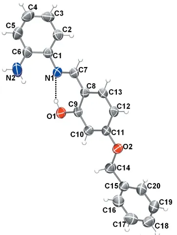
this to be double bond. In compound (I), however, the C9 O1 bond length of 1.3498 (19) A˚ indicates a single bond. Bond C7 C8 [1.395 (3) A˚ ] is a double bond in compound (II), whereas the corresponding bond in (I) [1.435 (3) A˚ ] is a single bond. Compound (I) adopts the enol–imine tautomeric
form and the configuration of the C7 N1 imine bond isE
with a length of 1.288 (3) A˚ . In contrast theo-hydroxy Schiff base of (II), has aZconfiguration about the C7 C8 double bond and the molecule adopts the keto–imine tautomeric form, with the N1—C7 bond length being 1.309 (2) A˚ . Neither molecule is planar: in (I), the central ring (C8–C13) is inclined to the two outer rings (C1–C6 and C15–C20) by 46.80 (10) and 78.19 (10), respectively, while for (II), the dihedral angles between these rings are 5.11 (9) and 58.42 (11), respectively. In compound (II), the C1—N1—C7 angle is 127.15 (17).
738
Ghichiet al. C20H18N2O2and C20H16ClNO3 Acta Cryst.(2018). E74, 737–742
[image:2.610.313.565.106.178.2]research communications
Figure 2
[image:2.610.43.299.219.342.2]The molecular structure of compound (II), with the atom labeling. Displacement ellipsoids are drawn at the 50% probability level. The intramolecular N—H O hydrogen bond is shown as a dashed line. Figure 1
[image:2.610.85.266.472.712.2]The molecular structure of compound (I), with the atom labelling. Displacement ellipsoids are drawn at the 50% probability level. The intramolecular O—H N hydrogen bond is shown as a dashed line.
Table 1
Hydrogen-bond geometry (A˚ ,) for (I).
Cg1 andCg3 are the centroids of the C1–C6 and C15–C20 rings respectively.
D—H A D—H H A D A D—H A
O1—H1 N1 0.82 1.90 2.629 (2) 147
N2—H2A O2i 0.86 2.43 3.211 (3) 151
C14—H14B Cg1ii 0.97 2.74 3.704 (3) 171 C16—H16 Cg1iii 0.93 2.96 3.792 (3) 150
C18—H18 Cg3iv 0.93 2.94 3.620 (2) 131
Symmetry codes: (i) x;y1;zþ1
2; (ii) x;y;z; (iii) x;y;zþ 1 2; (iv)
xþ1 2;yþ
1 2;zþ
1 2.
Table 2
Hydrogen-bond geometry (A˚ ,) for (II).
Cg3 is the centroid of the C15–C20 ring.
D—H A D—H H A D A D—H A
N1—H1 O1 0.86 (2) 1.93 (2) 2.637 (2) 139 (2) N1—H1 O2 0.86 (2) 2.27 (2) 2.620 (2) 104.5 (18) O2—H2 O1i 0.80 (3) 1.84 (3) 2.619 (2) 165 (3)
C7—H7 Cl1ii 0.98 (2) 2.84 (2) 3.7971 (18) 164.5 (17)
C14—H14A Cg3iii 0.97 2.71 3.569 (3) 148
[image:2.610.318.567.551.707.2]3. Supramolecular features
In the crystal of (I), strong N2—H2A O2ihydrogen bonds,
Table 1, form zigzag chains of molecules along the b-axis direction, Fig. 3. Weaker C—H and offset– stacking
interactions also contribute to the packing (Fig. 4)
[Cg2 Cg2(x,y,z+1
2) = 3.8151 (11) A˚ ;Cg2 is the centroid
of the central ring]. The overall crystal packing for this structure is shown in Fig. 5.
For (II), strong O2—H2 O1i hydrogen bonds Table 2,
form inversion dimers that enclose R2
2(18) rings. These
combine with weaker C7—H7 Cl1 hydrogen bonds, which
also generate inversion dimers but withR22(14) motifs.
Inver-sion-related C14—H14A Cg3ii contacts lead to the
forma-tion of sheets of molecules parallel to (120), Fig. 6, which are stacked approximately along theb-axis direction. The overall packing for this structure is shown in Fig. 7.
4. Database survey
A search of the Cambridge Database (Version 5.39, updated February 2018; Groomet al.2016) for structures similar to (I) gave two hits, viz. (Z)-6-{2-[(E )-2,4-dihydroxybenzylidene- amino]phenylaminomethylene}-3-hydroxycyclohexa-2,4-dien-one (Fun et al., 2008) and (E )-5-(benzyloxy)-2-[(4-nitro-phenyl)carbonoimidoyl]phenol reported by us in 2015 (Ghichi
et al., 2015). More recently, we have described the very similar structure of (E )-5-benzyloxy-2-{[(4-chlorophenyl)imino]-methyl}phenol (Ghichiet al., 2018). A search for analogues of
(II) produced three related
phenylethylamino)methyl-research communications
Acta Cryst.(2018). E74, 737–742 Ghichiet al. C
[image:3.610.45.292.70.400.2]20H18N2O2and C20H16ClNO3
739
Figure 4
[image:3.610.313.561.71.154.2]C—H and–conatcts (dotted green lines) in the crystal structure of (I).
Figure 5
[image:3.610.313.564.361.726.2]Overall packing for (I) viewed along theb-axis direction.
Figure 6
[image:3.610.314.564.429.573.2]Sheets of molecules of (II) parallel to (120). Figure 3
[image:3.610.316.564.605.726.2]Zigzag chains of molecules of (I) along theb-axis direction. Hydrogen bonds are drawn as blue dashed lines.
Figure 7
[image:3.610.45.299.613.710.2]ene)cyclohexa-2,4-dien-1-ones (Chatziefthimiou et al., 2006) and our recent contribution also reported (E )-5-benzyloxy-2-({[2-(1H-indol-3-yl)ethyl]iminiumyl)methyl)phenolate, which is closely similar to (II). The structures of Schiff bases derived from hydroxyaryl aldehydes have been the subject of a general survey, in which a number of structural errors, often involving misplaced H atoms, were pointed out (Blaguset al., 2010).
5. DFT-optimized calculations
DFT quantum chemical calculations were performed on molecule (II) using the hybrid functional B3LYP (Beckeet al., 1993; Lee et al., 1988), and base 6–311+G (d). The DFT structure optimization of (II) was performed starting from the X-ray geometry. The DFT and X-ray stuctures are compared in Fig. 8. The calculated values of bond lengths (Table 3) compare well with experimental values with the largest bond-length deviation being less than 0.031 A˚ from those found in the crystal structure. The adoption of the keto–imine tauto-meric form is also predicted by these calculations. The study also shows that the HOMO and LUMO are localized in the plane extending from the chlorohydroxybenzene ring to the
central phenol ring. The electron distribution of the HOMO-1, HOMO, LUMO and LUMO+1 energy levels is shown in Fig. 9. The occupied orbitals are predominantly of-character as is
the LUMO, while LUMO+1 is mainly of -character. The
HOMO–LUMO gap is 0.12449 a.u, with frontier molecular
orbital energies, EHOMO and ELUMO of 5.622 and
2.234 eV, respectively.
6. Antioxidant activity
The antioxidant activity profiles of (I) and (II) were deter-mined using the copper(II)–neocuprine [CuII–Nc] (CUPRAC) process (Apaket al., 2004). The CUPRAC method (cupric ion reducing antioxidant capacity) follows the variation in the absorbance of the neocuproine (2,9-dimethyl-1,10-phenan-throline, Nc), copper+2complex Nc2–Cu+2In the presence of
an antioxidant, the copper–neocuproine complex is reduced and this reaction is followed and quantified spectro-photometrically at a wavelength of 450 nm. The results indi-cate that the percentage (%) inhibition (IC50) in the CUPRAC
assay is small for both compounds in comparison to that for
740
Ghichiet al. C20H18N2O2and C20H16ClNO3 Acta Cryst.(2018). E74, 737–742
[image:4.610.309.566.74.437.2]research communications
Table 3
Experimental and calculated bond lengths (A˚ ) for compound (II).
Bond X-ray B3LYP/6–311+G(d)
N1—C1 1.406 (2) 1.399
N1—C7 1.309 (2) 1.340
O1—C9 1.277 (2) 1.254
O2—C2 1.351 (2) 1.364
O3—C11 1.363 (2) 1.355
O3—C14 1.432 (3) 1.439
C1—C2 1.403 (2) 1.410
C1—C6 1.389 (2) 1.398
C2—C3 1.384 (3) 1.389
C3—C4 1.381 (3) 1.394
C5—C11 1.742 (2) 1.759
C7—C8 1.395 (3) 1.385
C9—C10 1.418 (3) 1.411
C10—C11 1.373 (3) 1.373
C12—C13 1.350 (3) 1.358
C14—C15 1.504 (3) 1.504
C16—C17 1.392 (4) 1.393
[image:4.610.45.295.91.276.2]C19—C20 1.387 (3) 1.393
Figure 8
Comparison of the structures of (II) obtained from (a) the X-ray determination and (b) the DFT calculations.
Figure 9
[image:4.610.44.297.593.714.2]butylated hydroxytoluene (BHT) that was used as a positive control. In Table 4 the values shown are the means of three separate measurements.
7. Synthesis and crystallization
Compound (I)
1,2-Diaminobenzene (1 equiv.) and 4-benzyloxy-2-hy-droxybenzaldehyde (1 equiv.) in ethanol (15–20 ml) were refluxed for 1 h, the solvent was evaporated in vacuo. The residue was recrystallized from ethanol, yielding yellow block-like crystals on slow evaporation of the solvent. The purity of the compound was determined from its NMR spectrum (250 MHz, CDCl3). The azomethine proton appears in the
8.5–8.6 p.p.m.range, while the imine bond is characterized in
the 13C NMR spectrum with the imine C and the C atom
bound to the OH group appearing in the 161.58–163.20 p.p.m.range.1H NMR:= 6.6–7.6 (m, 12H;H-ar),= 13.5 (s, 1H;OH),= 4 (s, 1H;NH2),= 5.1 (s, 1H;CH2–O).13C NMR:
70.22, 127.66, 127.73, 128.32, 128.8, 140.66, 161.58, 163.02, 163.2.
Compound (II)
2-Amino-4-chloroyphenol (1 equiv.) and 4-benzyloxy-2-hydroxybenzaldehyde (1 equiv.) in ethanol (20 ml) were refluxed for 30–60 min, the solvent was evaporated invacuo. The residue was recrystallized from ethanol, yielding orange block-like crystals on slow evaporation of the solvent. The purity of the compound was detemined by its NMR spectrum (250 MHz, CDCl3).
1
H NMR:= 6.5–7.7 (m, 11H;H-ar),= 8.5–8.6 (s, 1H; OH),= 5.1 (s, 1H;CH2–O).
13
C NMR: 55.6, 128.2, 128.7, 133.3, 136.4, 141.4, 159.69, 162.82, 163.77.
8. Refinement
Crystal data, data collection and structure refinement details are summarized in Table 5. In compound (I), the hydroxyl H atom was located in a difference-Fourier map and initially freely refined. In the final cycles of refinements it was
posi-tioned geometrically (O—H = 0.82 A˚ ) and refined with
Uiso(H) = 1.5Ueq(O). In compound (II), the H atoms on N1, C7
and O2 were located in a difference-Fourier and refined freely. For both compounds, the other C-bound H atoms were
posi-research communications
Acta Cryst.(2018). E74, 737–742 Ghichiet al. C
[image:5.610.48.566.94.152.2]20H18N2O2and C20H16ClNO3
741
Table 4
Cupric ion reducing antioxidant capacity of compounds (I) and (II).
Percentage (%) Inhibition
3.125mg 6.25mg 12.5mg 25mg 50mg 100mg 200mg A0.50 (g/ml)
Compound (I) 0.280.01 0.460.00 0.760.03 1.550.04 2.600.14 3.810.15 4.330.04 7.40.21 Compound (II) 0.300.00 0.460.01 0.780.01 1.120.07 1.840.19 2.340.12 4.390.04 6.100.26
BHT 0.190.01 0.330.04 0.660.07 1.030.07 1.480.09 2.040.14 2.320.28 9.620.87
Table 5
Experimental details.
(I) (II)
Crystal data
Chemical formula C20H18N2O2 C20H16ClNO3
Mr 318.36 353.79
Crystal system, space group Monoclinic,C2/c Triclinic,P1
Temperature (K) 293 293
a,b,c(A˚ ) 35.1343 (12), 7.2564 (2), 13.1450 (5) 5.9590 (2), 7.8710 (3), 17.9743 (6)
,,() 90, 95.553 (2), 90 98.381 (2), 93.817 (2), 90.294 (2)
V(A˚3) 3335.57 (19) 832.11 (5)
Z 8 2
Radiation type MoK MoK
(mm1) 0.08 0.25
Crystal size (mm) 0.030.020.01 0.030.020.01
Data collection
Diffractometer Bruker APEXII CCD Bruker APEXII CCD
No. of measured, independent and observed [I> 2(I)] reflections
18218, 3811, 1915 13513, 3052, 2490
Rint 0.072 0.025
(sin /)max(A˚ 1
) 0.650 0.606
Refinement
R[F2> 2(F2)],wR(F2),S 0.049, 0.134, 1.00 0.042, 0.133, 1.10
No. of reflections 3811 3052
No. of parameters 221 238
H-atom treatment H atoms treated by a mixture of independent and constrained refinement
H atoms treated by a mixture of independent and constrained refinement
max,min(e A˚
3) 0.17,0.15 0.21,0.21
[image:5.610.43.563.465.735.2]tioned geometrically (C—H = 0.97–0.97 A˚ ) and refined as riding withUiso(H) = 1.2Ueq(C).
Funding information
We are grateful to the Department of Higher Scientific
Research and CHEMS Research Unit, University of
Constantine1, Algeria, for funding this research project.
References
Alpaslan, G., Macit, M., Erdo¨nmez, A. & Bu¨yu¨kgu¨ngo¨r, O. (2011).
Struct. Chem.22, 681–690.
Apak, R., Gu¨c¸lu¨, K., O¨ zyu¨rek, M. & Karademir, S. E. (2004).J. Agric.
Food Chem.52, 7970–7981.
Becke, A. D. (1993).J. Chem. Phys.98, 5648–5652.
Blagus, A., Cincˇic´, D., Frisˇcˇic´, T., Kaitner, B. & Stilinovic´, V. (2010).
Maced. J. Chem. Chem. Eng.29, 117–138.
Bruker (2012). APEX2 and SAINT. Bruker AXS Inc., Madison,
Wisconsion, USA.
Chatziefthimiou, S. D., Lazarou, Y. G., Hadjoudis, E., Dziembowska,
T. & Mavridis, I. M. (2006).J. Phys. Chem. B,110, 23701–23709.
Dalapati, S., Alam, M. A., Jana, S. & Guchhait, N. (2011).J. Fluor.
Chem.132, 536–540.
Fun, H.-K., Kia, R., Mirkhani, V. & Zargoshi, H. (2008).Acta Cryst.
E64, o1790–o1791.
Fun, H.-K., Quah, C. K., Viveka, S., Madhukumar, D. J. & Prasad,
D. J. (2011).Acta Cryst.E67, o1932.
Ghichi, N., Benaouida, M. A., Benboudiaf, A. & Merazig, H. (2015).
Acta Cryst.E71, o1000–o1001.
Ghichi, N., Bensouici, C., Benboudiaf, A., DJebli, Y. & Merazig, H.
(2018).Acta Cryst.E74, 478–482.
Groom, C. R., Bruno, I. J., Lightfoot, M. P. & Ward, S. C. (2016).Acta
Cryst.B72, 171–179.
Keypour, H., Dehghani-Firouzabadi, A. A., Rezaeivala, M. &
Goudarziafshar, H. (2010).J. Iran. Chem. Soc.7, 820–824.
Khalil, R. A., Jalil, A. H. & Abd-Alrazzak, A. Y. (2009). J. Iran.
Chem. Soc.6, 345–352.
Khanmohammadi, H., Salehifard, M. & Abnosi, M. H. (2009).J. Iran.
Chem. Soc.6, 300–309.
Lee, C., Yang, W. & Parr, R. G. (1988).Phys. Rev. B,37, 785–789.
Sheldrick, G. M. (2008).Acta Cryst.A64, 112–122.
Sheldrick, G. M. (2015).Acta Cryst.C71, 3–8.
Silva, C. M. da, da Silva, D. L., Modolo, L. V., Alves, R. B., de
Resende, M., Martins, C. V. B., de Fa´tima, A. & Aˆ ngelo, (2011).J.
Adv. Res.2, 1–8.
Sun, Y., Wang, Y., Liu, Z., Huang, C. & Yu, C. (2012).Spectrochim.
Acta Part A,96, 42–50.
U¨ nver, H., Kendi, E., Gu¨ven, K. & Durlu, T. (2002).Z. Naturforsch.
Teil B,57, 685–690.
742
Ghichiet al. C20H18N2O2and C20H16ClNO3 Acta Cryst.(2018). E74, 737–742
supporting information
sup-1 Acta Cryst. (2018). E74, 737-742
supporting information
Acta Cryst. (2018). E74, 737-742 [https://doi.org/10.1107/S2056989018005662]
(E)-2-{[(2-Aminophenyl)imino]methyl}-5-(benzyloxy)phenol and
(Z)-3-benzyl-
oxy-6-{[(5-chloro-2-hydroxyphenyl)amino]methylidene}cyclohexa-2,4-dien-1-one
Nadir Ghichi, Ali Benboudiaf, Chawki Bensouici, Yacine DJebli and Hocine Merazig
Computing details
For both structures, data collection: APEX2 (Bruker, 2012); cell refinement: SAINT (Bruker, 2012); data reduction:
SAINT (Bruker, 2012); program(s) used to solve structure: SHELXS97 (Sheldrick, 2008); program(s) used to refine
structure: SHELXL2017 (Sheldrick, 2015); molecular graphics: SHELXTL (Sheldrick, 2008); software used to prepare
material for publication: SHELXTL (Sheldrick, 2008).
(E)-2-{[(2-Aminophenyl)imino]methyl}-5-(benzyloxy)phenol (I)
Crystal data
C20H18N2O2
Mr = 318.36
Monoclinic, C2/c
a = 35.1343 (12) Å
b = 7.2564 (2) Å
c = 13.1450 (5) Å
β = 95.553 (2)°
V = 3335.57 (19) Å3
Z = 8
F(000) = 1344
Dx = 1.268 Mg m−3
Mo Kα radiation, λ = 0.71073 Å
Cell parameters from 1907 reflections
θ = 2.9–21.9°
µ = 0.08 mm−1
T = 293 K
Block, yellow
0.03 × 0.02 × 0.01 mm
Data collection
Bruker APEXII CCD diffractometer
Detector resolution: 18.4 pixels mm-1
φ and ω scans
18218 measured reflections 3811 independent reflections
1915 reflections with I > 2σ(I)
Rint = 0.072
θmax = 27.5°, θmin = 3.4°
h = −45→45
k = −9→9
l = −16→17
Refinement
Refinement on F2
Least-squares matrix: full
R[F2 > 2σ(F2)] = 0.049
wR(F2) = 0.134
S = 1.00
3811 reflections 221 parameters 0 restraints
Primary atom site location: structure-invariant direct methods
Secondary atom site location: difference Fourier map
Hydrogen site location: mixed
H atoms treated by a mixture of independent and constrained refinement
w = 1/[σ2(F
o2) + (0.0527P)2 + 0.5669P]
where P = (Fo2 + 2Fc2)/3
(Δ/σ)max = 0.001
Δρmax = 0.17 e Å−3
supporting information
sup-2 Acta Cryst. (2018). E74, 737-742
Special details
Geometry. Bond distances, angles etc. have been calculated using the rounded fractional coordinates. All su's are estimated from the variances of the (full) variance-covariance matrix. The cell esds are taken into account in the estimation of distances, angles and torsion angles
Fractional atomic coordinates and isotropic or equivalent isotropic displacement parameters (Å2)
x y z Uiso*/Ueq
O1 0.00836 (4) −0.03108 (16) 0.12180 (11) 0.0578 (5)
O2 0.10314 (4) 0.43746 (16) 0.17382 (10) 0.0513 (5)
N1 −0.06331 (5) 0.0754 (2) 0.08936 (12) 0.0477 (6)
N2 −0.09669 (6) −0.1879 (3) 0.19901 (17) 0.0913 (9)
C1 −0.10273 (6) 0.0392 (2) 0.06599 (15) 0.0458 (7)
C2 −0.12530 (6) 0.1274 (3) −0.01145 (17) 0.0529 (8)
C3 −0.16347 (7) 0.0816 (3) −0.03355 (19) 0.0653 (9)
C4 −0.17934 (7) −0.0518 (3) 0.0237 (2) 0.0708 (10)
C5 −0.15720 (7) −0.1400 (3) 0.1008 (2) 0.0683 (10)
C6 −0.11880 (6) −0.1003 (3) 0.12233 (17) 0.0553 (8)
C7 −0.05090 (6) 0.2425 (3) 0.09311 (14) 0.0436 (7)
C8 −0.01110 (5) 0.2870 (2) 0.11319 (14) 0.0396 (6)
C9 0.01726 (6) 0.1499 (2) 0.12587 (14) 0.0411 (7)
C10 0.05547 (6) 0.1960 (2) 0.14368 (15) 0.0452 (7)
C11 0.06603 (5) 0.3795 (2) 0.15199 (14) 0.0410 (6)
C12 0.03863 (6) 0.5185 (2) 0.13959 (14) 0.0431 (7)
C13 0.00105 (6) 0.4708 (2) 0.12065 (14) 0.0421 (7)
C14 0.13205 (6) 0.2969 (3) 0.18376 (19) 0.0617 (9)
C15 0.16990 (6) 0.3854 (2) 0.21507 (18) 0.0490 (7)
C16 0.18486 (7) 0.3842 (3) 0.3151 (2) 0.0623 (9)
C17 0.22001 (7) 0.4634 (3) 0.3441 (2) 0.0693 (10)
C18 0.24053 (7) 0.5423 (3) 0.2725 (2) 0.0698 (10)
C19 0.22599 (7) 0.5434 (3) 0.1723 (2) 0.0715 (10)
C20 0.19090 (6) 0.4654 (3) 0.14359 (19) 0.0617 (9)
H1 −0.01490 −0.04310 0.11149 0.0870*
H2 −0.11464 0.21904 −0.04924 0.0640*
H2A −0.10663 −0.27019 0.23540 0.1100*
H2B −0.07285 −0.16065 0.21082 0.1100*
H3 −0.17822 0.14023 −0.08652 0.0780*
H4 −0.20504 −0.08226 0.01022 0.0850*
H5 −0.16836 −0.22861 0.13952 0.0820*
H7 −0.0691 (5) 0.349 (3) 0.0810 (13) 0.045 (5)*
H10 0.07401 0.10419 0.15006 0.0540*
H12 0.04583 0.64178 0.14416 0.0520*
H13 −0.01720 0.56385 0.11238 0.0510*
H14A 0.12613 0.20744 0.23476 0.0740*
H14B 0.13296 0.23331 0.11913 0.0740*
H16 0.17121 0.32937 0.36425 0.0750*
H17 0.22971 0.46292 0.41244 0.0830*
supporting information
sup-3 Acta Cryst. (2018). E74, 737-742
H19 0.23986 0.59698 0.12325 0.0860*
H20 0.18125 0.46665 0.07518 0.0740*
Atomic displacement parameters (Å2)
U11 U22 U33 U12 U13 U23
O1 0.0526 (9) 0.0348 (7) 0.0852 (11) −0.0032 (6) 0.0022 (8) 0.0001 (6)
O2 0.0397 (9) 0.0389 (7) 0.0745 (10) 0.0008 (6) 0.0021 (7) −0.0048 (6)
N1 0.0457 (11) 0.0460 (10) 0.0507 (11) −0.0054 (7) 0.0018 (8) 0.0016 (7)
N2 0.0912 (17) 0.0807 (15) 0.0962 (17) −0.0325 (12) −0.0211 (13) 0.0422 (12)
C1 0.0440 (13) 0.0420 (11) 0.0509 (13) −0.0023 (9) 0.0028 (10) −0.0036 (9)
C2 0.0527 (15) 0.0473 (11) 0.0581 (14) 0.0000 (9) 0.0020 (11) 0.0007 (10)
C3 0.0532 (16) 0.0590 (14) 0.0801 (18) 0.0082 (11) −0.0113 (13) −0.0071 (12)
C4 0.0454 (15) 0.0639 (15) 0.102 (2) −0.0072 (11) 0.0012 (14) −0.0144 (14)
C5 0.0598 (17) 0.0602 (14) 0.0848 (19) −0.0201 (11) 0.0058 (14) 0.0032 (12)
C6 0.0544 (15) 0.0470 (12) 0.0633 (15) −0.0100 (10) −0.0002 (12) 0.0042 (10)
C7 0.0474 (13) 0.0430 (11) 0.0403 (12) 0.0003 (9) 0.0041 (9) 0.0017 (8)
C8 0.0417 (12) 0.0403 (10) 0.0365 (11) −0.0009 (8) 0.0027 (9) 0.0003 (8)
C9 0.0476 (13) 0.0347 (10) 0.0411 (12) −0.0006 (8) 0.0056 (10) −0.0004 (8)
C10 0.0434 (13) 0.0369 (10) 0.0553 (13) 0.0036 (8) 0.0043 (10) −0.0015 (9)
C11 0.0401 (12) 0.0418 (10) 0.0415 (11) −0.0024 (8) 0.0056 (9) −0.0018 (8)
C12 0.0471 (13) 0.0344 (10) 0.0472 (12) −0.0014 (8) 0.0021 (10) −0.0014 (8)
C13 0.0457 (13) 0.0380 (10) 0.0422 (12) 0.0044 (8) 0.0021 (9) 0.0006 (8)
C14 0.0454 (14) 0.0441 (12) 0.0954 (19) 0.0041 (9) 0.0055 (12) −0.0047 (11)
C15 0.0398 (13) 0.0381 (10) 0.0683 (15) 0.0037 (8) 0.0017 (11) 0.0001 (9)
C16 0.0557 (16) 0.0601 (14) 0.0720 (18) 0.0053 (11) 0.0111 (13) 0.0067 (11)
C17 0.0664 (18) 0.0707 (15) 0.0673 (17) 0.0101 (13) −0.0113 (14) −0.0064 (12)
C18 0.0473 (15) 0.0571 (14) 0.102 (2) −0.0037 (11) −0.0074 (15) −0.0060 (13)
C19 0.0623 (17) 0.0643 (15) 0.088 (2) −0.0158 (12) 0.0079 (15) 0.0094 (13)
C20 0.0618 (16) 0.0554 (13) 0.0668 (16) −0.0088 (11) 0.0014 (13) 0.0057 (11)
Geometric parameters (Å, º)
O1—C9 1.3498 (19) C14—C15 1.498 (3)
O2—C11 1.374 (2) C15—C20 1.378 (3)
O2—C14 1.437 (3) C15—C16 1.368 (3)
N1—C1 1.414 (3) C16—C17 1.382 (3)
N1—C7 1.288 (3) C17—C18 1.366 (4)
O1—H1 0.8200 C18—C19 1.366 (4)
N2—C6 1.368 (3) C19—C20 1.376 (3)
C1—C2 1.385 (3) C2—H2 0.9300
C1—C6 1.405 (3) C3—H3 0.9300
N2—H2A 0.8600 C4—H4 0.9300
N2—H2B 0.8600 C5—H5 0.9300
C2—C3 1.385 (3) C7—H7 1.01 (2)
C3—C4 1.377 (3) C10—H10 0.9300
C4—C5 1.375 (4) C12—H12 0.9300
supporting information
sup-4 Acta Cryst. (2018). E74, 737-742
C7—C8 1.435 (3) C14—H14A 0.9700
C8—C9 1.407 (2) C14—H14B 0.9700
C8—C13 1.401 (2) C16—H16 0.9300
C9—C10 1.381 (3) C17—H17 0.9300
C10—C11 1.384 (2) C18—H18 0.9300
C11—C12 1.393 (2) C19—H19 0.9300
C12—C13 1.364 (3) C20—H20 0.9300
C11—O2—C14 116.75 (13) C17—C18—C19 119.6 (2)
C1—N1—C7 120.27 (16) C18—C19—C20 120.3 (2)
C9—O1—H1 109.00 C15—C20—C19 120.7 (2)
N1—C1—C2 123.57 (17) C1—C2—H2 119.00
C2—C1—C6 119.33 (19) C3—C2—H2 119.00
N1—C1—C6 117.04 (17) C2—C3—H3 120.00
H2A—N2—H2B 120.00 C4—C3—H3 120.00
C6—N2—H2B 120.00 C3—C4—H4 120.00
C1—C2—C3 121.1 (2) C5—C4—H4 120.00
C6—N2—H2A 120.00 C4—C5—H5 119.00
C2—C3—C4 119.3 (2) C6—C5—H5 119.00
C3—C4—C5 120.0 (2) N1—C7—H7 120.6 (11)
C4—C5—C6 121.7 (2) C8—C7—H7 116.7 (11)
N2—C6—C5 121.8 (2) C9—C10—H10 120.00
C1—C6—C5 118.5 (2) C11—C10—H10 120.00
N2—C6—C1 119.7 (2) C11—C12—H12 121.00
N1—C7—C8 122.64 (19) C13—C12—H12 121.00
C7—C8—C9 121.96 (15) C8—C13—H13 119.00
C7—C8—C13 120.81 (17) C12—C13—H13 119.00
C9—C8—C13 117.23 (17) O2—C14—H14A 110.00
O1—C9—C10 117.38 (16) O2—C14—H14B 110.00
C8—C9—C10 120.97 (14) C15—C14—H14A 110.00
O1—C9—C8 121.65 (17) C15—C14—H14B 110.00
C9—C10—C11 119.68 (16) H14A—C14—H14B 108.00
C10—C11—C12 120.70 (17) C15—C16—H16 120.00
O2—C11—C10 123.57 (15) C17—C16—H16 120.00
O2—C11—C12 115.72 (14) C16—C17—H17 120.00
C11—C12—C13 118.90 (14) C18—C17—H17 120.00
C8—C13—C12 122.50 (16) C17—C18—H18 120.00
O2—C14—C15 108.77 (16) C19—C18—H18 120.00
C14—C15—C16 120.6 (2) C18—C19—H19 120.00
C14—C15—C20 120.9 (2) C20—C19—H19 120.00
C16—C15—C20 118.5 (2) C15—C20—H20 120.00
C15—C16—C17 120.8 (2) C19—C20—H20 120.00
C16—C17—C18 120.1 (2)
C14—O2—C11—C10 2.8 (3) C13—C8—C9—O1 178.84 (17)
C14—O2—C11—C12 −178.09 (17) C13—C8—C9—C10 −0.8 (3)
C11—O2—C14—C15 −175.82 (17) C7—C8—C13—C12 −179.96 (18)
supporting information
sup-5 Acta Cryst. (2018). E74, 737-742
C7—N1—C1—C6 −139.06 (19) O1—C9—C10—C11 −177.83 (17)
C1—N1—C7—C8 −177.94 (17) C8—C9—C10—C11 1.8 (3)
N1—C1—C2—C3 177.54 (19) C9—C10—C11—O2 177.20 (17)
C6—C1—C2—C3 0.6 (3) C9—C10—C11—C12 −1.9 (3)
N1—C1—C6—N2 3.0 (3) O2—C11—C12—C13 −178.16 (16)
N1—C1—C6—C5 −179.52 (19) C10—C11—C12—C13 1.0 (3)
C2—C1—C6—N2 −179.9 (2) C11—C12—C13—C8 0.0 (3)
C2—C1—C6—C5 −2.4 (3) O2—C14—C15—C16 99.2 (2)
C1—C2—C3—C4 1.1 (3) O2—C14—C15—C20 −82.5 (2)
C2—C3—C4—C5 −1.0 (4) C14—C15—C16—C17 179.1 (2)
C3—C4—C5—C6 −0.8 (4) C20—C15—C16—C17 0.8 (3)
C4—C5—C6—N2 −180.0 (2) C14—C15—C20—C19 −178.73 (19)
C4—C5—C6—C1 2.5 (3) C16—C15—C20—C19 −0.4 (3)
N1—C7—C8—C9 2.8 (3) C15—C16—C17—C18 −0.8 (3)
N1—C7—C8—C13 −177.43 (18) C16—C17—C18—C19 0.4 (3)
C7—C8—C9—O1 −1.4 (3) C17—C18—C19—C20 −0.1 (3)
C7—C8—C9—C10 179.05 (18) C18—C19—C20—C15 0.1 (3)
Hydrogen-bond geometry (Å, º)
Cg1 and Cg3 are the centroids of the C1–C6 and C15–C20 rings respectively.
D—H···A D—H H···A D···A D—H···A
O1—H1···N1 0.82 1.90 2.629 (2) 147
N2—H2A···O2i 0.86 2.43 3.211 (3) 151
C14—H14B···Cg1ii 0.97 2.74 3.704 (3) 171
C16—H16···Cg1iii 0.93 2.96 3.792 (3) 150
C18—H18···Cg3iv 0.93 2.94 3.620 (2) 131
Symmetry codes: (i) −x, y−1, −z+1/2; (ii) −x, −y, −z; (iii) −x, y, −z+1/2; (iv) −x+1/2, y+1/2, −z+1/2.
(Z)-3-Benzyloxy-6-{[(5-chloro-2-hydroxyphenyl)amino]methylidene}cyclohexa-2,4-dien-1-one (II)
Crystal data
C20H16ClNO3
Mr = 353.79
Triclinic, P1
a = 5.9590 (2) Å
b = 7.8710 (3) Å
c = 17.9743 (6) Å
α = 98.381 (2)°
β = 93.817 (2)°
γ = 90.294 (2)°
V = 832.11 (5) Å3
Z = 2
F(000) = 368
Dx = 1.412 Mg m−3
Mo Kα radiation, λ = 0.71073 Å
Cell parameters from 5281 reflections
θ = 2.7–30.7°
µ = 0.25 mm−1
T = 293 K
Block, orange
0.03 × 0.02 × 0.01 mm
Data collection
Bruker APEXII CCD diffractometer
Detector resolution: 18.4 pixels mm-1
φ and ω scans
13513 measured reflections 3052 independent reflections
2490 reflections with I > 2σ(I)
Rint = 0.025
θmax = 25.5°, θmin = 2.6°
h = −6→7
k = −9→9
supporting information
sup-6 Acta Cryst. (2018). E74, 737-742
Refinement
Refinement on F2
Least-squares matrix: full
R[F2 > 2σ(F2)] = 0.042
wR(F2) = 0.133
S = 1.10
3052 reflections 238 parameters 0 restraints
Primary atom site location: structure-invariant direct methods
Secondary atom site location: difference Fourier map
Hydrogen site location: mixed
H atoms treated by a mixture of independent and constrained refinement
w = 1/[σ2(F
o2) + (0.0727P)2 + 0.2078P]
where P = (Fo2 + 2Fc2)/3 (Δ/σ)max < 0.001
Δρmax = 0.21 e Å−3
Δρmin = −0.21 e Å−3
Special details
Geometry. Bond distances, angles etc. have been calculated using the rounded fractional coordinates. All su's are estimated from the variances of the (full) variance-covariance matrix. The cell esds are taken into account in the estimation of distances, angles and torsion angles
Fractional atomic coordinates and isotropic or equivalent isotropic displacement parameters (Å2)
x y z Uiso*/Ueq
Cl1 0.93492 (8) 0.93423 (7) −0.13197 (3) 0.0533 (2)
O1 0.1309 (2) 0.6289 (2) 0.14736 (7) 0.0518 (5)
O2 0.1581 (3) 0.5583 (2) −0.05318 (9) 0.0546 (5)
O3 0.3158 (2) 0.7742 (2) 0.41212 (7) 0.0520 (5)
N1 0.4214 (3) 0.7241 (2) 0.05641 (8) 0.0382 (5)
C1 0.4780 (3) 0.7305 (2) −0.01782 (10) 0.0348 (5)
C2 0.3343 (3) 0.6417 (2) −0.07572 (10) 0.0381 (6)
C3 0.3805 (3) 0.6429 (3) −0.15011 (10) 0.0444 (6)
C4 0.5643 (3) 0.7329 (3) −0.16776 (10) 0.0433 (6)
C5 0.7028 (3) 0.8202 (2) −0.11011 (10) 0.0374 (6)
C6 0.6635 (3) 0.8205 (2) −0.03551 (10) 0.0371 (5)
C7 0.5388 (3) 0.7860 (2) 0.11873 (10) 0.0394 (6)
C8 0.4718 (3) 0.7766 (2) 0.19092 (10) 0.0380 (5)
C9 0.2627 (3) 0.6945 (2) 0.20317 (10) 0.0380 (5)
C10 0.2119 (3) 0.6906 (3) 0.27886 (10) 0.0426 (6)
C11 0.3535 (3) 0.7669 (3) 0.33778 (10) 0.0409 (6)
C12 0.5562 (3) 0.8502 (3) 0.32557 (11) 0.0481 (7)
C13 0.6123 (3) 0.8526 (3) 0.25418 (11) 0.0465 (6)
C14 0.1117 (4) 0.6974 (3) 0.43003 (11) 0.0572 (8)
C15 0.1044 (4) 0.7217 (3) 0.51439 (10) 0.0474 (7)
C16 −0.0740 (4) 0.8010 (3) 0.54911 (14) 0.0632 (8)
C17 −0.0843 (5) 0.8143 (3) 0.62686 (15) 0.0719 (9)
C18 0.0817 (5) 0.7470 (3) 0.66975 (12) 0.0631 (8)
C19 0.2599 (5) 0.6690 (4) 0.63599 (13) 0.0677 (9)
C20 0.2723 (4) 0.6576 (3) 0.55862 (12) 0.0621 (8)
H1 0.299 (4) 0.674 (3) 0.0639 (13) 0.057 (7)*
H2 0.080 (5) 0.509 (4) −0.0877 (18) 0.086 (10)*
H3 0.28678 0.58262 −0.18855 0.0530*
supporting information
sup-7 Acta Cryst. (2018). E74, 737-742
H6 0.75959 0.88004 0.00245 0.0450*
H7 0.686 (4) 0.840 (3) 0.1147 (11) 0.043 (5)*
H10 0.08097 0.63559 0.28872 0.0510*
H12 0.64933 0.90248 0.36612 0.0580*
H13 0.74667 0.90542 0.24615 0.0560*
H14A 0.10695 0.57595 0.41018 0.0690*
H14B −0.01698 0.75127 0.40768 0.0690*
H16 −0.18852 0.84600 0.52046 0.0760*
H17 −0.20480 0.86928 0.64988 0.0860*
H18 0.07282 0.75461 0.72159 0.0760*
H19 0.37329 0.62325 0.66483 0.0810*
H20 0.39585 0.60591 0.53619 0.0740*
Atomic displacement parameters (Å2)
U11 U22 U33 U12 U13 U23
Cl1 0.0448 (3) 0.0693 (4) 0.0476 (3) −0.0148 (2) 0.0136 (2) 0.0105 (2)
O1 0.0473 (8) 0.0697 (10) 0.0352 (7) −0.0287 (7) −0.0005 (6) −0.0002 (6)
O2 0.0469 (8) 0.0723 (11) 0.0437 (8) −0.0303 (8) −0.0049 (7) 0.0105 (7)
O3 0.0513 (8) 0.0735 (10) 0.0313 (7) −0.0160 (7) 0.0047 (6) 0.0075 (6)
N1 0.0356 (8) 0.0456 (9) 0.0330 (8) −0.0118 (7) 0.0042 (6) 0.0039 (6)
C1 0.0346 (9) 0.0376 (9) 0.0323 (9) −0.0039 (7) 0.0041 (7) 0.0046 (7)
C2 0.0340 (9) 0.0399 (10) 0.0397 (10) −0.0075 (8) −0.0029 (8) 0.0066 (7)
C3 0.0469 (11) 0.0494 (11) 0.0349 (10) −0.0082 (9) −0.0067 (8) 0.0037 (8)
C4 0.0487 (11) 0.0505 (11) 0.0310 (9) −0.0036 (9) 0.0038 (8) 0.0068 (8)
C5 0.0340 (9) 0.0404 (10) 0.0385 (10) −0.0029 (8) 0.0062 (7) 0.0066 (7)
C6 0.0343 (9) 0.0421 (10) 0.0335 (9) −0.0077 (8) 0.0011 (7) 0.0017 (7)
C7 0.0352 (10) 0.0454 (11) 0.0373 (10) −0.0108 (8) 0.0025 (8) 0.0052 (8)
C8 0.0353 (9) 0.0440 (10) 0.0345 (9) −0.0087 (8) 0.0025 (7) 0.0051 (7)
C9 0.0370 (9) 0.0407 (10) 0.0352 (9) −0.0089 (8) 0.0021 (7) 0.0028 (7)
C10 0.0388 (10) 0.0513 (11) 0.0379 (10) −0.0134 (8) 0.0057 (8) 0.0063 (8)
C11 0.0426 (10) 0.0484 (11) 0.0321 (9) −0.0043 (8) 0.0034 (8) 0.0068 (8)
C12 0.0414 (11) 0.0641 (13) 0.0370 (10) −0.0152 (9) −0.0041 (8) 0.0047 (9)
C13 0.0376 (10) 0.0631 (13) 0.0380 (10) −0.0178 (9) 0.0004 (8) 0.0066 (9)
C14 0.0551 (13) 0.0804 (16) 0.0358 (10) −0.0195 (11) 0.0051 (9) 0.0072 (10)
C15 0.0520 (12) 0.0559 (12) 0.0340 (10) −0.0134 (9) 0.0084 (9) 0.0035 (8)
C16 0.0670 (15) 0.0686 (15) 0.0584 (14) 0.0097 (12) 0.0141 (12) 0.0195 (11)
C17 0.0911 (19) 0.0635 (15) 0.0647 (15) 0.0071 (14) 0.0405 (15) 0.0050 (12)
C18 0.0877 (18) 0.0656 (15) 0.0342 (10) −0.0144 (13) 0.0125 (11) −0.0020 (10)
C19 0.0683 (16) 0.0918 (19) 0.0397 (12) −0.0086 (14) −0.0080 (11) 0.0046 (11)
C20 0.0496 (13) 0.0896 (18) 0.0434 (12) 0.0007 (12) 0.0051 (10) −0.0033 (11)
Geometric parameters (Å, º)
Cl1—C5 1.7423 (18) C14—C15 1.504 (3)
O1—C9 1.277 (2) C15—C20 1.380 (3)
O2—C2 1.351 (2) C15—C16 1.375 (3)
supporting information
sup-8 Acta Cryst. (2018). E74, 737-742
O3—C14 1.432 (3) C17—C18 1.370 (4)
N1—C1 1.406 (2) C18—C19 1.363 (4)
N1—C7 1.309 (2) C19—C20 1.387 (3)
O2—H2 0.80 (3) C3—H3 0.9300
C1—C2 1.403 (2) C4—H4 0.9300
C1—C6 1.389 (2) C6—H6 0.9300
N1—H1 0.86 (2) C7—H7 0.98 (2)
C2—C3 1.384 (3) C10—H10 0.9300
C3—C4 1.381 (3) C12—H12 0.9300
C4—C5 1.378 (3) C13—H13 0.9300
C5—C6 1.376 (3) C14—H14A 0.9700
C7—C8 1.395 (3) C14—H14B 0.9700
C8—C13 1.422 (3) C16—H16 0.9300
C8—C9 1.445 (2) C17—H17 0.9300
C9—C10 1.418 (3) C18—H18 0.9300
C10—C11 1.373 (3) C19—H19 0.9300
C11—C12 1.416 (3) C20—H20 0.9300
C12—C13 1.350 (3)
C11—O3—C14 117.35 (14) C15—C16—C17 120.4 (2)
C1—N1—C7 127.15 (17) C16—C17—C18 120.5 (2)
C2—O2—H2 113 (2) C17—C18—C19 119.6 (2)
N1—C1—C6 123.47 (16) C18—C19—C20 120.1 (2)
C2—C1—C6 119.86 (16) C15—C20—C19 121.1 (2)
N1—C1—C2 116.67 (16) C2—C3—H3 120.00
C1—N1—H1 119.4 (15) C4—C3—H3 120.00
C7—N1—H1 113.4 (15) C3—C4—H4 121.00
C1—C2—C3 119.57 (16) C5—C4—H4 121.00
O2—C2—C3 124.73 (17) C1—C6—H6 121.00
O2—C2—C1 115.70 (16) C5—C6—H6 121.00
C2—C3—C4 120.61 (17) N1—C7—H7 118.2 (12)
C3—C4—C5 118.97 (17) C8—C7—H7 117.5 (12)
Cl1—C5—C4 119.22 (14) C9—C10—H10 120.00
Cl1—C5—C6 118.73 (13) C11—C10—H10 120.00
C4—C5—C6 122.05 (17) C11—C12—H12 121.00
C1—C6—C5 118.93 (16) C13—C12—H12 120.00
N1—C7—C8 124.28 (17) C8—C13—H13 119.00
C9—C8—C13 119.22 (16) C12—C13—H13 119.00
C7—C8—C9 121.97 (16) O3—C14—H14A 110.00
C7—C8—C13 118.80 (16) O3—C14—H14B 110.00
O1—C9—C8 120.48 (16) C15—C14—H14A 110.00
O1—C9—C10 122.15 (16) C15—C14—H14B 110.00
C8—C9—C10 117.36 (16) H14A—C14—H14B 108.00
C9—C10—C11 120.87 (17) C15—C16—H16 120.00
C10—C11—C12 121.61 (17) C17—C16—H16 120.00
O3—C11—C12 113.29 (16) C16—C17—H17 120.00
O3—C11—C10 125.09 (17) C18—C17—H17 120.00
supporting information
sup-9 Acta Cryst. (2018). E74, 737-742
C8—C13—C12 121.89 (18) C19—C18—H18 120.00
O3—C14—C15 108.26 (17) C18—C19—H19 120.00
C14—C15—C16 121.1 (2) C20—C19—H19 120.00
C14—C15—C20 120.6 (2) C15—C20—H20 119.00
C16—C15—C20 118.32 (19) C19—C20—H20 119.00
C14—O3—C11—C10 −0.5 (3) C7—C8—C9—C10 179.38 (17)
C14—O3—C11—C12 178.36 (19) C13—C8—C9—O1 178.32 (17)
C11—O3—C14—C15 −179.52 (18) C13—C8—C9—C10 −1.6 (3)
C7—N1—C1—C2 174.54 (17) C7—C8—C13—C12 179.1 (2)
C7—N1—C1—C6 −6.3 (3) C9—C8—C13—C12 0.1 (3)
C1—N1—C7—C8 179.41 (17) O1—C9—C10—C11 −177.95 (19)
N1—C1—C2—O2 −0.6 (2) C8—C9—C10—C11 2.0 (3)
N1—C1—C2—C3 180.00 (18) C9—C10—C11—O3 177.97 (19)
C6—C1—C2—O2 −179.83 (16) C9—C10—C11—C12 −0.8 (3)
C6—C1—C2—C3 0.8 (3) O3—C11—C12—C13 −179.7 (2)
N1—C1—C6—C5 −179.28 (16) C10—C11—C12—C13 −0.8 (3)
C2—C1—C6—C5 −0.2 (2) C11—C12—C13—C8 1.2 (3)
O2—C2—C3—C4 179.61 (19) O3—C14—C15—C16 124.1 (2)
C1—C2—C3—C4 −1.1 (3) O3—C14—C15—C20 −58.9 (3)
C2—C3—C4—C5 0.7 (3) C14—C15—C16—C17 176.5 (2)
C3—C4—C5—Cl1 −179.94 (17) C20—C15—C16—C17 −0.5 (4)
C3—C4—C5—C6 0.0 (3) C14—C15—C20—C19 −175.6 (2)
Cl1—C5—C6—C1 179.67 (13) C16—C15—C20—C19 1.4 (4)
C4—C5—C6—C1 −0.2 (3) C15—C16—C17—C18 −0.7 (4)
N1—C7—C8—C9 0.5 (3) C16—C17—C18—C19 1.1 (4)
N1—C7—C8—C13 −178.51 (18) C17—C18—C19—C20 −0.2 (4)
C7—C8—C9—O1 −0.7 (3) C18—C19—C20—C15 −1.1 (4)
Hydrogen-bond geometry (Å, º)
Cg3 is the centroid of the C15–C20 ring.
D—H···A D—H H···A D···A D—H···A
N1—H1···O1 0.86 (2) 1.93 (2) 2.637 (2) 139 (2)
N1—H1···O2 0.86 (2) 2.27 (2) 2.620 (2) 104.5 (18)
O2—H2···O1i 0.80 (3) 1.84 (3) 2.619 (2) 165 (3)
C7—H7···Cl1ii 0.98 (2) 2.84 (2) 3.7971 (18) 164.5 (17)
C14—H14A···Cg3iii 0.97 2.71 3.569 (3) 148