catena
-Poly[[diaquabis(
l
3-5-carboxylato-1
H
-pyrazole-3-carboxylic
acid-j
3O
3:
O
3;
O
5)dilithium(I)] monohydrate]
Wojciech Starosta and Janusz Leciejewicz*
Institute of Nuclear Chemistry and Technology, ul.Dorodna 16, 03-195 Warszawa, Poland
Correspondence e-mail: j.leciejewicz@ichtj.waw.pl
Received 10 September 2013; accepted 24 September 2013
Key indicators: single-crystal X-ray study;T= 293 K; mean(C–C) = 0.002 A˚; disorder in main residue;Rfactor = 0.048;wRfactor = 0.139; data-to-parameter ratio = 14.5.
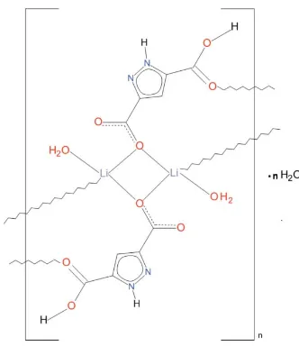
The basic structural unit of the title polymeric ribbon, {[Li2(C5H3N2O2)2(H2O)2]H2O}n, is a centrosymmetric
dinuc-lear complex in which the two LiI ions are bridged by two
carboxylato O atoms, to generate a centrosymmetric Li2O2
core. These are connected into a chain along [011] by carboxylic acid–carbonyl-O bonds. The tetrahedral
coordina-tion of the LiI cation is completed by an aqua ligand. The
carboxylic acid is involved in an intra-ribbon hydrogen bond. A solvate water molecule showing positional (50:50) disorder is observed. Polymeric ribbons along [011] are connected by
O—H O, N—H O and O—H N hydrogen bonds into a
three-dimensional architecture.
Related literature
For the structure of the pyrazole-3,5-dicarboxylic acid hydrate, see: Chinget al.(2000).
Experimental
Crystal data
[Li2(C5H3N2O2)2(H2O)2]H2O Mr= 378.12
Triclinic,P1 a= 7.2610 (15) A˚ b= 7.5835 (15) A˚ c= 8.5751 (17) A˚
= 68.38 (3)
= 89.07 (3)
= 63.66 (3)
V= 387.19 (13) A˚3 Z= 1
MoKradiation
= 0.15 mm1 T= 293 K
0.320.190.15 mm
Data collection
Kuma KM-4 four-circle diffractometer
Absorption correction: analytical (CrysAlis RED; Oxford Diffraction, 2008) Tmin= 0.963,Tmax= 0.983 2319 measured reflections
2139 independent reflections 1631 reflections withI> 2(I) Rint= 0.051
3 standard reflections every 200 reflections
intensity decay: 3.2%
Refinement
R[F2> 2(F2)] = 0.048 wR(F2) = 0.139 S= 1.04 2139 reflections 148 parameters 4 restraints
H atoms treated by a mixture of independent and constrained refinement
max= 0.36 e A˚
3
min=0.43 e A˚
3
Table 1
Selected bond lengths (A˚ ).
Li1—O1 1.948 (3) Li1—O4i
1.910 (3)
Li1—O1ii 1.930 (3) Li1—O5 1.981 (3)
[image:1.610.86.252.553.745.2]Symmetry codes: (i)x;y1;zþ1; (ii)xþ1;y;zþ1.
Table 2
Hydrogen-bond geometry (A˚ ,).
D—H A D—H H A D A D—H A
O3—H3 O2iii
0.82 1.73 2.5159 (16) 160 N1—H1 O5iv
0.84 (2) 2.02 (2) 2.8233 (17) 161 (2) O5—H52 O6 0.89 (3) 1.94 (3) 2.749 (3) 150 (3) O5—H52 O6v
0.89 (3) 2.01 (3) 2.851 (3) 157 (3) O5—H51 N2vi
0.93 (3) 1.89 (3) 2.810 (2) 169 (3) O5—H51 O3vi
0.93 (3) 2.60 (3) 3.1235 (16) 116 (2) O6—H62 O2iv
0.87 (2) 2.03 (3) 2.886 (3) 167 (7)
Symmetry codes: (iii) x;yþ1;z1; (iv) x;yþ1;zþ1; (v)
xþ1;yþ1;zþ1; (vi)xþ1;y1;zþ1.
Data collection: KM-4 Software(Kuma, 1996); cell refinement:
KM-4 Software; data reduction: DATAPROC (Kuma, 2001); program(s) used to solve structure: SHELXS97 (Sheldrick, 2008); program(s) used to refine structure:SHELXL97(Sheldrick, 2008); molecular graphics:SHELXTL(Sheldrick, 2008); software used to prepare material for publication:SHELXTL.
Supplementary data and figures for this paper are available from the IUCr electronic archives (Reference: KP2459).
References
Ching, N., Pan, L., Huang, X. & Li, J. (2000).Acta Cryst.C56, 1124–1125.
metal-organic compounds
Acta Cryst.(2013). E69, m593–m594 doi:10.1107/S1600536813026408 Starosta and Leciejewicz
m593
Acta Crystallographica Section E Structure Reports
Online
Kuma (1996).KM-4 Software. Kuma Diffraction Ltd, Wrocław, Poland. Kuma (2001).DATAPROC. Kuma Diffraction Ltd, Wrocław, Poland.
Oxford Diffraction (2008).CrysAlis RED. Oxford Diffraction Ltd, Yarnton, England.
supporting information
sup-1
Acta Cryst. (2013). E69, m593–m594
supporting information
Acta Cryst. (2013). E69, m593–m594 [doi:10.1107/S1600536813026408]
catena
-Poly[[diaquabis(
µ3
-5-carboxylato-1
H
-pyrazole-3-carboxylic
acid-κ
3O
3:
O
3;
O
5)dilithium(I)] monohydrate]
Wojciech Starosta and Janusz Leciejewicz
S1. Comment
The structural unit of the title complex is a centrosymmetric dinuclear moiety composed of two LiI ions bridged by two
bidentate carboxylato O atoms, each donated by a symmetry related ligand (Fig. 1). The ligand acts in µ3 bridging mode
since apart from the bidentate O1 atom, the O4 atom of its second carboxylate group is chelated to a Li(vi) ion in the
adjacent dimer. In this way a LiI ion is coordinated by the bridging O1 and O1(ii) atoms, the O4(i) from the adjacent dimer
and an aqua O5 atom resulting in a distorted tetrahedral geometry. The Li—O bond distances (Table 1) which fall in the
range between 1.930 (2) Å and 1.980 (3) Å are typical of LiI complexes with carboxylate and water ligands. The pyrazole
ring is planar with r.m.s. of 0.0009 (1) Å; the carboxylate group C6/O1/O2 and C7/O3/O4 make with it dihedral angles of
2.4 (1)° and 5.5 (1)°, respectively. The carboxylate O2 atom is chelating inactive, the O3 remains protonated and
participates as a donor in the short hydrogen bond of 2.516 (2) Å to O2vi in an adjacent dimer. Bond distances and bond
angles within the pyrazole ring do not differ from those reported in the structure of the parent acid (Ching et al., 2000).
The plane of the Li1,O1,Li(ii),O1(ii) dimer core makes a dihedral angle of 36.1° with the ligand plane. The dimeric units
linked by carboxylate O4 atoms form molecular ribbons . A solvate water molecule O6 with 50% site occupancy is
present in the asymmetric cell resulting in one molecule per a dimer. Moreover, this water molecule shows 0.5/0.5
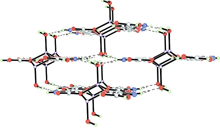
positional disorder. The ribbons are held together by a system of hydrogen bonds involving coordinated and crystal water
molecules the carboxylate/carboxylato groups and pyrazole N ring atoms (Fig. 2, Table 2).
S2. Experimental
1 mmol of pyrazole-3,5-dicarboxylic acid hydrate and ca2 mmol s of lithium hydroxide dissolved in 50 mL of hot,
doubly distilled water were boiled under reflux with stirring for six hours and then left to crystallize at room temperature.
Colourless single-crystal blocks with two differet shapes deposited after a week. One of the crystals was selected, washed
with cold ethanol and dried in the air.
S3. Refinement
Hydrogen atoms belonging to water molecules, the carboxylate group and hetero-ring N atom were located in a
Figure 1
A fragment of a molecular ribbon showing a dinuclear structural building unit of the title complex with atom labelling
scheme and 50% probability displacement ellipsoids. Symmetry code: (i) x, y - 1,z + 1; (ii) -x + 1, -y, -z + 1; (iii): -x + 1,
-y + 1, -z.
Figure 2
The packing of molecular ribbons via hydrogen bonds viewed along their propagation direction.
catena-Poly[[diaquabis(µ3-5-carboxylato-1H-pyrazole-3-carboxylic acid-κ3O3:O3;O5)dilithium(I)] monohydrate]
Crystal data
[Li2(C5H3N2O2)2(H2O)2]·H2O Mr = 378.12
Triclinic, P1 Hall symbol: -P 1 a = 7.2610 (15) Å b = 7.5835 (15) Å c = 8.5751 (17) Å α = 68.38 (3)° β = 89.07 (3)° γ = 63.66 (3)° V = 387.19 (13) Å3
Z = 1 F(000) = 194 Dx = 1.622 Mg m−3
Mo Kα radiation, λ = 0.71073 Å Cell parameters from 25 reflections θ = 6–15°
[image:4.610.128.482.288.499.2]supporting information
sup-3
Acta Cryst. (2013). E69, m593–m594
Data collection
Kuma KM-4 four-circle diffractometer
Radiation source: fine-focus sealed tube Graphite monochromator
profile data from ω/2θ scan Absorption correction: analytical
(CrysAlis RED; Oxford Diffraction, 2008) Tmin = 0.963, Tmax = 0.983
2319 measured reflections
2139 independent reflections 1631 reflections with I > 2σ(I) Rint = 0.051
θmax = 30.1°, θmin = 2.6° h = −9→9
k = 0→9 l = −11→11
3 standard reflections every 200 reflections intensity decay: 3.2%
Refinement
Refinement on F2 Least-squares matrix: full R[F2 > 2σ(F2)] = 0.048 wR(F2) = 0.139 S = 1.04 2139 reflections 148 parameters 4 restraints
Primary atom site location: structure-invariant direct methods
Secondary atom site location: difference Fourier map
Hydrogen site location: inferred from neighbouring sites
H atoms treated by a mixture of independent and constrained refinement
w = 1/[σ2(Fo2) + (0.0979P)2 + 0.0513P] where P = (Fo2 + 2Fc2)/3
(Δ/σ)max < 0.001 Δρmax = 0.36 e Å−3 Δρmin = −0.43 e Å−3
Special details
Geometry. All e.s.d.'s (except the e.s.d. in the dihedral angle between two l.s. planes) are estimated using the full covariance matrix. The cell e.s.d.'s are taken into account individually in the estimation of e.s.d.'s in distances, angles and torsion angles; correlations between e.s.d.'s in cell parameters are only used when they are defined by crystal symmetry. An approximate (isotropic) treatment of cell e.s.d.'s is used for estimating e.s.d.'s involving l.s. planes.
Refinement. Refinement of F2 against ALL reflections. The weighted R-factor wR and goodness of fit S are based on F2, conventional R-factors R are based on F, with F set to zero for negative F2. The threshold expression of F2 > σ(F2) is used only for calculating R-factors(gt) etc. and is not relevant to the choice of reflections for refinement. R-factors based on F2 are statistically about twice as large as those based on F, and R- factors based on ALL data will be even larger.
Fractional atomic coordinates and isotropic or equivalent isotropic displacement parameters (Å2)
x y z Uiso*/Ueq Occ. (<1)
O1 0.31476 (15) 0.19235 (16) 0.41828 (13) 0.0317 (3)
O3 −0.04302 (15) 1.06450 (16) −0.29666 (13) 0.0330 (3)
H3 −0.0066 1.1326 −0.3779 0.050*
N2 −0.12574 (16) 0.83399 (17) −0.01238 (13) 0.0229 (2)
O4 0.30113 (16) 0.84618 (17) −0.21398 (14) 0.0352 (3)
O2 −0.02499 (16) 0.33302 (17) 0.43139 (13) 0.0361 (3)
N1 −0.11472 (16) 0.68180 (16) 0.13403 (13) 0.0219 (2)
C5 0.08191 (17) 0.51952 (18) 0.19965 (15) 0.0208 (3)
C4 0.20777 (18) 0.56837 (19) 0.08850 (15) 0.0236 (3)
H4 0.3512 0.4884 0.0977 0.028*
C3 0.07157 (18) 0.76495 (18) −0.04109 (14) 0.0209 (3)
C7 0.12037 (19) 0.8962 (2) −0.19328 (15) 0.0230 (3)
C6 0.12709 (19) 0.33371 (19) 0.36231 (15) 0.0218 (3)
Li1 0.4462 (4) −0.0115 (4) 0.6522 (3) 0.0310 (5)
O6 0.4576 (4) 0.5520 (4) 0.5472 (4) 0.0493 (6) 0.50
H1 −0.225 (4) 0.700 (4) 0.173 (3) 0.046 (6)*
H52 0.531 (5) 0.254 (4) 0.683 (4) 0.070 (8)*
H51 0.630 (5) 0.063 (5) 0.832 (4) 0.078 (9)*
H62 0.328 (4) 0.602 (6) 0.559 (9) 0.14 (3)* 0.50
H61 0.508 (15) 0.57 (3) 0.624 (19) 0.38 (11)* 0.50
Atomic displacement parameters (Å2)
U11 U22 U33 U12 U13 U23
O1 0.0179 (4) 0.0260 (5) 0.0233 (4) 0.0000 (4) 0.0017 (3) 0.0056 (4)
O3 0.0216 (5) 0.0280 (5) 0.0247 (5) −0.0075 (4) 0.0030 (4) 0.0095 (4) N2 0.0175 (5) 0.0181 (5) 0.0188 (5) −0.0056 (4) 0.0027 (3) 0.0038 (4) O4 0.0215 (5) 0.0304 (5) 0.0329 (5) −0.0091 (4) 0.0097 (4) 0.0044 (4) O2 0.0212 (5) 0.0292 (5) 0.0296 (5) −0.0066 (4) 0.0068 (4) 0.0106 (4) N1 0.0148 (5) 0.0178 (5) 0.0188 (5) −0.0047 (4) 0.0032 (3) 0.0034 (4) C5 0.0156 (5) 0.0166 (5) 0.0182 (5) −0.0050 (4) 0.0025 (4) 0.0019 (4) C4 0.0150 (5) 0.0183 (5) 0.0218 (5) −0.0034 (4) 0.0040 (4) 0.0023 (4) C3 0.0173 (5) 0.0175 (5) 0.0178 (5) −0.0067 (4) 0.0038 (4) 0.0014 (4) C7 0.0210 (6) 0.0189 (5) 0.0195 (5) −0.0082 (4) 0.0047 (4) 0.0006 (4) C6 0.0172 (5) 0.0173 (5) 0.0177 (5) −0.0048 (4) 0.0018 (4) 0.0022 (4) Li1 0.0219 (10) 0.0268 (11) 0.0278 (11) −0.0074 (9) 0.0077 (8) 0.0006 (9) O5 0.0173 (4) 0.0320 (5) 0.0309 (5) −0.0070 (4) 0.0031 (4) 0.0012 (4) O6 0.0359 (13) 0.0414 (14) 0.0517 (15) −0.0200 (11) 0.0027 (10) 0.0030 (11)
Geometric parameters (Å, º)
O1—C6 1.2578 (16) C5—C6 1.4816 (17)
O1—Li1i 1.929 (3) C4—C3 1.3935 (17)
Li1—O1 1.948 (3) C4—H4 0.9300
O3—C7 1.2958 (17) C3—C7 1.4698 (16)
O3—H3 0.8200 Li1—O4iii 1.910 (3)
N2—N1 1.3298 (14) Li1—O1i 1.930 (3)
N2—C3 1.3436 (16) Li1—O5 1.981 (3)
O4—C7 1.2240 (16) Li1—Li1i 2.679 (5)
O4—Li1ii 1.910 (3) O5—H52 0.89 (3)
O2—C6 1.2458 (15) O5—H51 0.93 (3)
N1—C5 1.3513 (16) O6—O6iv 1.296 (6)
N1—H1 0.84 (2) O6—H62 0.87 (2)
C5—C4 1.3758 (16) O6—H61 0.86 (2)
C6—O1—Li1i 141.01 (13) O3—C7—C3 113.68 (11)
C6—O1—Li1 128.30 (12) O2—C6—O1 126.09 (12)
Li1i—O1—Li1 87.41 (12) O2—C6—C5 116.69 (11)
C7—O3—H3 109.5 O1—C6—C5 117.22 (12)
N1—N2—C3 104.63 (10) O4iii—Li1—O1i 114.50 (14)
C7—O4—Li1ii 136.31 (12) O4iii—Li1—O1 118.27 (14)
supporting information
sup-5
Acta Cryst. (2013). E69, m593–m594
N2—N1—H1 117.8 (16) O4iii—Li1—O5 111.34 (13)
C5—N1—H1 129.7 (16) O1i—Li1—O5 114.31 (14)
N1—C5—C4 106.99 (10) O1—Li1—O5 104.38 (13)
N1—C5—C6 120.84 (11) O4iii—Li1—Li1i 130.02 (19)
C4—C5—C6 132.17 (11) O1i—Li1—Li1i 46.58 (9)
C5—C4—C3 104.29 (10) O1—Li1—Li1i 46.02 (8)
C5—C4—H4 127.9 O5—Li1—Li1i 118.50 (16)
C3—C4—H4 127.9 Li1—O5—H52 111.3 (17)
N2—C3—C4 111.59 (11) Li1—O5—H51 106.9 (16)
N2—C3—C7 120.02 (11) H52—O5—H51 101 (2)
C4—C3—C7 128.37 (11) O6iv—O6—H62 126 (6)
O4—C7—O3 125.27 (12) O6iv—O6—H61 133 (7)
O4—C7—C3 121.04 (12) H62—O6—H61 100 (3)
Symmetry codes: (i) −x+1, −y, −z+1; (ii) x, y+1, z−1; (iii) x, y−1, z+1; (iv) −x+1, −y+1, −z+1.
Hydrogen-bond geometry (Å, º)
D—H···A D—H H···A D···A D—H···A
O3—H3···O2ii 0.82 1.73 2.5159 (16) 160
N1—H1···O5v 0.84 (2) 2.02 (2) 2.8233 (17) 161 (2)
O5—H52···O6 0.89 (3) 1.94 (3) 2.749 (3) 150 (3)
O5—H52···O6iv 0.89 (3) 2.01 (3) 2.851 (3) 157 (3)
O5—H51···N2vi 0.93 (3) 1.89 (3) 2.810 (2) 169 (3)
O5—H51···O3vi 0.93 (3) 2.60 (3) 3.1235 (16) 116 (2)
O6—H62···O2v 0.87 (2) 2.03 (3) 2.886 (3) 167 (7)
phosphonium chloride monohydrate](data:image/gif;base64,R0lGODlhAQABAIAAAP///wAAACH5BAEAAAAALAAAAAABAAEAAAICRAEAOw==)