Crystal structure of 2-cyano-
N
-(furan-2-ylmethyl)-3-(3-nitrophenyl)propanamide
Shivanna Subhadramma,aBudanur Papaiah Siddaraju,b Naveen Chandra,cJanardhanan Saravanandand Dasararaju Gayathrie*
a
Department of Physics, Dr M. G. R. Educational and Research Institute University, Maduravoyal, Chennai, India,bDepartment of Engineering Chemistry, Cauvery Institute of Technology, Sundhahalli, Mandya, India,cDepartment of Chemistry, Post-Graduate and Research Centre, St Joseph’s College (Autonomous), Bangalore 560 027, India,dDepartment of Pharmaceutical Chemistry, PES College of Pharmacy, Hanumanthnagar, Bangalore 560 050, India, andeCentre of Advanced Study in Crystallography and Biophysics, University of Madras, Guindy Campus, Chennai 600 025, India. *Correspondence e-mail: drdgayathri@gmail.com
Received 24 June 2015; accepted 4 July 2015
Edited by H. Stoeckli-Evans, University of Neuchaˆtel, Switzerland
In the title compound, C15H11N3O4, the acetamide group is inclined to the furan ring by 66.5 (1). The dihedral angle
between the furan ring and the benzene ring is 66.8 (1). In the
crystal, molecules are linked by pairs of N—H N hydrogen bonds, forming inversion dimers with anR22(12) ring motif. The dimers are linkedviatwo pairs of C—H O hydrogen bonds to the same acceptor oxygen atom, enclosingR21(6) ring motifs, forming chains along the [101] direction.
Keywords:crystal structure; furan; acetamide; N—H N hydrogen bonds; inversion dimers.
CCDC reference:1410650
1. Related literature
For examples of biological properties of furan derivatives, see: Anupam et al. (2011). For the biological activities of some heterocyclic derivatives containing the acetamide moiety, see: Fallah-Tafti et al.(2011); Shamset al.(2011). For the crystal structure of the similar compound 2-cyano-N -furfuryl-3-(2-furyl)acrylamide, see: Pome´s Herna´ndezet al.(1996).
2. Experimental 2.1. Crystal data
C15H11N3O4 Mr= 297.27
Triclinic,P1 a= 7.4358 (3) A˚ b= 9.4165 (5) A˚ c= 10.3934 (5) A˚ = 90.938 (2)
= 96.910 (2)
= 105.872 (2)
V= 693.98 (6) A˚3 Z= 2
MoKradiation = 0.11 mm1 T= 293 K
0.300.200.20 mm
2.2. Data collection
Bruker Kappa APEXII CCD diffractometer
Absorption correction: multi-scan (SADABS; Bruker, 2004) Tmin= 0.942,Tmax= 0.983
12708 measured reflections 2442 independent reflections 2055 reflections withI> 2(I) Rint= 0.027
2.3. Refinement
R[F2> 2(F2)] = 0.039 wR(F2) = 0.116 S= 1.02 2442 reflections
200 parameters
H-atom parameters constrained
max= 0.22 e A˚ 3
min=0.16 e A˚ 3
Table 1
Hydrogen-bond geometry (A˚ ,).
D—H A D—H H A D A D—H A
N3—H3A N2i
0.86 2.27 3.056 (2) 152
C5—H5 O3ii
0.93 2.49 3.337 (2) 151
C7—H7 O3ii
0.93 2.49 3.362 (2) 156
Symmetry codes: (i)xþ1;yþ1;zþ2; (ii)x;yþ1;zþ1.
Data collection:APEX2(Bruker, 2004); cell refinement:APEX2and SAINT(Bruker, 2004); data reduction:SAINTandXPREP(Bruker, 2004); program(s) used to solve structure:SHELXS2014(Sheldrick, 2008); program(s) used to refine structure:SHELXL2014(Sheldrick, 2015); molecular graphics:PLATON(Spek, 2009); software used to prepare material for publication:SHELXL2014andPLATON.
Acknowledgements
We thank Dr Babu Varghese for the XRD data collection at the Sophisticated Analytical Instrument Facility (SAIF), Indian Institute of Technology, Madras.
Supporting information for this paper is available from the IUCr electronic archives (Reference: SU5161).
data reports
o568
doi:10.1107/S2056989015012918 Acta Cryst.(2015).E71, o568–o569References
Anupam, V., Pandeya, S. N. & Shweta, S. (2011).Int. J. Res. Ayurveda Pharm. 2, 1110–1116.
Bruker (2004).APEX2,SAINT,XPREPandSADABS. Bruker AXS Inc., Madison, Wisconsin, USA.
Fallah-Tafti, A., Foroumadi, A., Tiwari, R., Shirazi, A. N., Hangauer, D. G., Bu, Y., Akbarzadeh, T., Parang, K. & Shafiee, A. (2011).Eur. J. Med. Chem.46, 4853–4858.
Pome´s Herna´ndez, R., Duque Rodrı´guez, J., Novoa de Armas, H. & Toscano, R. A. (1996).Acta Cryst.C52, 203–205.
Shams, H. Z., Mohareb, R. M., Helal, M. H. & Mahmoud, A. (2011). Molecules,16, 52–73.
supporting information
sup-1
Acta Cryst. (2015). E71, o568–o569
supporting information
Acta Cryst. (2015). E71, o568–o569 [https://doi.org/10.1107/S2056989015012918]
Crystal structure of 2-cyano-
N
-(furan-2-ylmethyl)-3-(3-nitrophenyl)-propanamide
Shivanna Subhadramma, Budanur Papaiah Siddaraju, Naveen Chandra, Janardhanan Saravanan
and Dasararaju Gayathri
S1. Comment
Furan is one of the most important five-membered heterocyclic ring systems and its derivatives are well known to
possess various biological properties, such as antibacterial, antitumor, anti-inflammatory, antifungal, anticonvulsant, and
analgesic (Anupam et al., 2011). Acetamide derivatives possess a wide range of pharmacological properties (Fallah-Tafti
et al., 2011; Shams et al., 2011). In view of the biological importance of furan and acetamide derivatives, we have
synthesized the title compound and report herein on its crystal structure.
In the title compound, Fig. 1, the acetamide group is inclined to the furan ring by 66.5 (1)°. Torsion angles N3—C9—
C8—C15 [-5.4 (2)°] and O3—C9—C8—C15 [174.6 (2)°] indicate that the acetonitrile and acetamide moieties are almost
planar. The dihedral angle between the furan ring and the benzene ring is 66.8 (1)°. The bond lengths and bond angles are
comparable with those reported for a similar structure, viz. 2-cyano-N-furfuryl-3-(2-furyl)acrylamide (Pomés Hernández
et al., 1996).
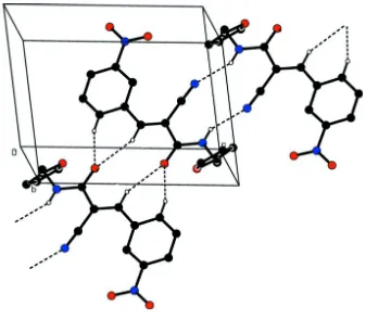
In the crystal (Table 1 and Fig. 2), molecules are linked by pairs of N-H···N hydrogen bonds forming inversion dimers
with an R22(12) ring motif. The dimers are linked via two pairs of bifurcated C-H···O hydrogen bonds, enclosing R21(6)
ring motifs, forming chains along direction [101].
S2. Experimental
An equimolar mixture of furfuryl amine and ethyl cyano acetate was mixed in a conical flask and the mixture was heated
under microwave irradiation at 700 W for 3 min with an interval of 20 s each time. The mixture was then poured into a
beaker and cooled giving a solid that was washed with ethanol. The furfuryl cyano acetamide product so obtained was
treated with an equi-molar ratio of 3-nitro benzaldehyde in the presence of glacial acetic acid and refluxed for 3 h. On
cooling, colourless crystals of the title compound were obtained.
S3. Refinement
Crystal data, data collection and structure refinement details are summarized in Table 2. The NH and C-bound H atoms
were included in calculated positions and refined using a riding model: N—H = 0.86 Å, C—H = 0.93 - 0.97 Å with
Figure 1
The molecular structure of the title compound, with atom labelling. Displacement ellipsoids are drawn at the 30%
probability level.
Figure 2
A view along the b axis of the crystal packing of the title compound. Hydrogen bonds are shown as dashed lines (see
[image:4.610.140.477.322.616.2]supporting information
sup-3
Acta Cryst. (2015). E71, o568–o569
2-Cyano-N-(furan-2-ylmethyl)-3-(3-nitrophenyl)propanamide
Crystal data
C15H11N3O4 Mr = 297.27 Triclinic, P1
a = 7.4358 (3) Å
b = 9.4165 (5) Å
c = 10.3934 (5) Å
α = 90.938 (2)°
β = 96.910 (2)°
γ = 105.872 (2)°
V = 693.98 (6) Å3
Z = 2
F(000) = 308
Dx = 1.423 Mg m−3
Mo Kα radiation, λ = 0.71073 Å Cell parameters from 6452 reflections
θ = 2.8–25.6°
µ = 0.11 mm−1 T = 293 K Block, colourless 0.30 × 0.20 × 0.20 mm
Data collection
Bruker Kappa APEXII CCD diffractometer
Radiation source: fine-focus sealed tube Graphite monochromator
ω and φ scan
Absorption correction: multi-scan (SADABS; Bruker, 2004)
Tmin = 0.942, Tmax = 0.983
12708 measured reflections 2442 independent reflections 2055 reflections with I > 2σ(I)
Rint = 0.027
θmax = 25.0°, θmin = 2.0° h = −8→8
k = −11→10
l = −12→12
Refinement
Refinement on F2
Least-squares matrix: full
R[F2 > 2σ(F2)] = 0.039 wR(F2) = 0.116 S = 1.02 2442 reflections 200 parameters 0 restraints
Primary atom site location: structure-invariant direct methods
Secondary atom site location: difference Fourier map
Hydrogen site location: inferred from neighbouring sites
H-atom parameters constrained
w = 1/[σ2(F
o2) + (0.059P)2 + 0.2005P]
where P = (Fo2 + 2Fc2)/3
(Δ/σ)max < 0.001
Δρmax = 0.22 e Å−3
Δρmin = −0.16 e Å−3
Extinction correction: SHELXL2014 (Sheldrick, 2015), Fc*=kFc[1+0.001xFc2λ3/sin(2θ)]-1/4
Extinction coefficient: 0.030 (5)
Special details
Geometry. All e.s.d.'s (except the e.s.d. in the dihedral angle between two l.s. planes) are estimated using the full covariance matrix. The cell e.s.d.'s are taken into account individually in the estimation of e.s.d.'s in distances, angles and torsion angles; correlations between e.s.d.'s in cell parameters are only used when they are defined by crystal symmetry. An approximate (isotropic) treatment of cell e.s.d.'s is used for estimating e.s.d.'s involving l.s. planes.
Fractional atomic coordinates and isotropic or equivalent isotropic displacement parameters (Å2)
x y z Uiso*/Ueq
C1 0.5353 (2) 0.28891 (17) 0.58091 (15) 0.0437 (4)
H1 0.5862 0.3239 0.6654 0.052*
C2 0.6211 (2) 0.20823 (17) 0.51113 (16) 0.0450 (4) C3 0.5534 (3) 0.15471 (19) 0.38555 (17) 0.0548 (5)
H3 0.6160 0.1015 0.3400 0.066*
H4 0.3401 0.1470 0.2450 0.069* C5 0.2988 (2) 0.26268 (18) 0.39767 (15) 0.0479 (4)
H5 0.1879 0.2798 0.3586 0.057*
C6 0.3703 (2) 0.31818 (16) 0.52382 (14) 0.0389 (4) C7 0.2662 (2) 0.40380 (16) 0.58752 (14) 0.0392 (4)
H7 0.1617 0.4177 0.5360 0.047*
C8 0.2965 (2) 0.46539 (16) 0.70769 (14) 0.0380 (4) C9 0.1628 (2) 0.54718 (17) 0.74909 (14) 0.0404 (4) C10 0.0773 (2) 0.67695 (19) 0.92842 (16) 0.0486 (4) H10A 0.0750 0.6564 1.0194 0.058* H10B −0.0509 0.6412 0.8851 0.058* C11 0.1429 (2) 0.83840 (19) 0.91721 (15) 0.0474 (4) C12 0.2518 (5) 0.9468 (3) 0.9961 (2) 0.1020 (10) H12 0.3096 0.9378 1.0788 0.122* C13 0.2647 (5) 1.0791 (3) 0.9319 (3) 0.1089 (10) H13 0.3310 1.1739 0.9645 0.131* C14 0.1663 (3) 1.0432 (3) 0.8186 (3) 0.0790 (7) H14 0.1519 1.1094 0.7553 0.095* C15 0.4516 (2) 0.45908 (19) 0.80123 (14) 0.0447 (4) N1 0.7935 (2) 0.17794 (17) 0.57526 (18) 0.0592 (4) N2 0.5728 (2) 0.4565 (2) 0.87857 (14) 0.0667 (5) N3 0.19322 (18) 0.59563 (15) 0.87343 (12) 0.0439 (3) H3A 0.2851 0.5781 0.9231 0.053* O1 0.8792 (2) 0.1167 (2) 0.51177 (17) 0.0881 (5) O2 0.8406 (2) 0.2139 (2) 0.69036 (16) 0.0840 (5) O3 0.03619 (18) 0.56592 (15) 0.67145 (11) 0.0609 (4) O4 0.0876 (2) 0.89464 (16) 0.80594 (13) 0.0724 (4)
Atomic displacement parameters (Å2)
U11 U22 U33 U12 U13 U23
supporting information
sup-5
Acta Cryst. (2015). E71, o568–o569
N3 0.0474 (7) 0.0536 (8) 0.0364 (7) 0.0261 (6) −0.0006 (5) 0.0010 (6) O1 0.0840 (11) 0.1097 (13) 0.1024 (12) 0.0702 (10) 0.0338 (9) 0.0228 (9) O2 0.0720 (10) 0.1099 (13) 0.0829 (11) 0.0555 (9) −0.0105 (8) 0.0032 (9) O3 0.0626 (8) 0.0875 (9) 0.0446 (6) 0.0495 (7) −0.0111 (6) −0.0084 (6) O4 0.0791 (9) 0.0714 (9) 0.0649 (8) 0.0266 (7) −0.0123 (7) 0.0114 (7)
Geometric parameters (Å, º)
C1—C2 1.367 (2) C9—N3 1.3354 (19)
C1—C6 1.396 (2) C10—N3 1.4564 (19)
C1—H1 0.9300 C10—C11 1.475 (2)
C2—C3 1.376 (2) C10—H10A 0.9700
C2—N1 1.472 (2) C10—H10B 0.9700
C3—C4 1.377 (3) C11—C12 1.315 (3)
C3—H3 0.9300 C11—O4 1.346 (2)
C4—C5 1.380 (2) C12—C13 1.408 (4)
C4—H4 0.9300 C12—H12 0.9300
C5—C6 1.390 (2) C13—C14 1.296 (4)
C5—H5 0.9300 C13—H13 0.9300
C6—C7 1.461 (2) C14—O4 1.358 (3)
C7—C8 1.336 (2) C14—H14 0.9300
C7—H7 0.9300 C15—N2 1.139 (2)
C8—C15 1.432 (2) N1—O1 1.210 (2)
C8—C9 1.509 (2) N1—O2 1.220 (2)
C9—O3 1.2170 (18) N3—H3A 0.8600
C2—C1—C6 119.19 (15) N3—C10—C11 114.03 (14)
C2—C1—H1 120.4 N3—C10—H10A 108.7
C6—C1—H1 120.4 C11—C10—H10A 108.7
C1—C2—C3 123.02 (15) N3—C10—H10B 108.7 C1—C2—N1 117.60 (15) C11—C10—H10B 108.7 C3—C2—N1 119.38 (15) H10A—C10—H10B 107.6 C2—C3—C4 117.80 (15) C12—C11—O4 109.03 (18) C2—C3—H3 121.1 C12—C11—C10 132.98 (17) C4—C3—H3 121.1 O4—C11—C10 117.98 (15) C3—C4—C5 120.64 (16) C11—C12—C13 107.3 (2)
C3—C4—H4 119.7 C11—C12—H12 126.4
C5—C4—H4 119.7 C13—C12—H12 126.4
C4—C5—C6 121.02 (15) C14—C13—C12 106.8 (2)
C4—C5—H5 119.5 C14—C13—H13 126.6
C6—C5—H5 119.5 C12—C13—H13 126.6
C5—C6—C1 118.31 (14) C13—C14—O4 109.9 (2) C5—C6—C7 117.18 (13) C13—C14—H14 125.0 C1—C6—C7 124.50 (13) O4—C14—H14 125.0 C8—C7—C6 130.56 (13) N2—C15—C8 177.71 (16)
C8—C7—H7 114.7 O1—N1—O2 123.45 (16)
C6—C7—H7 114.7 O1—N1—C2 118.39 (17)
C7—C8—C9 119.37 (13) C9—N3—C10 122.71 (13) C15—C8—C9 117.24 (12) C9—N3—H3A 118.6 O3—C9—N3 123.55 (14) C10—N3—H3A 118.6 O3—C9—C8 120.49 (13) C11—O4—C14 106.99 (16) N3—C9—C8 115.97 (12)
C6—C1—C2—C3 0.6 (3) C15—C8—C9—N3 −5.4 (2) C6—C1—C2—N1 −179.20 (14) N3—C10—C11—C12 −94.5 (3) C1—C2—C3—C4 −1.1 (3) N3—C10—C11—O4 84.68 (19) N1—C2—C3—C4 178.69 (15) O4—C11—C12—C13 0.6 (3) C2—C3—C4—C5 0.5 (3) C10—C11—C12—C13 179.9 (2) C3—C4—C5—C6 0.5 (3) C11—C12—C13—C14 −0.9 (4) C4—C5—C6—C1 −1.0 (3) C12—C13—C14—O4 0.9 (4) C4—C5—C6—C7 178.99 (15) C1—C2—N1—O1 −174.37 (16) C2—C1—C6—C5 0.5 (2) C3—C2—N1—O1 5.9 (2) C2—C1—C6—C7 −179.50 (14) C1—C2—N1—O2 6.8 (2) C5—C6—C7—C8 177.38 (16) C3—C2—N1—O2 −172.96 (17) C1—C6—C7—C8 −2.6 (3) O3—C9—N3—C10 −0.3 (3) C6—C7—C8—C15 1.2 (3) C8—C9—N3—C10 179.74 (14) C6—C7—C8—C9 −179.14 (15) C11—C10—N3—C9 −88.52 (19) C7—C8—C9—O3 −5.1 (2) C12—C11—O4—C14 −0.1 (3) C15—C8—C9—O3 174.61 (15) C10—C11—O4—C14 −179.50 (16) C7—C8—C9—N3 174.88 (14) C13—C14—O4—C11 −0.5 (3)
Hydrogen-bond geometry (Å, º)
D—H···A D—H H···A D···A D—H···A
N3—H3A···N2i 0.86 2.27 3.056 (2) 152
C5—H5···O3ii 0.93 2.49 3.337 (2) 151
C7—H7···O3ii 0.93 2.49 3.362 (2) 156