1096
https://doi.org/10.1107/S2056989019009253 Acta Cryst.(2019). E75, 1096–1101research communications
Received 10 June 2019 Accepted 27 June 2019
Edited by E. V. Boldyreva, Russian Academy of Sciences, Russia
Keywords:crystal structure; fructosamine; Amadori rearrangement; cycloleucine; hydrogen bonding; Hirshfeld surface analysis.
CCDC reference:1583255
Supporting information:this article has supporting information at journals.iucr.org/e
Multicentered hydrogen bonding in 1-[(1-deoxy-
b
-
D-fructopyranos-1-yl)azaniumyl]cyclopentane-carboxylate (‘D-fructose-cycloleucine’)
Valeri V. Mossine,a* Charles L. Barnesband Thomas P. Mawhinneyc
a
Department of Biochemistry, University of Missouri, Columbia, MO 65211, USA,bDepartment of Chemistry, University of Missouri, Columbia, MO 65211, USA, andcDepartment of Biochemistry, University of Missouri, Columbia, MO 65211, U.S.A.. *Correspondence e-mail: MossineV@missouri.edu
The title compound, C12H21NO7, (I), is conformationally unstable; the
predominant form present in its solution is the -pyranose form (74.3%), followed by the- and-furanoses (12.1 and 10.2%, respectively),-pyranose (3.4%), and traces of the acyclic carbohydrate tautomer. In the crystalline state, the carbohydrate part of (I) adopts the2C5-pyranose conformation, and the
amino acid portion exists as a zwitterion, with the side chain cyclopentane ring assuming the E9 envelope conformation. All heteroatoms are involved in
hydrogen bonding that forms a system of antiparallel infinite chains of fused
R3 3
(6) andR3 3
(8) rings. The molecule features extensive intramolecular hydrogen bonding, which is uniquely multicentered and involves the carboxylate, ammonium and carbohydrate hydroxy groups. In contrast, the contribution of intermolecular O H/H O contacts to the Hirshfeld surface is relatively low (38.4%), as compared to structures of other d-fructose-amino acids. The 1H
NMR data suggest a slow rotation around the C1—C2 bond in (I), indicating that the intramolecular heteroatom contacts survive in aqueous solution of the molecule as well.
1. Chemical context
d-Fructosamine derivatives are products of non-enzymatic
condensation reactions between d-glucose and biomolecules
containing free aliphatic amino groups, such as amino acids, proteins, aminophospholipids, or biogenic amines (Mossine & Mawhinney, 2010). d-Fructosamines are thus present in all
living systems and in foods. For instance, in healthy humans, about 5% of plasma proteins are decorated with fructosamine residues, while dietary intake ofd-fructosamines, primarily in
the form of N"-(1-deoxy-d-fructos-1-yl)-l-lysine, has been
estimated at 1 g per day. Although the normal physiological functions ofd-fructosamines are not understood, a number of
bacterial, fungal, and mammalian carbohydrate-processing enzymes (Wu & Monnier, 2003; Van Schaftingenet al., 2012), transporters (Marty et al., 2016), and lectins (Mossine et al., 2008) can recognize d-fructosamine, thus implying the
parti-cipation of this structure in metabolic and signaling processes. Biomedical research has suggested the involvement of d
-fructosamines in the development of diabetic complications (Wu & Monnier, 2003), bacterial infections (Aliet al., 2014), and cancer (Malmstro¨met al., 2016). We and others (Mossine
et al., 2010; Rabinovich et al., 2006) have demonstrated the efficacy of syntheticd-fructosamine derivatives as blockers of
galectins, a family of tumor-associated lectins. In this context, several structure determinations of biologically active
samines have previously been undertaken (Mossine et al., 2007a,b, 2009, 2018).
As a part of our search for efficient blockers of galectins-1, 3 and4, we have preparedd-fructose-cycloleucine (I), a
structural analog of the galectin inhibitord-fructose-l-leucine
(Mossineet al., 2008). Here we report on the molecular and crystal structures of (I), with an emphasis on the hydrogen-bonding patterns in the structure.
2. Structural commentary
The molecular structure and atomic numbering are shown in Fig. 1. The title compound, (I), crystallizes in the monoclinic space groupP21, with two equivalent molecules per unit cell.
The molecule may be considered as a conjugate of a carbo-hydrate, 1-amino-1-deoxy-d-fructose, and an amino acid,
1-aminocyclopentane-1-carboxylic acid, which are joined through the common amino group. The-d-pyranose ring of
the carbohydrate portion exists in the 2C5 or 1C(D) chair
conformation, with puckering parametersQ= 0.5763 A˚ , = 172.71, and ’ = 248.80. These parameters correspond to a
conformation with the lowest energy possible for fructose (French et al., 1997). The bond distances and the valence angles are close to the average values for a number of crys-talline pyranose structures (Jeffrey & Taylor, 1980). In an aqueous solution of (I), the-d-pyranose anomer dominates
the tautomeric equilibrium (Fig. 2), at 74.3%, as follows from its 13C NMR spectrum (Table 1). The acyclic forms are not readily detectable because of their low populations; their presence is suggested based on literature evidence available for other fructosamine derivatives (Table 1). In the1H NMR spectrum of the major anomer, the vicinal proton–proton coupling constantsJ3,4= 9.8 Hz andJ4,5= 3.4 Hz indicate that
atom H4 is in a trans disposition to H3 and in a gauche
disposition to H5. Hence, the predominant conformation ofd
-fructose-cycloleucine in solution is also2C5 -d
-fructopyran-ose.
The amino acid portion of the molecule is in the zwitterionic form, with a positively charged tetrahedral secondary ammo-nium nitrogen atom and a negatively charged deprotonated carboxyl group. The side-chain cyclopentane ring is, by the atom numbering in Fig. 1, in theE9(envelope on C9) or Cs—
C-exo conformation, with puckering parameters Q = 0.4220 A˚ ,’= 254.94, and pseudorotational parameters (Rao
et al., 1981)P= 56.9and = 43.7for the C7—C8 bond.
[image:2.610.88.248.104.178.2]The ammonium group and all but one (O8) oxygen atoms are involved in intramolecular hydrogen bonding (Table 2). At the centre of this system are heteroatom contacts between the conjugated carbohydrate and the amino acid portions of the molecule, which involve the carboxylate atom O7, the ammonium atom H1A, the pyranose ring atom O5, and the anomeric hydroxyl group O1—H1O (Fig. 1). Although the value of N1—H1A O7 angle is 99.4, the distance N1 O7 is 2.702 (2) A˚ , short enough for this heteroatom contact to
Figure 2
Tautomeric equilibrium in aqueous solution ofd-fructose-cycloleucine at
[image:2.610.313.565.112.252.2]293 K and pH 6, as determined by13C NMR.
Table 1
Chemical shifts (p.p.m.) in the13C NMR spectrum of (I) and the anomeric distribution ofd-fructose-cycloleucine and structurally related molecules
in D2O at 293 K.
Carbon -pyranose -pyranose -furanose -furanose acyclic
C1 52.61 52.89 51.12 52.51
C2 99.11 98.34 104.69 101.84
C3 73.12 72.20 85.18 80.58
C4 74.75 72.33 78.71 77.12
C5 68.73 71.80 85.32 83.73
C6 65.78 66.66 63.63 64.76
C 75.99 76.30 76.14 76.24
% for (I) 3.4 74.3 10.2 12.1 < 0.5
% for fructosea 2.1 68.6 5.7 23.0 0.5
% for FruLeub
4 72 12 12 < 1
% for FruAibc 3.0 75.6 10.1 10.4 < 0.7
% for FruProa
4.2 64.8 12.9 16.9 1.2
[image:2.610.294.559.536.720.2]Notes: (a) Kaufmannet al.(2016); (b) Glinskyet al.(1996); (c) Mossineet al.(2018).
Figure 1
[image:2.610.45.318.583.707.2]qualify as a strong hydrogen bond. Then the central motif of the intramolecular hydrogen-bonded structure can be described in terms of a compact ringS2
2
(4)pattern represented by the four-atom O7 H1A O1—H1O O7 cycle. In the
1
H NMR spectrum of (I), two protons, H1Cand H1D, which are attached to C1, produce two distinct signals at 3.332 and 3.199 ppm, with J1C,1D= 12.8 Hz (Fig. 3). The
non-equiva-lence of these protons indicates restricted rotation around the C1—C2 and C1—N1 bonds, thus suggesting the intra-molecular hydrogen bonding retains this structure in solution.
3. Supramolecular features
The crystal packing of (I) features infinite chains of anti-parallel hydrogen bonds running along the a-axis direction
(Fig. 4). The basic hydrogen-bonding patterns are depicted in Fig. 5 and include two rings,R33(6)andR33(8), and a small finite
chain D22(4). Alternatively, the fused rings pattern can be
1098
Mossineet al. C12H21NO7 Acta Cryst.(2019). E75, 1096–1101
research communications
Figure 3
A portion of the1H NMR spectrum ofd-fructose-cycloleucine in D2O at
293 K containing four sets of signals for methylene protons H1C and H1D. The labeled set of two doublets belongs to the dominant
-d-fructopyranose anomer of (I). The smaller, unlabeled peaks are
unresolved signals of H1Cand H1Dbelonging to the-pyranose,- and
[image:3.610.47.295.69.223.2] [image:3.610.315.564.82.189.2]-furanose conformations of (I).
Table 2
Hydrogen-bond geometry (A˚ ,).
D—H A D—H H A D A D—H A
N1—H1B O8i 0.91 1.80 2.704 (2) 174
N1—H1A O3ii 0.91 2.00 2.784 (2) 143
N1—H1A O1 0.91 2.59 2.980 (2) 107
O4—H4O O3 0.85 (3) 2.41 (3) 2.746 (2) 105 (2) O4—H4O O7iii 0.85 (3) 2.09 (3) 2.845 (2) 149 (3) O1—H1O O7 0.80 (4) 1.97 (4) 2.769 (2) 178 (3) O3—H3O O7iv 0.84 (4) 1.99 (4) 2.796 (2) 162 (3) O2—H2O O4v 0.81 (3) 2.00 (3) 2.765 (2) 159 (3)
Symmetry codes: (i)xþ1;y;z; (ii)xþ2;y1
2;zþ1; (iii)xþ2;yþ 1
2;zþ1; (iv) xþ1;yþ1
[image:3.610.314.567.248.344.2]2;zþ1; (v)x1;y;z.
Table 3
Suspected hydrogen bonds and short C—H Acontacts (A˚ ,).
D—H A D—H H A D A D—H ASymmetry code
N1—H1A O7 0.91 2.40 2.702 (2) 99 N1—H1A O5 0.91 2.43 2.698 (2) 97 O2—H2O O1 0.81 (3) 2.60 (4) 2.845 (2) 99 C8—H8A O8 0.99 2.47 2.810 (3) 100
C9—H9B O1 0.99 2.66 3.618 (3) 162 x,y,z1 C11—H11A O3 0.99 2.65 3.416 (3) 134 x+ 2,y1
2,z+ 1 C4—H4 O4 1.00 2.69 3.364 (3) 124 x1,y,z
[image:3.610.331.540.418.711.2]C6—H6A O1 0.99 2.67 3.430 (3) 134 x+ 1,y,z
Figure 4
[image:3.610.46.298.507.705.2]The molecular packing in (I). A view of the unit-cell contents shown in projection down theaaxis. Color code for crystallographic axes: reda, greenb, bluec. Hydrogen bonds are shown as cyan dotted lines.
Figure 5
described in terms of two chains, C2 2
(4) and C3 3
(8). The ammonium proton H1A is involved in a rare five-centered hydrogen bond, involving three weakly directional intra-molecular contacts with O1, O5, and O7 (at distances of 2.59, 2.43, and 2.40 A˚ , respectively) and one intermolecular, shorter (2.00 A˚ distance) bond with O3. The carboxyl atom O7 is also involved in an unusual multicenter hydrogen bond, by coordinating four surrounding protons at reasonably short distances, 1.97–2.40 A˚ (Tables 2 and 3). This multicentered character of the short heteroatom contacts implies a
signifi-cant contribution of the electrostatic component (Taoet al., 2017) to the interaction, apparently between the positively charged ammonium group and the negatively charged carboxyl atom O7 (Fig. 6). Indeed, the C12—O7 bond [1.269 (3) A˚ ] is significantly longer than the C12—O8 distance [1.243 (3) A˚ ], suggesting a more polarized character of the former. This may be a consequence of highly differing heteroatom arrangements around the two carboxylate oxygen atoms in the crystal. One, O7, is surrounded by four heteroatoms (O1, O3, O4, N1) at distances qualifying for hydrogen bonds, while O8 has only one heteroatom, N1, located at a short distance.
The Hirshfeld surface analysis (Spackman & Jayatilaka, 2009) revealed that a major proportion of the intermolecular contacts in crystal structure of (I) is provided by non- or low-polar H H interactions (Fig. 7). Of note, there are three short interatomic contacts of the C—H O type (Table 3, Fig. 6) that involve the cyclopentane ring and which may be responsible for conformational stabilization of the ring. In contrast, a number of published cycloleucine structures feature disordered conformations as a result of the ring pucker pseudorotation (Mallikarjunan et al., 1972; Varughese & Chacko, 1978; Santiniet al.1988).
4. Database survey
Searches of SciFinder (2018) and the Cambridge Structural Database (2019 CSD release; Groom et al., 2016) by both structure and chemical names returned no previous structural description of N-(1-deoxy--d-fructopyranos-1-yl)-10
-amino-cyclopentane-10-carboxylic acid or d-fructose-cycloleucine;
thus the compound appears to be novel. Since the confor-mational instability of thed-fructosamine moiety determines
the chemical reactivities and biological activities ofd
-fructo-samine derivatives (Mossine & Mawhinney, 2010), we compared the structure of (I) with solved structures of other
d-fructose-amino acids. The most closely related structures are d-fructose-2-aminoisobutyric acid (CCDC 1583254; Mossine
et al., 2018), d-fructose-glycine (CCDC 1307697; Mossine,
Glinskyet al., 1995),d-fructose-l-proline [CCDC 628806 and
628807 (Tarnawskiet al., 2007), 631528 (Mossineet al., 2007a)], and d-fructose-l-histidine (CCDC 622419; Mossine et al.,
2007b). Although some fructosamine derivatives can crystal-lize as the-furanose,spiro-bicyclic hemiketal, or acyclicketo
tautomers (Mossine, Barnes et al., 1995, 2009), all of the above-listedd-fructose-amino acids adopt the2C5-pyranose
conformation and exist as zwitterions, with the intramolecular hydrogen-bonding central pattern localized around the ammonium group and involving the carboxylate and one hydroxyl group donated by the carbohydrate moiety. This hydrogen-bonded conjugation between the amino acid zwit-terion bridge and the-pyranose provides for conformational stability around the C1—C2 bond in solutions ofd
[image:4.610.45.279.72.228.2]-fructose-amino acids. The staggeredgauche–transconformation of the N1—C1—C2—O5 torsion, such as in (I), has also been observed in CCDC 1583254 (moleculeA; Mossineet al., 2018), CCDC 631528 (Mossine et al., 2007a), and CCDC 622419
Figure 6
Views of the Hirshfeld surface for (I) mapped over: (a) the electrostatic potential in the range0.156 to +0.261 a.u. with the red and blue colors representing the distribution of the negative and positive electrostatic potential, respectively; (b) thedefunction, in the range 0.674 to 2.424 A˚ ,
[image:4.610.46.297.478.703.2]calculated for the external contact atoms in the crystal. The molecular fragments involved in short C—H O interactions are shown; these allegedly stabilize the cyclopentane ring conformation in crystalline (I).
Figure 7
(Mossineet al., 2007b), while thetrans–gaucheconformation was observed in four other structures of d-fructose-amino
acids (Table 4). However, none of these structures, except (I), features the cyclic motif of intramolecular multicentered hydrogen bonding (Fig. 1), which is supported by a unique direct interaction between the carbohydrate anomeric hydroxyl donor, O1—H1O, and the carboxylate acceptor, O7. In total, there are six intramolecular short heteroatom contacts in the structure of (I), more than in any other d
-fructose-amino acid structure known to date. Such effect of the ‘internalization’ of hydrogen bonding in (I) is also revealed in a comparative analysis of the fingerprint plots (Fig. 7) that are based on the calculations of Hirshfeld surfaces (Spackman & Jayatilaka, 2009) and delineated into the O H/ H O intermolecular contacts in the crystal structure of (I). The relative abundance of these contacts in structures of
d-fructose-amino acids decreases with an increase in the
number of intramolecular hydrogen bonds; this trend is clearly revealed by the data presented in Table 4. The significant difference between the carboxylate C—O lengths of 0.026 A˚ in (I) is comparable to the respective bond-length differences noted in other fructose-amino acid structures, including CCDC 1583254 (0.022 A˚ in moleculeB; Mossineet al., 2018) and CCDC 622419 (0.021 A˚ ; Mossine et al., 2007b). In the latter two structures, the carboxylate oxygen atoms are involved in close heteroatom contacts unequally, although not to the extent observed in (I).
5. Synthesis and crystallization
Cycloleucine (2.6 g, 0.02 mol), d-glucose (9 g, 0.05 mol), and
sodium acetate (0.82 g, 0.01 mol) were dissolved in 100 mL of a methanol/glycerol (3:1) mixture and refluxed for 3 h. The reaction progress was monitored by TLC on silica. The reac-tion mixture was diluted with 900 mL of water and passed through a column charged with 80 mL of Amberlite IRN-77 (H+-form). The target compound was then eluted with 0.2M
pyridine, and fractions containing pure (I) were pooled and evaporated. The residue was redissolved in 100 mL of water, decolorized with 0.5 g of charcoal and evaporated to a syrup.
The latter was dissolved in 30 mL of ethanol and made nearly cloudy with the dropwise addition of acetone. Crystallization occurred within a week at room temperature. Yield 3.4 g (58%, based on the starting cycloleucine).
Major -pyranose tautomer peaks (ppm) in 13C NMR spectrum in D2O: 179.82 (C12); 98.34 (C2); 76.30 (C7); 72.33
(C4); 72.20 (C3); 71.80 (C5); 66.66 (C6); 52.89 (C1); 37.44, 37.40 (C8, C11); 28.00, 27.98 (C9, C10). See Table 1 for minor peaks assignments in the spectrum. Major signals (ppm) and resolved coupling constants (Hz) in the1H NMR spectrum: 4.035 (dd, H6B); 4.017 (m, H5); 3.896 (dd, H4); 3.774 (d, H3); 3.771 (dd, H6A); 3.332 (d, H1D); 3.199 (d, H1C); 2.220 (m, 2H11); 1.955 (m, 2H8); 1.83 (m, 2H9 + 2H10);J1C,1D=12.8;
J3,4= 9.8;J4,5= 3.4;J5,6A= 1.3;J6A,6B=12.9.
6. Refinement details
Crystal data, data collection and structure refinement details are summarized in Table 5. Hydroxyl H atoms were located in difference-Fourier maps and were allowed to refine freely. Other H atoms were placed at calculated positions and treated as riding, with N—H = 0.91 A˚ , C—H = 0.99 A˚ (methylene) or 1.00 A˚ (methine) and with Uiso(H) = 1.2Ueq (methine or
methylene). As a result of the unrealistic value obtained for the Flack absolute structure parameter [0.4 (4) for 1097 quotients; Parsonset al., 2013], the absolute configuration of the pyranose ring system (2R,3S,4R,5R) was assigned on the basis of the known configuration for the starting compound
d-glucose (McNaught, 1996).
Acknowledgements
The authors thank Dr Shaokai Jiang for assistance with acquiring the NMR spectra.
Funding information
Funding for this research was provided by: University of Missouri Agriculture Experiment Station Chemical Labora-tories; National Institute of Food and Agriculture (grant No. MO-HABC0002).
1100
Mossineet al. C12H21NO7 Acta Cryst.(2019). E75, 1096–1101
[image:5.610.45.569.116.238.2]research communications
Table 4
Conformation, intramolecular hydrogen bonding around the amino group, and contributions of the intermolecular O H/H O contacts to the Hirshfeld surfaces inN-(-d-fructopyranos-1-yl)-amino acids.
Hydrogen-bond selection criteria:D A< 3.0 A˚ ; H D< 2.7 A˚ ;D—H A> 95 .
Structure N—C1—C2—O5
torsion (
), conformation
Intramolecular hydrogen bonds around the amino group
No. of intra/intermolecular hydrogen bonds
O H/H O contacts on Hirshfeld surface (%)
Fru-cycloLeu, (I) +53.3gt N1—H1A O1 (106); N1—H1A O5 (97); N1—H1A O7 (99
); O1—H1O O7 (178 )
6/5 38.5
FruGlya
+165.5tg N1—H1A O2 (140); N1—H1A O7 (104) 2/6 51.6
FruAib (moleculeA)b +64.7gt N1—H1B O5 (110
); N1—H1B O7 (107
) 3/5 44.0
FruAib (moleculeB)b
+176.8tg N1—H1A O2 (145
); N1—H1A O7 (100
) 3/5 45.9
FruProH2O
d
+75.8gt N1—H1 O1 (109
); N1—H1 O7 (125
) 3/6 49.2
FruPro2H2O
d
+176.8tg N1—H1 O2 (140
); N1—H1 O7 (113
) 3/6 49.3
FruProMeOHd +174.4tg N1—H1 O2 (139
); N1—H1 O7 (114
) 4/5 40.2
FruHisH2O
e
+60.7gt N1—H1B O5 (100
); N1—H1B O7 (102 ); N1—H1A O1 (108)
5/7 41.2
References
Ali, M. M., Newsom, D. L., Gonza´lez, J. F., Sabag-Daigle, A., Stahl, C., Steidley, B., Dubena, J., Dyszel, J. L., Smith, J. N., Dieye, Y., Arsenescu, R., Boyaka, P. N., Krakowka, S., Romeo, T., Behrman, E. J., White, P. & Ahmer, B. M. M. (2014). PLoS Pathog. 10, e1004209.
Bruker. (1998). SMART and SAINT-Plus. Bruker AXS Inc., Madison, Wisconsin, USA.
French, A. D., Dowd, M. K. & Reilly, P. J. (1997). J. Mol. Struct. Theochem,395–396, 271–287.
Glinsky, G. V., Mossine, V. V., Price, J. E., Bielenberg, D., Glinsky, V. V., Ananthaswamy, H. N. & Feather, M. S. (1996).Clin. Exp. Metastasis,14, 253–267.
Groom, C. R., Bruno, I. J., Lightfoot, M. P. & Ward, S. C. (2016).Acta Cryst.B72, 171–179.
Jeffrey, G. A. & Taylor, R. (1980).J. Comput. Chem.1, 99–109. Kaufmann, M., Meissner, P. M., Pelke, D., Mu¨gge, C. & Kroh, L. W.
(2016).Carbohydr. Res.428, 87–99.
Krause, L., Herbst-Irmer, R., Sheldrick, G. M. & Stalke, D. (2015).J. Appl. Cryst.48, 3–10.
Macrae, C. F., Bruno, I. J., Chisholm, J. A., Edgington, P. R., McCabe, P., Pidcock, E., Rodriguez-Monge, L., Taylor, R., van de Streek, J. & Wood, P. A. (2008).J. Appl. Cryst.41, 466–470.
Mallikarjunan, M., Chacko, K. K. & Zand, R. (1972).J. Cryst. Mol. Struct.2, 53–66.
Malmstro¨m, H., Wa¨ndell, P. E., Holzmann, M. J., A¨ rnlo¨v, J., Jungner, I., Hammar, N., Walldius, G. & Carlsson, A. C. (2016).Nutr. Metab. Cardiovasc. Dis.26, 1120–1128.
Marty, L., Vigouroux, A., Aumont-Nicaise, M., Dessaux, Y., Faure, D. & More´ra, S. (2016).J. Biol. Chem.291, 22638–22649.
McNaught, A. D. (1996).Pure Appl. Chem.68, 1919–2008.
Mossine, V. V., Barnes, C. L., Chance, D. L. & Mawhinney, T. P. (2009).Angew. Chem. Int. Ed.48, 5517–5520.
Mossine, V. V., Barnes, C. L., Glinsky, G. V. & Feather, M. S. (1995). Carbohydr. Lett.1, 355–362.
Mossine, V. V., Barnes, C. L. & Mawhinney, T. P. (2007a). J. Carbohydr. Chem.26, 249–266.
Mossine, V. V., Barnes, C. L. & Mawhinney, T. P. (2007b).Carbohydr. Res.342, 131–138.
Mossine, V. V., Barnes, C. L. & Mawhinney, T. P. (2018).Acta Cryst. E74, 72–77.
Mossine, V. V., Glinsky, G. V., Barnes, C. L. & Feather, M. S. (1995). Carbohydr. Res.266, 5–14.
Mossine, V. V., Glinsky, V. V. & Mawhinney, T. P. (2008). InGalectins, edited by A. A. Klyosov, D. Platt, D. & Z. J. Witczak, pp. 235–270. Hoboken, NJ: John Wiley & Sons.
Mossine, V. V., Glinsky, V. V. & Mawhinney, T. P. (2010).Nutrition and Metabolism, edited by M. C. Thomas & J. Forbes, J., pp. 170–179. London: Royal Society of Chemistry.
Mossine, V. V. & Mawhinney, T. P. (2010).Adv. Carbohydr. Chem. Biochem.64, 291–402.
Parsons, S., Flack, H. D. & Wagner, T. (2013).Acta Cryst.B69, 249– 259.
Rabinovich, G. A., Cumashi, A., Bianco, G. A., Ciavardelli, D., Iurisci, I., D’Egidio, M., Piccolo, E., Tinari, N., Nifantiev, N. & Iacobelli, S. (2006).Glycobiology,16, 210–220.
Rao, S. T., Westhof, E. & Sundaralingam, M. (1981).Acta Cryst.A37, 421–425.
Santini, A., Barone, V., Bavoso, A., Benedetti, E., Di Blasio, B., Fraternali, F., Lelj, F., Pavone, V., Pedone, C., Crisma, M., Bonora, G. M. & Toniolo, C. (1988).Int. J. Biol. Macromol.10, 292–299. SciFinder (2018). Chemical Abstracts Service: Columbus, OH, 2010;
RN 58-08-2.
Sheldrick, G. M. (2008).Acta Cryst.A64, 112–122. Sheldrick, G. M. (2015).Acta Cryst.C71, 3–8.
Spackman, M. A. & Jayatilaka, D. (2009).CrystEngComm,11, 19–32. Tao, Y., Zou, W., Jia, J., Li, W. & Cremer, D. (2017).J. Chem. Theory
Comput.13, 55–76.
Tarnawski, M., S´lepokura, K., Lis, T., Kulis´-Orzechowska, R. & Szelepin, B. (2007).Carbohydr. Res.342, 1264–1270.
Van Schaftingen, E., Collard, F., Wiame, E. & Veiga-da-Cunha, M. (2012).Amino Acids,42, 1143–1150.
Varughese, K. I. & Chacko, K. K. (1978).Cryst. Struct. Commun.7, 149–152.
Westrip, S. P. (2010).J. Appl. Cryst.43, 920–925.
[image:6.610.44.292.87.400.2]Wu, X. & Monnier, V. M. (2003).Arch. Biochem. Biophys.419, 16–24.
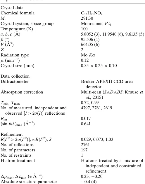
Table 5
Experimental details.
Crystal data
Chemical formula C12H21NO7
Mr 291.30
Crystal system, space group Monoclinic,P21
Temperature (K) 100
a,b,c(A˚ ) 5.8052 (3), 11.9540 (6), 9.6135 (5)
(
) 95.506 (1)
V(A˚3) 664.05 (6)
Z 2
Radiation type MoK
(mm1) 0.12
Crystal size (mm) 0.550.250.10
Data collection
Diffractometer Bruker APEXII CCD area
detector
Absorption correction Multi-scan (SADABS; Krauseet al., 2015)
Tmin,Tmax 0.72, 0.99
No. of measured, independent and observed [I> 2(I)] reflections
4797, 2761, 2619
Rint 0.017
(sin/ )max(A˚
1
) 0.641
Refinement
R[F2> 2(F2)],wR(F2),S 0.029, 0.073, 1.03
No. of reflections 2761
No. of parameters 197
No. of restraints 1
H-atom treatment H atoms treated by a mixture of independent and constrained refinement
max,min(e A˚
3) 0.23,0.20
Absolute structure parameter 0.4 (4)
Computer programs:SMARTandSAINT-Plus(Bruker, 1998),SHELXS97(Sheldrick, 2008), SHELXL2017/1 (Sheldrick, 2015), Mercury (Macrae et al., 2008), CIFTAB
supporting information
sup-1
Acta Cryst. (2019). E75, 1096-1101
supporting information
Acta Cryst. (2019). E75, 1096-1101 [https://doi.org/10.1107/S2056989019009253]
Multicentered hydrogen bonding in 1-[(1-deoxy-
β
-
D-fructopyranos-1-yl)aza-niumyl]cyclopentanecarboxylate (`
D-fructose-cycloleucine
′
)
Valeri V. Mossine, Charles L. Barnes and Thomas P. Mawhinney
Computing details
Data collection: SMART (Bruker, 1998); cell refinement: SAINT-Plus (Bruker, 1998); data reduction: SAINT-Plus
(Bruker, 1998); program(s) used to solve structure: SHELXS97 (Sheldrick, 2008); program(s) used to refine structure:
SHELXL2017/1 (Sheldrick, 2015); molecular graphics: Mercury (Macrae et al., 2008); software used to prepare material
for publication: CIFTAB (Sheldrick, 2008) and publCIF (Westrip, 2010).
1-[(1-Deoxy-β-D-fructopyranos-1-yl)azaniumyl]cyclopentanecarboxylate
Crystal data
C12H21NO7
Mr = 291.30
Monoclinic, P21
a = 5.8052 (3) Å
b = 11.9540 (6) Å
c = 9.6135 (5) Å
β = 95.506 (1)°
V = 664.05 (6) Å3
Z = 2
F(000) = 312
Dx = 1.457 Mg m−3
Mo Kα radiation, λ = 0.71073 Å
Cell parameters from 3178 reflections
θ = 2.7–27.1°
µ = 0.12 mm−1
T = 100 K
Plate, colourless 0.55 × 0.25 × 0.10 mm
Data collection
Bruker APEXII CCD area detector diffractometer
ω scans
Absorption correction: multi-scan
(SADABS; Krause et al., 2015)
Tmin = 0.72, Tmax = 0.99
4797 measured reflections
2761 independent reflections 2619 reflections with I > 2σ(I)
Rint = 0.017
θmax = 27.1°, θmin = 2.1°
h = −7→7
k = −15→15
l = −12→10
Refinement
Refinement on F2
Least-squares matrix: full
R[F2 > 2σ(F2)] = 0.029
wR(F2) = 0.073
S = 1.03
2761 reflections 197 parameters 1 restraint
Hydrogen site location: mixed
H atoms treated by a mixture of independent and constrained refinement
w = 1/[σ2(F
o2) + (0.0364P)2 + 0.1566P]
where P = (Fo2 + 2Fc2)/3
(Δ/σ)max = 0.001
Δρmax = 0.23 e Å−3
Δρmin = −0.20 e Å−3
Absolute structure: Flack x determined using
1097 quotients [(I+)-(I-)]/[(I+)+(I-)] (Parsons et
al., 2013).
sup-2
Acta Cryst. (2019). E75, 1096-1101
Special details
Geometry. All esds (except the esd in the dihedral angle between two l.s. planes) are estimated using the full covariance matrix. The cell esds are taken into account individually in the estimation of esds in distances, angles and torsion angles; correlations between esds in cell parameters are only used when they are defined by crystal symmetry. An approximate (isotropic) treatment of cell esds is used for estimating esds involving l.s. planes.
Fractional atomic coordinates and isotropic or equivalent isotropic displacement parameters (Å2)
x y z Uiso*/Ueq
O1 0.7009 (3) 0.99423 (13) 0.44759 (17) 0.0210 (3)
O2 0.6482 (3) 1.23008 (14) 0.42123 (17) 0.0236 (4)
O3 0.9451 (3) 1.26855 (13) 0.68102 (17) 0.0223 (3)
O4 1.3447 (3) 1.16847 (15) 0.61241 (19) 0.0271 (4)
O5 1.0920 (3) 1.01394 (12) 0.41337 (15) 0.0200 (3)
O7 0.4680 (3) 0.86115 (14) 0.24446 (16) 0.0215 (3)
O8 0.2727 (3) 0.93833 (15) 0.05537 (16) 0.0283 (4)
N1 0.8644 (3) 0.95057 (15) 0.16816 (17) 0.0160 (3)
H1A 0.872660 0.899266 0.238389 0.019*
H1B 1.003891 0.951327 0.132317 0.019*
C1 0.8227 (4) 1.06402 (17) 0.2287 (2) 0.0189 (4)
H1C 0.661121 1.087516 0.201456 0.023*
H1D 0.927252 1.119552 0.191299 0.023*
C2 0.8665 (4) 1.05974 (17) 0.3875 (2) 0.0182 (4)
C3 0.8630 (4) 1.17629 (17) 0.4547 (2) 0.0177 (4)
H3 0.986207 1.222671 0.417043 0.021*
C4 0.9241 (4) 1.16278 (17) 0.6125 (2) 0.0178 (4)
H4 0.799149 1.118808 0.652062 0.021*
C5 1.1540 (4) 1.10136 (18) 0.6441 (2) 0.0205 (4)
H5 1.173771 1.081867 0.745658 0.025*
C6 1.1571 (4) 0.99419 (18) 0.5592 (2) 0.0215 (5)
H6A 1.314355 0.961418 0.570981 0.026*
H6B 1.048878 0.939529 0.594849 0.026*
C7 0.6822 (3) 0.91363 (17) 0.0555 (2) 0.0164 (4)
C8 0.6746 (4) 0.99140 (19) −0.0720 (2) 0.0202 (4)
H8A 0.549752 1.047572 −0.069571 0.024*
H8B 0.824036 1.030727 −0.075537 0.024*
C9 0.6270 (4) 0.9129 (2) −0.1969 (2) 0.0270 (5)
H9A 0.461610 0.891584 −0.210590 0.032*
H9B 0.672248 0.947623 −0.283720 0.032*
C10 0.7793 (5) 0.8121 (2) −0.1546 (2) 0.0295 (5)
H10A 0.943566 0.827661 −0.167132 0.035*
H10B 0.728704 0.745231 −0.210171 0.035*
C11 0.7450 (4) 0.79532 (18) 0.0003 (2) 0.0220 (4)
H11A 0.888615 0.766805 0.052214 0.026*
H11B 0.618351 0.741477 0.011014 0.026*
C12 0.4522 (4) 0.90500 (18) 0.1238 (2) 0.0188 (4)
H4O 1.347 (5) 1.227 (3) 0.662 (3) 0.032 (8)*
supporting information
sup-3
Acta Cryst. (2019). E75, 1096-1101
H3O 0.814 (6) 1.296 (3) 0.684 (3) 0.037 (8)*
H2O 0.553 (6) 1.199 (3) 0.463 (3) 0.037 (9)*
Atomic displacement parameters (Å2)
U11 U22 U33 U12 U13 U23
O1 0.0221 (8) 0.0233 (8) 0.0179 (7) −0.0065 (6) 0.0033 (6) −0.0029 (6)
O2 0.0212 (9) 0.0230 (8) 0.0263 (9) 0.0047 (7) 0.0009 (7) 0.0015 (7)
O3 0.0165 (8) 0.0229 (7) 0.0272 (9) 0.0015 (6) 0.0009 (6) −0.0097 (6)
O4 0.0173 (8) 0.0290 (9) 0.0359 (9) −0.0021 (7) 0.0066 (7) −0.0129 (8)
O5 0.0191 (8) 0.0227 (8) 0.0180 (7) 0.0022 (6) 0.0014 (6) −0.0033 (6)
O7 0.0184 (7) 0.0269 (8) 0.0195 (8) −0.0031 (6) 0.0037 (6) 0.0034 (6)
O8 0.0144 (7) 0.0461 (11) 0.0244 (8) 0.0031 (7) 0.0019 (6) 0.0037 (8)
N1 0.0130 (8) 0.0193 (8) 0.0159 (8) −0.0007 (7) 0.0021 (6) 0.0008 (7)
C1 0.0215 (10) 0.0173 (10) 0.0180 (10) −0.0001 (8) 0.0023 (8) −0.0006 (8)
C2 0.0182 (10) 0.0194 (10) 0.0168 (10) −0.0008 (8) 0.0012 (8) −0.0004 (8)
C3 0.0164 (10) 0.0177 (10) 0.0193 (10) 0.0006 (8) 0.0026 (8) −0.0014 (8)
C4 0.0172 (10) 0.0176 (9) 0.0187 (10) −0.0011 (8) 0.0020 (8) −0.0033 (8)
C5 0.0182 (10) 0.0244 (11) 0.0188 (10) 0.0011 (9) 0.0017 (8) −0.0032 (9)
C6 0.0219 (11) 0.0214 (10) 0.0204 (11) 0.0048 (9) −0.0018 (8) −0.0010 (9)
C7 0.0144 (9) 0.0193 (9) 0.0155 (9) −0.0002 (8) 0.0013 (7) −0.0009 (8)
C8 0.0202 (11) 0.0227 (10) 0.0177 (10) −0.0020 (8) 0.0017 (8) 0.0029 (8)
C9 0.0289 (12) 0.0334 (12) 0.0183 (10) −0.0022 (10) 0.0005 (9) 0.0013 (9)
C10 0.0311 (13) 0.0353 (13) 0.0226 (12) 0.0036 (11) 0.0049 (9) −0.0055 (10)
C11 0.0239 (11) 0.0202 (10) 0.0216 (10) 0.0019 (8) 0.0009 (8) −0.0029 (8)
C12 0.0162 (10) 0.0205 (9) 0.0200 (10) −0.0024 (8) 0.0038 (8) −0.0028 (8)
Geometric parameters (Å, º)
O1—C2 1.406 (3) C3—H3 1.0000
O1—H1O 0.80 (4) C4—C5 1.528 (3)
O2—C3 1.412 (3) C4—H4 1.0000
O2—H2O 0.81 (3) C5—C6 1.520 (3)
O3—C4 1.425 (2) C5—H5 1.0000
O3—H3O 0.84 (4) C6—H6A 0.9900
O4—C5 1.423 (3) C6—H6B 0.9900
O4—H4O 0.85 (3) C7—C8 1.535 (3)
O5—C2 1.419 (3) C7—C12 1.547 (3)
O5—C6 1.436 (3) C7—C11 1.566 (3)
O7—C12 1.269 (3) C8—C9 1.529 (3)
O8—C12 1.243 (3) C8—H8A 0.9900
N1—C1 1.504 (3) C8—H8B 0.9900
N1—C7 1.506 (3) C9—C10 1.526 (3)
N1—H1A 0.9100 C9—H9A 0.9900
N1—H1B 0.9100 C9—H9B 0.9900
C1—C2 1.524 (3) C10—C11 1.535 (3)
C1—H1C 0.9900 C10—H10A 0.9900
sup-4
Acta Cryst. (2019). E75, 1096-1101
C2—C3 1.537 (3) C11—H11A 0.9900
C3—C4 1.533 (3) C11—H11B 0.9900
C2—O1—H1O 111 (2) C4—C5—H5 108.8
C3—O2—H2O 108 (2) O5—C6—C5 111.70 (17)
C4—O3—H3O 109 (2) O5—C6—H6A 109.3
C5—O4—H4O 108 (2) C5—C6—H6A 109.3
C2—O5—C6 112.75 (15) O5—C6—H6B 109.3
C1—N1—C7 114.50 (16) C5—C6—H6B 109.3
C1—N1—H1A 108.6 H6A—C6—H6B 107.9
C7—N1—H1A 108.6 N1—C7—C8 111.15 (16)
C1—N1—H1B 108.6 N1—C7—C12 106.82 (16)
C7—N1—H1B 108.6 C8—C7—C12 114.79 (17)
H1A—N1—H1B 107.6 N1—C7—C11 109.76 (17)
N1—C1—C2 109.85 (16) C8—C7—C11 105.47 (17)
N1—C1—H1C 109.7 C12—C7—C11 108.79 (17)
C2—C1—H1C 109.7 C9—C8—C7 104.14 (18)
N1—C1—H1D 109.7 C9—C8—H8A 110.9
C2—C1—H1D 109.7 C7—C8—H8A 110.9
H1C—C1—H1D 108.2 C9—C8—H8B 110.9
O1—C2—O5 111.62 (17) C7—C8—H8B 110.9
O1—C2—C1 112.06 (17) H8A—C8—H8B 108.9
O5—C2—C1 104.54 (16) C10—C9—C8 102.65 (19)
O1—C2—C3 107.14 (17) C10—C9—H9A 111.2
O5—C2—C3 108.97 (17) C8—C9—H9A 111.2
C1—C2—C3 112.54 (17) C10—C9—H9B 111.2
O2—C3—C4 112.90 (17) C8—C9—H9B 111.2
O2—C3—C2 111.32 (18) H9A—C9—H9B 109.1
C4—C3—C2 108.05 (16) C9—C10—C11 103.64 (19)
O2—C3—H3 108.1 C9—C10—H10A 111.0
C4—C3—H3 108.1 C11—C10—H10A 111.0
C2—C3—H3 108.1 C9—C10—H10B 111.0
O3—C4—C5 107.48 (17) C11—C10—H10B 111.0
O3—C4—C3 111.41 (17) H10A—C10—H10B 109.0
C5—C4—C3 111.20 (17) C10—C11—C7 105.47 (18)
O3—C4—H4 108.9 C10—C11—H11A 110.6
C5—C4—H4 108.9 C7—C11—H11A 110.6
C3—C4—H4 108.9 C10—C11—H11B 110.6
O4—C5—C6 108.13 (17) C7—C11—H11B 110.6
O4—C5—C4 111.66 (18) H11A—C11—H11B 108.8
C6—C5—C4 110.70 (17) O8—C12—O7 126.9 (2)
O4—C5—H5 108.8 O8—C12—C7 117.89 (19)
C6—C5—H5 108.8 O7—C12—C7 115.22 (18)
C7—N1—C1—C2 135.27 (18) C2—O5—C6—C5 −59.7 (2)
C6—O5—C2—O1 −53.6 (2) O4—C5—C6—O5 −71.6 (2)
C6—O5—C2—C1 −174.91 (17) C4—C5—C6—O5 51.0 (2)
supporting information
sup-5
Acta Cryst. (2019). E75, 1096-1101
N1—C1—C2—O1 −67.8 (2) C1—N1—C7—C12 −62.6 (2)
N1—C1—C2—O5 53.2 (2) C1—N1—C7—C11 179.67 (16)
N1—C1—C2—C3 171.33 (17) N1—C7—C8—C9 142.64 (17)
O1—C2—C3—O2 −64.5 (2) C12—C7—C8—C9 −96.0 (2)
O5—C2—C3—O2 174.57 (17) C11—C7—C8—C9 23.7 (2)
C1—C2—C3—O2 59.1 (2) C7—C8—C9—C10 −40.9 (2)
O1—C2—C3—C4 60.0 (2) C8—C9—C10—C11 42.1 (2)
O5—C2—C3—C4 −60.9 (2) C9—C10—C11—C7 −27.1 (2)
C1—C2—C3—C4 −176.39 (17) N1—C7—C11—C10 −117.74 (19)
O2—C3—C4—O3 −61.7 (2) C8—C7—C11—C10 2.1 (2)
C2—C3—C4—O3 174.70 (18) C12—C7—C11—C10 125.71 (19)
O2—C3—C4—C5 178.39 (18) N1—C7—C12—O8 139.8 (2)
C2—C3—C4—C5 54.8 (2) C8—C7—C12—O8 16.1 (3)
O3—C4—C5—O4 −52.0 (2) C11—C7—C12—O8 −101.8 (2)
C3—C4—C5—O4 70.2 (2) N1—C7—C12—O7 −41.9 (2)
O3—C4—C5—C6 −172.52 (17) C8—C7—C12—O7 −165.61 (19)
C3—C4—C5—C6 −50.3 (2) C11—C7—C12—O7 76.5 (2)
Hydrogen-bond geometry (Å, º)
D—H···A D—H H···A D···A D—H···A
N1—H1B···O8i 0.91 1.80 2.704 (2) 174
N1—H1A···O3ii 0.91 2.00 2.784 (2) 143
N1—H1A···O1 0.91 2.59 2.980 (2) 107
O4—H4O···O3 0.85 (3) 2.41 (3) 2.746 (2) 105 (2)
O4—H4O···O7iii 0.85 (3) 2.09 (3) 2.845 (2) 149 (3)
O1—H1O···O7 0.80 (4) 1.97 (4) 2.769 (2) 178 (3)
O3—H3O···O7iv 0.84 (4) 1.99 (4) 2.796 (2) 162 (3)
O2—H2O···O4v 0.81 (3) 2.00 (3) 2.765 (2) 159 (3)