Received 21 February 2017 Accepted 27 February 2017
Edited by M. Weil, Vienna University of Technology, Austria
Keywords:crystal structure; hydrogen bonding; 4-cyanopyridine; iron; thiocyanate.
CCDC reference:1534965
Supporting information:this article has supporting information at journals.iucr.org/e
Crystal structure of
diaquabis(4-cyanopyridine-jN
)bis(thiocyanato-
jN
)iron(II) 4-cyanopyridine
disolvate
Aleksej Jochim,* Inke Jess and Christian Na¨ther
Institut fu¨r Anorganische Chemie, Christian-Albrechts-Universita¨t Kiel, Max-Eyth Strasse 2, D-24118 Kiel, Germany. *Correspondence e-mail: ajochim@ac.uni-kiel.de
The asymmetric unit of the title compound, [Fe(NCS)2(C6H4N2)2(H2O)2]
-2C6H4N2, comprises one Fe II
cation occupying an inversion centre as well as one thiocyanate anion, one water molecule and two 4-cyanopyridine molecules in general positions. The iron cations are coordinated by two N-bonded thiocyanate anions, two (pyridine)N-bonded 4-cyanopyridine ligands and two water molecules into discrete complexes. The resulting coordination polyhedron can be described as a slightly distorted octahedron. The discrete complexes are
connected through centrosymmetric pairs of (pyridine)C—H N(cyano)
hydrogen bonds into chains that are further linked into a three-dimensional
network through intermolecular O—H N hydrogen bonds involving the
4-cyanopyridine solvent molecules.
1. Chemical context
Thiocyanate anions are versatile ligands that can coordinate in different modes to metal cations. In most cases the anionic ligands are terminally N-bonded to the metal cation but there are also several examples for a-1,3bridging mode (Werneret
al., 2015; Boeckmann & Na¨ther, 2012; Palion-Gazda et al., 2015). The latter coordination is of special interest if the compounds contain paramagnetic metal cations because then cooperative magnetic properties can be expected (Palion-Gazda et al., 2015). In this context, we have reported on several compounds with one- or two-dimensional structures based on Mn, Fe, Co or Ni as metals, thiocyanate ligands and different N-donor co-ligands that show different magnetic properties (Suckertet al., 2016; Ramset al., 2017; Boeckmann
et al., 2012). Whereas compounds with a terminal coordination of the anionic ligands can usually be synthesized straightfor-wardly, compounds with bridging ligands are sometimes difficult to obtain from solution. Therefore, we have devel-oped an alternative procedure which is based on thermal decomposition of precursors with a terminal NCS coordina-tion that frequently transform into the desired polymeric compounds on heating. In the course of our investigations on the synthesis of coordination polymers with iron as metal, thiocyanate ligands and 4-cyanopyridine as co-ligands, we obtained the title compound which was identified by single crystal X-ray diffraction. Unfortunately, all samples were always contaminated with a second unknown crystalline phase, preventing any further investigations.
2. Structural commentary
The asymmetric unit of [Fe(NCS)2(C6H4N2)2(H2O)2]
-2C6H4N2 contains one FeII cation that is located on an
inversion centre, one thiocyanate anion, one water molecule and two 4-cyanopyridine molecules (Fig. 1). Discrete centro-symmetric [Fe(NCS)2(C6H4N2)2(H2O)2] complexes are
formed, in which the FeIIcations are octahedrally coordinated by two N-bonded thiocyanate anions, two (pyridine)N-bonded 4-cyanopyridine ligands and two water molecules, each of them in atrans-position (Fig. 1). The disparate bond lengths are similar to those in related thiocyanate compounds. The
distortion of the octahedron is also reflected by the deviation of the bond angles from ideal values. The structure contains additional 4-cyanopyridine solvate molecules that are located in the cavities of the structure.
3. Supramolecular features
The discrete complexes are linked into chains parallel to [101]
by centrosymmetric pairs of intermolecular C—H N
hydrogen bonds between the cyano group of the coordinating 4-cyanopyridine ligand and one of the pyridine H atoms (Fig. 2, Table 1). These chains are further linked by the 4-cyano-pyridine solvate molecules through intermolecular O—H N hydrogen bonding. One water H atom is hydrogen-bonded to the N atom of the cyano group and the other H atom to the pyridine N atom of another 4-cyanopyridine solvate molecule. Since all water H atoms are involved in hydrogen bonding, each of the complexes is surrounded by four 4-cyanopyridine
ligands, of which two are hydrogen-bonded via the cyano
group, whereas the other two are hydrogen-bonded via the
pyridine N atom (Fig. 3, Table 1). This arrangement leads to a three-dimensional network structure. It is noted that there are additional short contacts between the thiocyanate anions and the pyridine H atoms of the coordinating 4-cyanopyridine ligand of a neighbouring complex, which is indicative of weak
C—H S hydrogen bonding (Table 1).
464
Jochimet al. [Fe(NCS) [image:2.610.45.293.80.319.2] [image:2.610.310.566.83.176.2]2(C6H4N2)2(H2O)2]2C6H4N2 Acta Cryst.(2017). E73, 463–466
research communications
Table 1
Hydrogen-bond geometry (A˚ ,).
D—H A D—H H A D A D—H A
C12—H12 N12i 0.95 2.52 3.437 (3) 162
C14—H14 S1ii 0.95 3.01 3.960 (2) 177
O1—H1 N22iii 0.84 2.00 2.8380 (19) 177
O1—H2 N21iv 0.84 1.89 2.7159 (19) 168
Symmetry codes: (i) x;yþ1;zþ1; (ii) x;yþ3 2;z
1 2; (iii) xþ1;yþ1;zþ2; (iv)x1;yþ3
2;zþ 1 2.
Figure 1
The discrete complex and the solvent molecule of the title compound with labeling and displacement ellipsoids drawn at the 50% probability level. [Symmetry code: (i) 1x, 1y, 2z.]
Figure 2
Part of the crystal structure of the title compound in a view along theb
[image:2.610.46.300.510.703.2] [image:2.610.312.564.538.700.2]4. Database survey
In the Cambridge Structure Database (Version 5.38, last update 2016; Groomet al., 2016), five structures of coordina-tion polymers with 4-cyanopyridine and thiocyanate as ligands are reported, in which the metal cations are solely connected
through -1,3 bridging thiocyanate anions. Two of these
compounds contain copper, two cadmium and one is a bi-metallic compound in which copper and mercury are present. The two copper-containing compounds are built up of chains, in which the cations are either tetrahedrally (Linet al., 2004) or octahedrally (Machura et al., 2013a) coordinated. In the bimetallic compound the cations are linked into a three-dimensional structure (Machura et al., 2013b), whereas the two cadmium-containing compounds exhibit either one-dimensional or three-one-dimensional coordination networks (Chenet al., 2002).
5. Synthesis and crystallization
Iron(II) chloride tetrahydrate, potassium thiocyanate and 4-cyanopyridine were obtained from Alfa Aesar and used without further purification.
29.8 mg iron(II) chloride tetrahydrate (0.15 mmol) and 29.2 mg KSCN (0.30 mmol) were reacted with 62.5 mg 4-cyanopyridine (0.60 mmol) in 1.5 ml water at room tempera-ture. After two days, single crystals suitable for structure analysis were obtained. The batch contained a small amount of an additional crystalline phase that could not be identified.
6. Refinement
Crystal data, data collection and structure refinement details are summarized in Table 2. Hydrogen atoms of the water molecule were located from a difference map, and C-bound hydrogen atoms were refined in calculated positions [C—H = 0.95 A˚ and O—H = 0.84 A˚] withUiso(H) = 1.2Ueq(C) [1.5 for
Ueq(O)] using a riding model (O—H hydrogen atoms were
allowed to rotate but not to tip).
Acknowledgements
This project was supported by the Deutsche Forschungsge-meinschaft (Project No. NA 720/5–1) and the State of Schleswig-Holstein. We thank Professor Dr Wolfgang Bensch for access to his experimental facilities.
Funding information
Funding for this research was provided by: Deutsche Forschungsgemeinschaft (award No. NA 720/5–1); State of Schleswig-Holstein.
References
Boeckmann, J. & Na¨ther, C. (2012).Polyhedron,31, 587–595. Boeckmann, J., Wriedt, M. & Na¨ther, C. (2012).Chem. Eur. J.18,
5284–5289.
Brandenburg, K. (2014).DIAMOND. Crystal Impact GbR, Bonn, Germany.
Chen, W., Liu, F. & You, X. (2002).Bull. Chem. Soc. Jpn,75, 1559– 1560.
Groom, C. R., Bruno, I. J., Lightfoot, M. P. & Ward, S. C. (2016).Acta Cryst.B72, 171–179.
Lin, P., Henderson, R. A., Harrington, R. W., Clegg, W., Wu, C. D. & Wu, X. T. (2004).Inorg. Chem.43, 181–188.
Machura, B., S´witlicka, A., Mrozin´ski, J., Kalin´ska, B. & Kruszynski, R. (2013a).Polyhedron,52, 1276–1286.
Machura, B., S´witlicka, A., Zwolin´ski, P., Mrozin´ski, J., Kalin´ska, B. & Kruszynski, R. (2013b).J. Solid State Chem.197, 218–227. Palion-Gazda, J., Machura, B., Lloret, F. & Julve, M. (2015).Cryst.
[image:3.610.44.296.69.237.2] [image:3.610.313.565.90.366.2]Growth Des.15, 2380–2388.
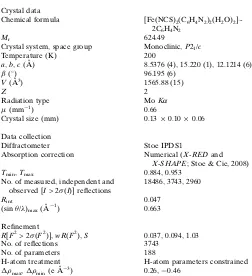
Table 2
Experimental details.
Crystal data
Chemical formula [Fe(NCS)2(C6H4N2)2(H2O)2 ]-2C6H4N2
Mr 624.49
Crystal system, space group Monoclinic,P21/c
Temperature (K) 200
a,b,c(A˚ ) 8.5376 (4), 15.220 (1), 12.1214 (6)
() 96.195 (6)
V(A˚3) 1565.88 (15)
Z 2
Radiation type MoK
(mm1) 0.66
Crystal size (mm) 0.130.100.06
Data collection
Diffractometer Stoe IPDS1
Absorption correction Numerical (X-REDand
X-SHAPE; Stoe & Cie, 2008)
Tmin,Tmax 0.884, 0.953
No. of measured, independent and observed [I> 2(I)] reflections
18486, 3743, 2960
Rint 0.047
(sin/)max(A˚
1) 0.663
Refinement
R[F2> 2(F2)],wR(F2),S 0.037, 0.094, 1.03
No. of reflections 3743
No. of parameters 188
H-atom treatment H-atom parameters constrained
max,min(e A˚
3) 0.26,0.46
Computer programs:X-AREA(Stoe & Cie, 2008),SHELXS97andXP(Sheldrick, 2008),
SHELXL2014 (Sheldrick, 2015), DIAMOND (Brandenburg, 2014) and publCIF
(Westrip, 2010). Figure 3
Rams, M., Tomkowicz, Z., Bo¨hme, M., Plass, W., Suckert, S., Werner, J., Jess, I. & Na¨ther, C. (2017).Phys. Chem. Chem. Phys.19, 3232– 3243.
Sheldrick, G. M. (2008).Acta Cryst.A64, 112–122. Sheldrick, G. M. (2015).Acta Cryst.A71, 3–8.
Stoe & Cie (2008).X-AREA,X-RED andX-SHAPE. Stoe & Cie, Darmstadt, Germany.
Suckert, S., Rams, M., Bo¨hme, M., Germann, L. S., Dinnebier, R. E., Plass, W., Werner, J. & Na¨ther, C. (2016).Dalton Trans.45, 18190– 18201.
Werner, J., Tomkowicz, Z., Reinert, T. & Na¨ther, C. (2015).Eur. J. Inorg. Chem.pp. 3066–3075.
Westrip, S. P. (2010).J. Appl. Cryst.43, 920–925.
466
Jochimet al. [Fe(NCS)sup-1 Acta Cryst. (2017). E73, 463-466
supporting information
Acta Cryst. (2017). E73, 463-466 [https://doi.org/10.1107/S205698901700322X]
Crystal structure of diaquabis(4-cyanopyridine-κ
N
)bis(thiocyanato-κ
N
)iron(II)
4-cyanopyridine disolvate
Aleksej Jochim, Inke Jess and Christian Näther
Computing details
Data collection: X-AREA (Stoe & Cie, 2008); cell refinement: X-AREA (Stoe & Cie, 2008); data reduction: X-AREA (Stoe
& Cie, 2008); program(s) used to solve structure: SHELXS97 (Sheldrick, 2008); program(s) used to refine structure:
SHELXL2014 (Sheldrick, 2015); molecular graphics: XP (Sheldrick, 2008) and DIAMOND (Brandenburg, 2014);
software used to prepare material for publication: publCIF (Westrip, 2010).
Diaquabis(4-cyanopyridine-κN)bis(thiocyanato-κN)iron(II) 4-cyanopyridine disolvate
Crystal data
[Fe(NCS)2(C6H4N2)2(H2O)2]·2C6H4N2
Mr = 624.49
Monoclinic, P21/c
a = 8.5376 (4) Å
b = 15.220 (1) Å
c = 12.1214 (6) Å
β = 96.195 (6)°
V = 1565.88 (15) Å3
Z = 2
F(000) = 640
Dx = 1.324 Mg m−3
Mo Kα radiation, λ = 0.71073 Å
Cell parameters from 18864 reflections
θ = 3.8–56.3°
µ = 0.66 mm−1
T = 200 K
Block, yellow
0.13 × 0.10 × 0.06 mm
Data collection
Stoe IPDS-1 diffractometer Phi scans
Absorption correction: numerical
(X-RED and X-SHAPE; Stoe & Cie, 2008)
Tmin = 0.884, Tmax = 0.953
18486 measured reflections
3743 independent reflections 2960 reflections with I > 2σ(I)
Rint = 0.047
θmax = 28.1°, θmin = 2.7°
h = −11→10
k = −20→20
l = −16→16
Refinement
Refinement on F2
Least-squares matrix: full
R[F2 > 2σ(F2)] = 0.037
wR(F2) = 0.094
S = 1.03
3743 reflections 188 parameters 0 restraints
Hydrogen site location: mixed H-atom parameters constrained
w = 1/[σ2(F
o2) + (0.0581P)2 + 0.1102P]
where P = (Fo2 + 2Fc2)/3
(Δ/σ)max = 0.001
Δρmax = 0.26 e Å−3
Δρmin = −0.46 e Å−3
Extinction correction: SHELXL2014 (Sheldrick, 2015),
Fc*=kFc[1+0.001xFc2λ3/sin(2θ)]-1/4
supporting information
sup-2 Acta Cryst. (2017). E73, 463-466
Special details
Geometry. All esds (except the esd in the dihedral angle between two l.s. planes) are estimated using the full covariance matrix. The cell esds are taken into account individually in the estimation of esds in distances, angles and torsion angles; correlations between esds in cell parameters are only used when they are defined by crystal symmetry. An approximate (isotropic) treatment of cell esds is used for estimating esds involving l.s. planes.
Fractional atomic coordinates and isotropic or equivalent isotropic displacement parameters (Å2)
x y z Uiso*/Ueq
Fe1 0.5000 0.5000 1.0000 0.02513 (11)
N1 0.58788 (18) 0.62971 (9) 0.99472 (12) 0.0358 (3)
C1 0.62673 (19) 0.69914 (10) 0.96961 (13) 0.0305 (3)
S1 0.68128 (7) 0.79604 (3) 0.93149 (5) 0.05165 (16)
N11 0.42359 (17) 0.50738 (9) 0.81712 (11) 0.0317 (3)
C11 0.2830 (2) 0.47429 (11) 0.77885 (14) 0.0351 (4)
H11 0.2267 0.4414 0.8283 0.042*
C12 0.2158 (2) 0.48550 (11) 0.67119 (15) 0.0390 (4)
H12 0.1150 0.4617 0.6472 0.047*
C13 0.2990 (2) 0.53234 (13) 0.59914 (14) 0.0399 (4)
C14 0.4458 (2) 0.56588 (13) 0.63609 (15) 0.0425 (4)
H14 0.5055 0.5976 0.5877 0.051*
C15 0.5029 (2) 0.55172 (12) 0.74578 (14) 0.0381 (4)
H15 0.6036 0.5747 0.7718 0.046*
C16 0.2332 (3) 0.54478 (18) 0.48511 (18) 0.0570 (6)
N12 0.1795 (3) 0.5537 (2) 0.39540 (17) 0.0837 (8)
O1 0.28045 (13) 0.55033 (7) 1.02877 (9) 0.0320 (2)
H1 0.2201 0.5134 1.0535 0.048*
H2 0.2728 0.5951 1.0684 0.048*
N21 1.2086 (2) 0.80713 (12) 0.64837 (15) 0.0523 (4)
C21 1.0626 (3) 0.7796 (2) 0.6202 (2) 0.0774 (9)
H21 1.0072 0.8031 0.5546 0.093*
C22 0.9863 (3) 0.71950 (19) 0.67912 (19) 0.0685 (8)
H22 0.8813 0.7018 0.6554 0.082*
C23 1.0672 (2) 0.68573 (11) 0.77413 (14) 0.0347 (4)
C24 1.2197 (2) 0.71283 (11) 0.80597 (15) 0.0378 (4)
H24 1.2775 0.6904 0.8713 0.045*
C25 1.2856 (2) 0.77362 (13) 0.73992 (17) 0.0445 (4)
H25 1.3907 0.7923 0.7609 0.053*
C26 0.9913 (2) 0.62137 (12) 0.83758 (14) 0.0361 (4)
N22 0.9307 (2) 0.56987 (11) 0.88643 (14) 0.0460 (4)
Atomic displacement parameters (Å2)
U11 U22 U33 U12 U13 U23
Fe1 0.02351 (17) 0.02120 (16) 0.03167 (17) −0.00047 (11) 0.00745 (11) 0.00057 (11)
N1 0.0402 (9) 0.0231 (6) 0.0448 (8) −0.0057 (5) 0.0075 (6) 0.0016 (5)
C1 0.0277 (8) 0.0303 (8) 0.0329 (8) −0.0006 (6) 0.0004 (6) −0.0007 (6)
sup-3 Acta Cryst. (2017). E73, 463-466
N11 0.0305 (7) 0.0324 (7) 0.0328 (7) 0.0002 (5) 0.0053 (5) −0.0015 (5)
C11 0.0343 (9) 0.0308 (8) 0.0408 (9) −0.0028 (6) 0.0061 (7) −0.0035 (6)
C12 0.0341 (9) 0.0375 (9) 0.0446 (9) 0.0010 (7) 0.0001 (7) −0.0080 (7)
C13 0.0397 (10) 0.0458 (10) 0.0338 (8) 0.0094 (8) 0.0022 (7) −0.0055 (7)
C14 0.0393 (10) 0.0550 (11) 0.0342 (8) 0.0011 (8) 0.0086 (7) 0.0046 (8)
C15 0.0325 (9) 0.0472 (10) 0.0353 (8) −0.0032 (7) 0.0071 (7) 0.0014 (7)
C16 0.0451 (12) 0.0819 (16) 0.0433 (11) 0.0035 (11) 0.0015 (9) −0.0003 (10)
N12 0.0618 (14) 0.140 (2) 0.0460 (11) −0.0095 (14) −0.0079 (9) 0.0126 (13)
O1 0.0272 (6) 0.0284 (5) 0.0422 (6) −0.0004 (4) 0.0117 (5) −0.0031 (4)
N21 0.0464 (10) 0.0518 (10) 0.0607 (10) −0.0051 (8) 0.0144 (8) 0.0215 (8)
C21 0.0541 (15) 0.111 (2) 0.0643 (15) −0.0162 (14) −0.0084 (11) 0.0537 (15)
C22 0.0417 (12) 0.105 (2) 0.0554 (13) −0.0231 (12) −0.0112 (10) 0.0401 (13)
C23 0.0313 (9) 0.0379 (9) 0.0351 (8) −0.0039 (7) 0.0048 (6) 0.0051 (6)
C24 0.0338 (10) 0.0367 (9) 0.0421 (9) −0.0014 (7) 0.0005 (7) 0.0066 (7)
C25 0.0341 (10) 0.0406 (10) 0.0593 (11) −0.0058 (7) 0.0068 (8) 0.0074 (8)
C26 0.0315 (9) 0.0419 (9) 0.0351 (8) −0.0026 (7) 0.0039 (6) 0.0020 (7)
N22 0.0397 (9) 0.0490 (9) 0.0508 (9) −0.0068 (7) 0.0122 (7) 0.0104 (7)
Geometric parameters (Å, º)
Fe1—O1i 2.0888 (11) C14—H14 0.9500
Fe1—O1 2.0888 (11) C15—H15 0.9500
Fe1—N1 2.1153 (13) C16—N12 1.141 (3)
Fe1—N1i 2.1153 (13) O1—H1 0.8400
Fe1—N11i 2.2451 (14) O1—H2 0.8400
Fe1—N11 2.2451 (14) N21—C21 1.325 (3)
N1—C1 1.158 (2) N21—C25 1.329 (3)
C1—S1 1.6286 (16) C21—C22 1.368 (3)
N11—C15 1.337 (2) C21—H21 0.9500
N11—C11 1.338 (2) C22—C23 1.377 (3)
C11—C12 1.378 (3) C22—H22 0.9500
C11—H11 0.9500 C23—C24 1.380 (3)
C12—C13 1.382 (3) C23—C26 1.442 (2)
C12—H12 0.9500 C24—C25 1.382 (3)
C13—C14 1.382 (3) C24—H24 0.9500
C13—C16 1.447 (3) C25—H25 0.9500
C14—C15 1.383 (3) C26—N22 1.140 (2)
O1i—Fe1—O1 180.0 C14—C13—C16 120.42 (19)
O1i—Fe1—N1 90.55 (5) C13—C14—C15 117.86 (17)
O1—Fe1—N1 89.45 (5) C13—C14—H14 121.1
O1i—Fe1—N1i 89.45 (5) C15—C14—H14 121.1
O1—Fe1—N1i 90.55 (5) N11—C15—C14 123.35 (17)
N1—Fe1—N1i 180.0 N11—C15—H15 118.3
O1i—Fe1—N11i 88.63 (5) C14—C15—H15 118.3
O1—Fe1—N11i 91.37 (5) N12—C16—C13 179.0 (3)
N1—Fe1—N11i 90.59 (5) Fe1—O1—H1 114.2
supporting information
sup-4 Acta Cryst. (2017). E73, 463-466
O1i—Fe1—N11 91.37 (5) H1—O1—H2 104.5
O1—Fe1—N11 88.63 (5) C21—N21—C25 117.42 (17)
N1—Fe1—N11 89.41 (5) N21—C21—C22 124.4 (2)
N1i—Fe1—N11 90.59 (5) N21—C21—H21 117.8
N11i—Fe1—N11 180.0 C22—C21—H21 117.8
C1—N1—Fe1 166.44 (14) C21—C22—C23 117.5 (2)
N1—C1—S1 178.74 (15) C21—C22—H22 121.2
C15—N11—C11 117.64 (15) C23—C22—H22 121.2
C15—N11—Fe1 123.39 (12) C22—C23—C24 119.69 (17)
C11—N11—Fe1 118.54 (11) C22—C23—C26 119.08 (17)
N11—C11—C12 123.23 (17) C24—C23—C26 121.22 (15)
N11—C11—H11 118.4 C23—C24—C25 117.94 (17)
C12—C11—H11 118.4 C23—C24—H24 121.0
C11—C12—C13 118.18 (17) C25—C24—H24 121.0
C11—C12—H12 120.9 N21—C25—C24 123.03 (18)
C13—C12—H12 120.9 N21—C25—H25 118.5
C12—C13—C14 119.72 (17) C24—C25—H25 118.5
C12—C13—C16 119.85 (19) N22—C26—C23 179.1 (2)
Symmetry code: (i) −x+1, −y+1, −z+2.
Hydrogen-bond geometry (Å, º)
D—H···A D—H H···A D···A D—H···A
C12—H12···N12ii 0.95 2.52 3.437 (3) 162
C14—H14···S1iii 0.95 3.01 3.960 (2) 177
O1—H1···N22i 0.84 2.00 2.8380 (19) 177
O1—H2···N21iv 0.84 1.89 2.7159 (19) 168
![Figure 1The discrete complex and the solvent molecule of the title compound withlabeling and displacement ellipsoids drawn at the 50% probability level.[Symmetry code: (i) 1 � x, 1 � y, 2 � z.]](https://thumb-us.123doks.com/thumbv2/123dok_us/409928.538523/2.610.45.293.80.319/discrete-molecule-compound-withlabeling-displacement-ellipsoids-probability-symmetry.webp)