Crystal structure of (
E
)-3-(4-hydroxy-
benzyl)-4-{[4-(methylsulfanyl)benzyl-idene]amino}-1
H
-1,2,4-triazole-5(4
H
)-thione
P. S. Manjula,aB. K. Sarojini,bB. Narayana,cK. Byrappad and S. Madan Kumare*
aDepartment of Chemistry, PA College of Engineering, Nadupadavu 574 153, D.K., Mangaluru, India,bDepartment of Industrial Chemistry, Mangalagangotri, Mangalore University, Mangaluru 574 199, India,cDepartment of Chemistry, Mangalagangotri, Mangalore University, Mangaluru 574 199, India,dDepartment of Materials Science, Mangalagangotri, Mangalore University, Mangaluru 574 199, India, andePURSE Lab, Mangalagangotri, Mangalore University, Mangaluru 574 199, India. *Corre-spondence e-mail: madanmx@mangaloreuniversity.ac.in
Received 4 November 2015; accepted 18 November 2015
Edited by H. Stoeckli-Evans, University of Neuchaˆtel, Switzerland
In the title compound, C17H16N4OS2, the triazole and
methylthiobenzylidene rings are nearly coplanar, making a dihedral angle of 6.52 (12). An intramolecular C—H S
hydrogen bond forms an S(6) ring motif. The hydroxybenzyl ring is almost normal to the triazole and methylthiobenzyl-idene rings, making dihedral angles of 78.56 (12) and 84.79 (11), respectively. In the crystal, molecules are linked
through O—H N and N—H O hydrogen bonds, forming layers parallel to the acplane. The layers are linkedviaC— H N hydrogen bonds, forming a three-dimensional struc-ture. In addition, a short – interaction is observed [inter-centroid distance = 3.764 (3) A˚ ], involving inversion-related methylthiobenzylidene rings.
Keywords:crystal structure; triazole; thione; methylthiobenzylidene; hydrogen bonding;–interactions.
CCDC reference:1437595
1. Related literature
For the structure of a related compound, see: Manjula et al.
(2015).
2. Experimental
2.1. Crystal data
C17H16N4OS2
Mr= 356.46
Monoclinic,P21=c
a= 7.739 (5) A˚
b= 28.161 (16) A˚
c= 7.945 (4) A˚
= 100.407 (11)
V= 1703.0 (17) A˚3
Z= 4
MoKradiation
= 0.32 mm1
T= 293 K
0.570.340.24 mm
2.2. Data collection
Rigaku Saturn724+ diffractometer Absorption correction: numerical
(NUMABS; Rigaku, 1999)
Tmin= 0.895,Tmax= 0.954
8210 measured reflections 3017 independent reflections 2200 reflections withI> 2(I)
Rint= 0.031
2.3. Refinement
R[F2> 2(F2)] = 0.047
wR(F2) = 0.107
S= 1.06 3017 reflections
217 parameters
H-atom parameters constrained
max= 0.16 e A˚3
min=0.17 e A˚3
Table 1
Hydrogen-bond geometry (A˚ ,).
D—H A D—H H A D A D—H A
C10—H10 S1 0.93 2.52 3.267 (3) 138 O1—H1 N2i
0.82 2.03 2.806 (3) 159 N1—H1A O1ii
0.86 1.98 2.816 (3) 164 C17—H17C N4iii
0.96 2.62 3.472 (4) 148
Symmetry codes: (i)x1;yþ3 2;z
1
2; (ii)xþ1;y;zþ1; (iii)x;yþ1;zþ1.
Data collection:CrystalClear(Rigaku, 2011); cell refinement: Crys-talClear; data reduction: CrystalClear; program(s) used to solve structure:SHELXS97(Sheldrick, 2008); program(s) used to refine structure: SHELXL2014 (Sheldrick, 2015); molecular graphics: Mercury(Macraeet al., 2008); software used to prepare material for publication:OLEX2(Dolomanovet al., 2009).
data reports
o982
Manjulaet al. doi:10.1107/S2056989015021994 Acta Cryst.(2015).E71, o982–o983Acknowledgements
The authors thank DST–PURSE, Mangalore University, Mangaluru, for providing the single-crystal X-ray diffraction facility. BKS and PSM gratefully acknowledge the Depart-ment of Chemistry, P. A. College of Engineering for providing research facilities.
Supporting information for this paper is available from the IUCr electronic archives (Reference: SU5236).
Dolomanov, O. V., Bourhis, L. J., Gildea, R. J., Howard, J. A. K. & Puschmann, H. (2009).J. Appl. Cryst.42, 339–341.
Macrae, C. F., Bruno, I. J., Chisholm, J. A., Edgington, P. R., McCabe, P., Pidcock, E., Rodriguez-Monge, L., Taylor, R., van de Streek, J. & Wood, P. A. (2008).J. Appl. Cryst.41, 466–470.
Manjula, P. S., Sarojini, B. K., Narayana, B., Byrappa, K. & Madan Kumar, S. (2015).Acta Cryst.E71, o912–o913.
Rigaku. (1999).NUMABS. Rigaku Corporation, Tokyo, Japan. Rigaku (2011).CrystalClear.Rigaku Corporation, Tokyo, Japan. Sheldrick, G. M. (2008).Acta Cryst.A64, 112–122.
supporting information
sup-1
Acta Cryst. (2015). E71, o982–o983
supporting information
Acta Cryst. (2015). E71, o982–o983 [https://doi.org/10.1107/S2056989015021994]
Crystal structure of (
E
)-3-(4-hydroxybenzyl)-4-{[4-(methylsulfanyl)benzyl-idene]amino}-1
H
-1,2,4-triazole-5(4
H
)-thione
P. S. Manjula, B. K. Sarojini, B. Narayana, K. Byrappa and S. Madan Kumar
S1. Comment
The title compound was synthesized, crystallized and its crystal structure is presented as part of our work on
3-methyl-1H-1,2,4-triazole-5(4H)-thione derivatives (Manjula et al., 2015).
The molecular structure of the title compound is as shown in Fig 1. The methylsulfanylbenzylidene and triazole rings
are almost coplanar with a dihedral angle of 6.52 (12) °. The hydroxybenzyl ring makes dihedral angles of 78.56 (12) °
and 84.79 (11) ° with the triazole and methylthiobenzylidene rings, respectively. An intramolecular interaction of the type
C10—H10···S1 is observed (Fig. 1 and Table 1).
In the crystal, the molecules are connected through O1—H1···N2 and N1—H1A···O1 hydrogen bonds (Table 1) forming
layers parallel to (010). The layers are linked by C17—H17C···N4 hydrogen bonds forming a three-dimensional structure
(Fig. 2 and Table 1) . In addition, a parallel slipped π–π (Cg···Cgi) interaction is observed with an inter-centroid distance
of 3.764 (3) Å [Cg is the centroid of ring C11—C16; inter-planar distance = 3.500 (1) Å; slippage 1.384 Å; symmetry
code: (i) -x, -y+1, -z+1].
S2. Experimental
The synthesis of title compound, (3), is illustrated in Fig. 3. A suspension of 4-(methylthio)benzaldehyde (2) (0.01 mol)
in ethanol (15 ml) was added to 4-amino-3-(4-hydroxybenzyl)-1H-1,2,4-triazole-5(4H)-thione (1) (0.01 mol) and heated
until a clear solution was obtained. To this a few drops of conc. H2SO4 were added as a catalyst and the mixture was
refluxed for 36 h on a water bath. The precipitate formed was filtered and recrystallized from methanol to give the titled
compound. Single crystals were obtained by recrystallization from acetic acid (m.p. 469—471 K).
S3. Refinement
Crystal data, data collection and structure refinement details are summarized in Table 2. The H atoms were fixed
geometrically (O-H = 0.82 Å, N-H = 0.86 Å, and C—H = 0.93–0.97 Å) and allowed to ride on their parent atoms with
Figure 1
A view of the molecular structure of the title compound, with atom labelling. Displacement ellipsoids are drawn at the
supporting information
sup-3
[image:5.610.139.474.69.310.2]Acta Cryst. (2015). E71, o982–o983
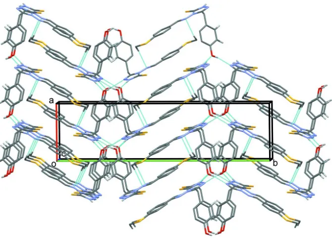
Figure 2
A viewed along the c axis of the crystal packing of the title compound. Hydrogen bonds are drawn as a dashed lines (see
[image:5.610.105.479.366.529.2]Table 1), and H atoms not involved in hydrogen bonding have been omitted for clarity.
Figure 3
Reaction scheme.
(E)-3-(4-Hydroxybenzyl)-4-{[4-(methylsulfanyl)benzylidene]amino}-1H-1,2,4-triazole-5(4H)-thione
Crystal data
C17H16N4OS2
Mr = 356.46
Monoclinic, P21/c
a = 7.739 (5) Å
b = 28.161 (16) Å
c = 7.945 (4) Å
β = 100.407 (11)°
V = 1703.0 (17) Å3
Z = 4
F(000) = 744
Dx = 1.390 Mg m−3
Mo Kα radiation, λ = 0.71075 Å Cell parameters from 2021 reflections
θ = 3.0–25.3°
µ = 0.32 mm−1
T = 293 K Prism, yellow
Rigaku Saturn724+ diffractometer
Detector resolution: 7.111 pixels mm-1
profile data from ω–scans Absorption correction: numerical
(NUMABS; Rigaku, 1999)
Tmin = 0.895, Tmax = 0.954
8210 measured reflections
3017 independent reflections 2200 reflections with I > 2σ(I)
Rint = 0.031
θmax = 25.3°, θmin = 3.0°
h = −9→9
k = −33→33
l = −9→6
Refinement
Refinement on F2
Least-squares matrix: full
R[F2 > 2σ(F2)] = 0.047
wR(F2) = 0.107
S = 1.06 3017 reflections 217 parameters 0 restraints
Primary atom site location: structure-invariant direct methods
Hydrogen site location: inferred from neighbouring sites
H-atom parameters constrained
w = 1/[σ2(F
o2) + (0.0424P)2 + 0.239P]
where P = (Fo2 + 2Fc2)/3
(Δ/σ)max < 0.001
Δρmax = 0.16 e Å−3
Δρmin = −0.17 e Å−3
Special details
Geometry. All e.s.d.'s (except the e.s.d. in the dihedral angle between two l.s. planes) are estimated using the full covariance matrix. The cell e.s.d.'s are taken into account individually in the estimation of e.s.d.'s in distances, angles and torsion angles; correlations between e.s.d.'s in cell parameters are only used when they are defined by crystal symmetry. An approximate (isotropic) treatment of cell e.s.d.'s is used for estimating e.s.d.'s involving l.s. planes.
Fractional atomic coordinates and isotropic or equivalent isotropic displacement parameters (Å2)
x y z Uiso*/Ueq
S1 0.53353 (10) 0.58782 (3) 1.04260 (8) 0.0612 (2) S2 0.00577 (11) 0.40779 (3) 0.16264 (10) 0.0797 (3) O1 −0.2083 (2) 0.71862 (6) 0.21145 (19) 0.0526 (5)
H1 −0.2399 0.7431 0.2531 0.063*
N1 0.6203 (2) 0.67074 (7) 0.9170 (2) 0.0482 (5)
H1A 0.6603 0.6818 1.0173 0.058*
N2 0.6227 (3) 0.69644 (7) 0.7707 (2) 0.0469 (5) N3 0.5039 (2) 0.62570 (6) 0.7141 (2) 0.0386 (5) N4 0.4256 (2) 0.59134 (7) 0.5984 (2) 0.0410 (5) C1 0.5514 (3) 0.62736 (9) 0.8925 (3) 0.0414 (6) C2 0.5495 (3) 0.66794 (8) 0.6491 (3) 0.0403 (6) C3 0.5120 (3) 0.68017 (9) 0.4640 (3) 0.0490 (6)
H3A 0.5476 0.6539 0.3991 0.059*
H3B 0.5815 0.7077 0.4448 0.059*
C4 0.3195 (3) 0.69085 (8) 0.3991 (3) 0.0401 (6) C5 0.2392 (3) 0.72987 (8) 0.4562 (3) 0.0482 (6)
H5 0.3045 0.7500 0.5360 0.058*
C6 0.0633 (3) 0.73960 (8) 0.3971 (3) 0.0465 (6)
H6 0.0115 0.7661 0.4371 0.056*
supporting information
sup-5
Acta Cryst. (2015). E71, o982–o983
C8 0.0426 (3) 0.67050 (8) 0.2227 (3) 0.0494 (6)
H8 −0.0234 0.6499 0.1447 0.059*
C9 0.2177 (3) 0.66139 (8) 0.2818 (3) 0.0470 (6)
H9 0.2689 0.6348 0.2418 0.056*
C10 0.3768 (3) 0.55258 (9) 0.6531 (3) 0.0459 (6)
H10 0.3949 0.5468 0.7702 0.055*
C11 0.2918 (3) 0.51667 (8) 0.5332 (3) 0.0414 (6) C12 0.2277 (3) 0.47597 (9) 0.5961 (3) 0.0497 (6)
H12 0.2421 0.4717 0.7138 0.060*
C13 0.1424 (3) 0.44141 (9) 0.4884 (4) 0.0539 (7)
H13 0.1007 0.4142 0.5337 0.065*
C14 0.1194 (3) 0.44748 (9) 0.3131 (3) 0.0497 (6) C15 0.1865 (3) 0.48812 (9) 0.2491 (3) 0.0524 (7)
H15 0.1737 0.4923 0.1314 0.063*
C16 0.2715 (3) 0.52218 (9) 0.3575 (3) 0.0477 (6)
H16 0.3157 0.5491 0.3125 0.057*
C17 −0.0520 (4) 0.35993 (11) 0.2856 (4) 0.0858 (10)
H17A −0.1153 0.3365 0.2109 0.103*
H17B 0.0526 0.3459 0.3497 0.103*
H17C −0.1246 0.3714 0.3629 0.103*
Atomic displacement parameters (Å2)
U11 U22 U33 U12 U13 U23
S1—C1 1.655 (2) C6—H6 0.9300
S2—C14 1.752 (3) C6—C7 1.377 (3)
S2—C17 1.769 (3) C7—C8 1.375 (3)
O1—H1 0.8200 C8—H8 0.9300
O1—C7 1.374 (3) C8—C9 1.376 (3)
N1—H1A 0.8600 C9—H9 0.9300
N1—N2 1.372 (2) C10—H10 0.9300
N1—C1 1.333 (3) C10—C11 1.462 (3)
N2—C2 1.304 (3) C11—C12 1.378 (3)
N3—N4 1.396 (2) C11—C16 1.386 (3)
N3—C1 1.399 (3) C12—H12 0.9300
N3—C2 1.368 (3) C12—C13 1.382 (3)
N4—C10 1.258 (3) C13—H13 0.9300
C2—C3 1.487 (3) C13—C14 1.383 (3)
C3—H3A 0.9700 C14—C15 1.390 (3)
C3—H3B 0.9700 C15—H15 0.9300
C3—C4 1.517 (3) C15—C16 1.375 (3)
C4—C5 1.379 (3) C16—H16 0.9300
C4—C9 1.383 (3) C17—H17A 0.9600
C5—H5 0.9300 C17—H17B 0.9600
C5—C6 1.385 (3) C17—H17C 0.9600
C14—S2—C17 104.78 (14) C7—C8—H8 120.0
C7—O1—H1 109.5 C7—C8—C9 120.0 (2)
N2—N1—H1A 122.4 C9—C8—H8 120.0
C1—N1—H1A 122.4 C4—C9—H9 119.2
C1—N1—N2 115.23 (18) C8—C9—C4 121.6 (2)
C2—N2—N1 103.43 (19) C8—C9—H9 119.2
N4—N3—C1 133.95 (19) N4—C10—H10 119.9
C2—N3—N4 117.69 (18) N4—C10—C11 120.2 (2) C2—N3—C1 108.36 (19) C11—C10—H10 119.9 C10—N4—N3 119.70 (19) C12—C11—C10 119.3 (2) N1—C1—S1 126.50 (18) C12—C11—C16 118.4 (2) N1—C1—N3 101.73 (19) C16—C11—C10 122.3 (2) N3—C1—S1 131.78 (19) C11—C12—H12 119.2 N2—C2—N3 111.2 (2) C11—C12—C13 121.6 (2)
N2—C2—C3 124.8 (2) C13—C12—H12 119.2
N3—C2—C3 123.9 (2) C12—C13—H13 120.1
C2—C3—H3A 109.0 C12—C13—C14 119.8 (2)
C2—C3—H3B 109.0 C14—C13—H13 120.1
C2—C3—C4 112.74 (18) C13—C14—S2 124.5 (2)
H3A—C3—H3B 107.8 C13—C14—C15 118.8 (2)
C4—C3—H3A 109.0 C15—C14—S2 116.8 (2)
C4—C3—H3B 109.0 C14—C15—H15 119.5
C5—C4—C3 121.3 (2) C16—C15—C14 120.9 (2)
supporting information
sup-7
Acta Cryst. (2015). E71, o982–o983
C9—C4—C3 121.0 (2) C11—C16—H16 119.8
C4—C5—H5 119.3 C15—C16—C11 120.5 (2)
C4—C5—C6 121.3 (2) C15—C16—H16 119.8
C6—C5—H5 119.3 S2—C17—H17A 109.5
C5—C6—H6 120.1 S2—C17—H17B 109.5
C7—C6—C5 119.9 (2) S2—C17—H17C 109.5
C7—C6—H6 120.1 H17A—C17—H17B 109.5
O1—C7—C6 122.5 (2) H17A—C17—H17C 109.5 O1—C7—C8 118.0 (2) H17B—C17—H17C 109.5 C8—C7—C6 119.5 (2)
S2—C14—C15—C16 177.44 (19) C2—C3—C4—C5 65.8 (3) O1—C7—C8—C9 177.6 (2) C2—C3—C4—C9 −113.4 (2) N1—N2—C2—N3 −0.7 (2) C3—C4—C5—C6 −179.9 (2) N1—N2—C2—C3 176.7 (2) C3—C4—C9—C8 179.6 (2) N2—N1—C1—S1 178.79 (16) C4—C5—C6—C7 0.0 (3) N2—N1—C1—N3 −0.8 (2) C5—C4—C9—C8 0.3 (3) N2—C2—C3—C4 −103.6 (3) C5—C6—C7—O1 −177.9 (2) N3—N4—C10—C11 178.90 (18) C5—C6—C7—C8 1.0 (3) N3—C2—C3—C4 73.4 (3) C6—C7—C8—C9 −1.4 (3) N4—N3—C1—S1 0.2 (4) C7—C8—C9—C4 0.7 (4) N4—N3—C1—N1 179.7 (2) C9—C4—C5—C6 −0.7 (3) N4—N3—C2—N2 −179.30 (18) C10—C11—C12—C13 178.4 (2) N4—N3—C2—C3 3.4 (3) C10—C11—C16—C15 −178.2 (2) N4—C10—C11—C12 −175.2 (2) C11—C12—C13—C14 −0.3 (4) N4—C10—C11—C16 4.2 (3) C12—C11—C16—C15 1.1 (3) C1—N1—N2—C2 0.9 (3) C12—C13—C14—S2 −177.14 (19) C1—N3—N4—C10 2.8 (3) C12—C13—C14—C15 1.4 (4) C1—N3—C2—N2 0.2 (3) C13—C14—C15—C16 −1.2 (4) C1—N3—C2—C3 −177.1 (2) C14—C15—C16—C11 −0.1 (4) C2—N3—N4—C10 −177.9 (2) C16—C11—C12—C13 −1.0 (4) C2—N3—C1—S1 −179.21 (18) C17—S2—C14—C13 −3.9 (2) C2—N3—C1—N1 0.3 (2) C17—S2—C14—C15 177.60 (19)
Hydrogen-bond geometry (Å, º)
D—H···A D—H H···A D···A D—H···A
C10—H10···S1 0.93 2.52 3.267 (3) 138
O1—H1···N2i 0.82 2.03 2.806 (3) 159
N1—H1A···O1ii 0.86 1.98 2.816 (3) 164
C17—H17C···N4iii 0.96 2.62 3.472 (4) 148