EGFR
Mutation of Tumor and Serum in Gefitinib-Treated
Patients with Chemotherapy-Naive Non–small Cell
Lung Cancer
Hideharu Kimura, MD, Kazuo Kasahara, MD, Kazuhiko Shibata, MD, Takashi Sone, MD,
Akihiro Yoshimoto, MD, Toshiyuki Kita, MD, Yukari Ichikawa, MD, Yuko Waseda, MD,
Kazuyoshi Watanabe, MD, Hiroki Shiarasaki, MD, Yoshihisa Ishiura, MD, Masayuki Mizuguchi, MD,
Yasuto Nakatsumi, MD, Tatsuhiko Kashii, MD, Masashi Kobayashi, MD, Hideo Kunitoh, MD,
Tomohide Tamura, MD, Kazuto Nishio, MD, Masaki Fujimura, MD, and Shinji Nakao, MD
Background:The authors evaluate the efficacy and safety of ge-fitinib monotherapy in chemotherapy-naive patients with advanced non–small-cell lung cancer (NSCLC). A secondary endpoint is to evaluate the relationship between clinical manifestations and epi-dermal growth factor receptor (EGFR) mutation status.
Methods:Japanese chemotherapy-naive NSCLC patients were en-rolled. They had measurable lesions, Eastern Cooperative Oncology Group performance status of 0 to 2, and adequate organ and bone marrow function. Patients received 250 mg of oral gefitinib daily.
EGFR mutations in exon 18, 19, and 21 of DNA extracted from tumor and serum were analyzed by genomic polymerase chain reaction and direct sequence.
Results:All 30 patients were eligible for the assessment of efficacy and safety. An objective response and stable disease were observed in 10 patients (33.3%) and nine patients (30.0%), respectively. The median time to progression was 3.3 months and the median overall survival was 10.6 months. The 1-year survival rate was 43.3%. Grade 3 toxicities were observed in seven patients.EGFRmutation was observed in four of 13 (30.8%) tumors, and two of them achieved partial response. In serum samples, three of 10 patients withEGFRmutations in the serum before treatment had a response to gefitinib.EGFR mutation was observed in 10 of 27 and signifi-cantly more frequently observed in the posttreatment samples from patients with a partial response or stable disease than in those from patients with progressive disease (p⫽0.006).
Conclusions: Gefitinib monotherapy in chemotherapy-naive NSCLC patients was active, with acceptable toxicities. These results warrant further evaluation of gefitinib monotherapy as a first-line therapy. TheEGFRmutation in serum DNA may be a biomarker for monitoring the response to gefitinib during treatment.
Key Words: Non–small-cell lung cancer, Gefitinib, Epidermal growth factor receptor, Mutation, Serum DNA.
(J Thorac Oncol.2006;1: 260–267)
N
on–small-cell lung cancer (NSCLC) is the leading cause of cancer death in Japan and throughout the world.1Unfortunately, the majority of patients with NSCLC present with locally advanced or metastatic disease at the time of diagnosis. Although chemotherapy has produced modest sur-vival benefits in advanced NSCLC patients, the outcome of chemotherapy for NSCLC remains unsatisfactory.
Protein tyrosine kinases play important roles in the pathogenesis of malignant tumors.2 Among them, epidermal
growth factor receptor (EGFR) tyrosine kinase has been implicated in the initiation and progression of NSCLC.3–5
The overexpression of EGFR is frequent in NSCLC.6
Mono-clonal antibodies and low-molecular-weight compounds that inhibit the EGFR signaling pathway have been developed and shown to have antitumor effects. Gefitinib (Iressa, Astra-Zenca, London, England) is an orally active EGFR type tyrosine kinase inhibitor. In four phase I studies, tumor shrinkage or stabilization after gefitinib monotherapy was observed in some patients with NSCLC. In two phase II trials, Iressa Dose Evaluation in Advanced Lung cancer (IDEAL) 1 and 2, gefitinib monotherapy was shown to have a substantial effect in NSCLC patients treated previously with chemother-apy.7,8In these trials, patients of Asian origin and who had
never been smokers had a statistically significant improve-ment in overall survival. In spite of encouraging results in the IDEAL trials, two large-scale, phase III, randomized trials, Iressa NSCLC Trial Assessing Combination Treatment, failed to show any survival benefit for the use of gefitinib.9,10
Patients in a large-scale phase III trial comparing gefitinib
From Respiratory Medicine, Kanazawa University Hospital, Ishikawa; In-ternal Medicine, Kouseiren Takaoka Hospital, Takaoka; InIn-ternal Medi-cine, Shinminato Municipal Hospital, Shinminato, Japan. Internal Med-icine, Fukuiken Saiseikai Hospital; Respiratory MedMed-icine, Toyama City Hospital, Toyama; Respiratory Medicine, Ishikawa Prefectoral Hospital, Kanazawa; Respiratory Medicine, Kanazawa Municipal Hospital; Ka-nazawa; and National Cancer Center Hospital, and National Cancer Center Research Institute, Tokyo, Japan.
Address for correspondence: Kazuo Kasahara, MD, Takara-machi 13-1, Kanazawa, Ishikawa, Japan; email: kasa1237@med3.m.kanazawa-u.ac.jp.
Copyright © 2006 by the International Association for the Study of Lung Cancer
and placebo in advanced NSCLC with prior chemotherapy demonstrated in preliminary analysis a tendency to have improvement in overall survival but did not have a statisti-cally significant improvement in overall survival.11There are
many issues that need to be addressed with regard to the clinical application of gefitinib; one of the most important issues is the efficacy of gefitinib monotherapy in patients with chemotherapy-naive NSCLC,12and another is to establish a
way to predict response to gefitinib.
Recently, it has been suggested that mutations in the EGFR tyrosine kinase domain play a critical role in deter-mining tumor response to gefitinib in NSCLC patients.13,14
The mutations consisted of small, in-frame deletions or sub-stitutions clustered around the adenosine triphosphate– bind-ing site in exons 18, 19, and 21 of the EGFR. After these reports, some investigators supported the belief that EGFR
mutation is one of the strong determinants of tumor response to gefitinib.15–17 Tumors with EGFR mutations tend to be
more common in adenocarcinomas, female patients, non-smokers, and those of Asian origin. In most of those studies, tumor samples that were resected by operations were used. Because it is often difficult to obtain a tumor sample from an inoperable NSCLC patient, it is necessary to establish a method for detecting mutant EGFR from a patient sample other than from tumor specimens.
Polymerase chain reaction (PCR) technology for the amplification of small amounts of DNA has made it possible to identify the same alterations typically observed in DNA from serum samples from NSCLC patients.18,19Serum DNA
may provide a noninvasive and repeatable source of geno-typic information that could influence treatment and progno-sis, especially in advanced NSCLC patients who have re-ceived gefitinib therapy. We essentially consider that it is possible to detect the EGFR mutation in serum DNA. We hypothesized that serum DNA may provide useful informa-tion on EGFRmutations in lung cancer patients.
As described above, the usefulness of gefitinib mono-therapy is controversial and that in patients without pretreat-ment is unclear. BecauseEGFRmutations have been shown to be strongly associated with the response of NSCLC pa-tients to gefitinib treatment, the analysis ofEGFRmutations is necessary to evaluate the clinical benefit of gefitinib. We therefore conducted a multicenter phase II trial for these patients. The primary objective was to evaluate the objective response rate, and secondary objectives were to estimate the disease control rate, disease-related symptom improvement rate, safety, time to progression (TTP), and overall survival (OS). In addition, as a correlative study, we planned to detect
EGFRmutations in serum samples from NSCLC patients and evaluate the relationship between the EGFR mutation and clinical manifestations in NSCLC patients receiving gefitinib treatment.
PATIENTS AND METHODS Patient Eligibility
Patients who had histologically or cytologically proven stage IIIb or IV NSCLC and no previous chemotherapy were enrolled into this trial. Radiotherapy for metastatic lesions
until 3 weeks before entry was allowed on condition that these lesions were not assessed for tumor response. Patients in whom recurrence occurred after surgery were also eligible. Patient eligibility criteria included at least one measurable lesion, age of 20 years or older, Eastern Cooperative Oncol-ogy Group performance status (PS) of 0 to 2, and life expectancy of greater than or equal to12 weeks. Adequate organ and bone marrow function was necessary, defined as leukocyte counts greater than or equal to 3.0 ⫻ 106/liter,
neutrophil counts greater than or equal to 1.5 ⫻ 106/ liter, platelet counts greater than or equal to 100⫻ 109/liter,
hemoglobin levels greater than or equal to 8.5 g/dl, alanine aminotransferase or aspartate aminotransferase levels less than or equal to two times the upper limit of the reference range (⬍100 IU/liter in the presence of liver metastases), serum bilirubin levels less than or equal to 1.5 mg/dl, serum creatinine levels less than or equal to1.5 mg/dl, and PaO 2
levels greater than or equal to 65 mmHg. Patients with any of the following were excluded: active double cancer; severe complications such as myocardial infarction within 3 months before entry or uncontrolled diabetes; symptomatic brain or bone metastasis; diarrhea more severe than grade 2 according to National Cancer Institute Common Toxicity Criteria ver-sion 2; systemic administration of steroids to treat skin diseases; pleural, pericardial, or peritoneal effusion requiring treatment; and pregnancy or lactation. All patients were required to give informed consent.
Treatment
Patients were treated with gefitinib 250 mg orally once per day. Treatment was discontinued when the disease pro-gressed, intolerable toxicities appeared, the patients requested withdrawal, or disease-related symptoms worsened without tumor response after 8 weeks of gefitinib monotherapy. These patients received chemotherapeutic treatment after gefitinib therapy. The chemotherapy regimen consisted of platinum (cisplatin or carboplatin) plus new agents (paclitaxel, do-cetaxel, gemcitabine, vinorelbine, or irinotecan) in patients aged 74 years or younger and vinorelbine monotherapy in patients aged 75 years or older. If symptomatic bone or brain metastasis occurred during gefitinib monotherapy, patients received radiotherapy after gefitinib treatment.
Efficacy and Drug-Related Adverse Events
Tumor size was assessed with computed tomography or magnetic resonance imaging scans every 4 weeks from the start to cessation of protocol treatment, using Response Eval-uation Criteria in Solid Tumors guidelines.20Disease control
was judged when patients achieved the best response of complete response, partial response (PR), or stable disease (SD), which was confirmed and sustained for 4 weeks. TTP was measured as the period from the start of the treatment to an identifiable time of disease progression. OS was measured from the start of the treatment until death or the last follow-up. The Kaplan-Meier method was used to calculate these measures.
Drug adverse events were recorded and graded accord-ing to National Cancer Institute Common Toxicity Criteria
version 2.0. Changes in physical and laboratory findings were assessed at least every 2 weeks.
Serum Sample Collection and DNA Extraction
Blood samples from patients were collected before and 14 days after the initiation of gefitinib administration. Sepa-rated serum was stocked at – 80°C until use. DNA extraction from the serum samples was performed using a nonorganic method (Oncor, Gaithersburg, MD). Serum DNA was puri-fied using Qiamp Blood Kit (Qiagen, Hilden, Germany), with the following protocol modifications. One column was used repeatedly until the whole sample had been processed. The extracted DNA was stocked at –20°C until use.
Tissue Sample Collection and DNA Extraction
Tumor specimens were obtained on protocols approved by the institutional review board. Twenty paraffin blocks of tumor material, obtained from 15 patients for diagnosis be-fore treatment, were collected retrospectively. Eleven tumor samples were collected from primary cancer by means of transbronchial lung biopsy, one was resected by operation, and nine were from metastatic sites (four from bone, three from lymph nodes, one from the brain, and one from the colon). All specimens underwent histologic examination to confirm the diagnosis of NSCLC. DNA extraction from tumor samples was performed using the TaKaRa DEXPAT kit (TaKaRa Biomedicals, Shiga, Japan).
PCR Amplification
PCR was performed in 25-l volumes using 15l of template DNA, 0.75 units of Ampli Taq Gold DNA polymer-ase (Perkin-Elmer, Roche Molecular Systems, Inc., Branch-burg, NJ), 2.5 l of PCR buffer, 0.8 mM dNTP, 0.5M of each primer, and different concentrations of MgCl2,
depend-ing on the polymorphic marker. A set of designed primers was used to amplify exon 19 of EGFR (upper primer, 5=-CAGCCCCCAGCAATATCAGCCTTAGGT-3=; lower primer, 5=- CACTAGAGCTAGAAAGGGAAAGACATA-3=). Thirty cycles of amplification were performed using a thermal cycler (Perkin-Elmer, Foster City, CA) (95°C for 45 seconds, 55.5°C for 30 seconds, 72°C for 30 seconds, fol-lowed by incubation at 72°C for 10 minutes). The bands were visualized using a 2100 bioanalyzer, DNA 500 Labchip kit (Aglient Technologies, Waldbronn, Germany). If no PCR products were detected by the first PCR, an additional 20 cycles of PCR was carried out and the sample was revisual-ized. To confirm the deletional mutation in exon 19, and to detect the mutation in exons 18 and 21 ofEGFR, PCR was performed again using another primer set as described pre-viously.13
Sequencing
Amplification and sequencing were performed in du-plicate for each sample using an ABI prism 310 (Applied Biosystems). The sequences were compared with the Gen-Bank-archived human sequence for EGFR (accession no. AY588246).
Trial Design and Statistical Methods
The trial was a two-stage multicenter phase II study. The primary endpoint was response rate, and secondary endpoints were disease control rate, safety, TTP, and OS. As a correlative study, EGFR mutations in tumor and serum samples were analyzed. The protocol and consent form were approved by the institutional review board of each participat-ing hospital. Initially, 15 patients were recruited to the study. If one of these patients responded to treatment with gefitinib monotherapy, an additional 10 patients were recruited. If five or more of these 25 patients responded to therapy, treatment with gefitinib was concluded to be effective. According to Simon’s minimax design,21our study, with a sample size of
25, had an 80% power to support the hypothesis that the true objective response rate was greater than 30% and a 5% significance to deny the hypothesis that the true objective response rate was less than 10%. Assuming a nonevaluability rate of less than 20%, we projected an accrual of 30 patients. In analysis ofEGFRmutation in serum samples, the categor-ical variables were compared using the Fisher’s exact test. A value ofp⬍0.05 was considered significant. The statistical analyses were performed using the StatView software pack-age, version 5.0 (SAS Institute, Inc., Cary, NC).
RESULTS Patients
From October of 2002 to August of 2003, 30 patients were enrolled into the study. Patient characteristics are sum-marized in Table 1. The most common histologic subtype was adenocarcinoma (25 patients [83.3%]). Three patients had undergone surgery and three had received radiotherapy to bone or brain metastases. Twenty patients were current or previous smokers. Twenty-six patients (86.7%) had good PS (0 –1) and 86.7% of enrolled patients had stage IV disease. A total of 43 sites of metastatic lesions in 26 patients were diagnosed. Thirteen of the 26 patients had more than one metastatic lesion. All four patients with stage IIIb disease had pleural effusion and were ineligible for radiotherapy.
Efficacy
All patients were assessable for tumor response (Table 2). Complete response was not observed. Ten patients achieved PR, nine had SD as their best response, and 11 patients had progressive disease (PD). The objective response rate was 33.3% (95% confidence interval, 16.2– 49.8%) and the disease control rate was 63.3% (95% confidence interval, 46.0 – 80.5%). All responders had adenocarcinoma. Of the responders, four were male patients and six were female patients. None of the prognostic factors such as gender (male versus female), PS (0 –1 versus 2), smoking (never-smoker versus smoker), histology (adenocarcinoma versus nonade-nocarcinoma), clinical stage (IIIb versus IV), and prior treat-ment (yes versus no) was significantly associated with tumor responses (Table 2). Disease control was observed in 19 patients (eight men and 11 women). A significantly higher disease control rate was observed in female patients (p ⫽
0.018) and nonsmokers (p⫽0.049). The other factors did not affect the disease control rate (Table 2).
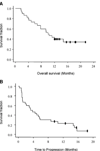
TTP and OS
At a median follow-up of 12 months, 20 patients had died and 26 patients were refractory or had become resistant to gefitinib monotherapy. Median TTP was 3.3 months (range, 0.3–19.6 months) and median OS was 10 months (range, 1.7–21.4 months) (Figure 1. Duration of response for patients with partial response was 5.8 months. OS and TTP were not affected by histologic type, smoking, PS, stage, or prior treatment. However, there was a significant difference in survival in gender (median survival time,⬎12 months in female patients versus 7.7 months in male patients; log-rank test,p⬍ 0.04; Wilcoxon test,p⬍ 0.04).
Tolerability
Table 3 shows drug-related adverse events. Twenty-six patients (86.7%) experienced drug-related adverse events, most of which were mild. Frequent adverse events included diarrhea, skin rash, and elevated transaminases. Twenty-two patients experienced skin toxicities, such as acne, pruritus, and rash. Grade 3 skin toxicities were observed in two
patients, but these resolved spontaneously during treatment. Diarrhea was observed in 12 patients (40.0%) and was con-trolled with antidiarrheal agents such as loperamide. One patient developed grade 3 diarrhea, which required temporal interruption of therapy. Two patients developed drug-related pneumonitis; both were treated with steroid therapy, antibi-otics, and oxygen inhalation and recovered within a few weeks. These patients were smokers and had not received thoracic radiotherapy. No patients experienced hematologic toxicities.
Postgefitinib Treatment
Twenty-five patients became resistant or were refrac-tory to gefitinib monotherapy. Eight of these patients received neither chemotherapy nor radiotherapy because of deteriora-tion of PS in four patients and withdrawal of informed consent to chemotherapy in three patients. One patient un-derwent palliative surgery and two received radiotherapy for symptomatic brain metastases. Fifteen patients received che-motherapy as postgefitinib treatment (platinum-based chemo-therapy in 14 patients and vinorelbine monochemo-therapy in one patient). Five patients achieved PR and four showed SD by the second-line chemotherapy.
EGFR Mutations in Tumor Samples
Twenty tumor samples were obtained from 15 patients retrospectively. Sequencing of exons 18, 19, and 21 inEGFR
was performed in 12 of 20 samples under the same PCR conditions. EGFR mutations were detected in four tumor TABLE 1. Patient Characteristics
Characteristic Value No. of patients 30 Age (yr) Median 64 Range 44–87 Gender Male 18 Female 12 Histology Adenocarcinoma 25 Squamous-cell carcinoma 3 Large-cell carcinoma 2 Stage IIIB 4 IV 26 Metastatic sites Pulmonary 16 Bone 12 Brain 11 Others 4
ECOG performance status
0 20 1 6 2 4 Prior treatment Yes 6 Operation 6 Radiation 3 No 24 Smoking Yes 20 Pack-years (mean⫾SD) 51⫾39 No 10
ECOG, Eastern Cooperative Oncology Group.
TABLE 2. Response to Gefitinib Monotherapy and Prognostic Factors* No. PR SD PD RR (%) p Value DCR (%) p Value Total 30 10 9 11 33.3 63.3 Prognostic factors Gender Male 18 4 4 10 22.2 0.14 44.4 0.018 Female 12 6 5 1 50.0 91.7 Smoking habit Smoker 20 5 5 10 25 0.231 50 0.049 Nonsmoker 10 5 4 1 50 90 Histologic type Adenocarcinoma 25 10 8 7 40 0.139 72 0.327 Nonadenocarcinoma 5 0 2 3 0 40 PS 0–1 26 8 8 10 30.8 0.584 61.5 0.999 2 4 2 1 1 50 75 Clinical stage IIIb 4 2 1 1 50 0.584 75 0.999 IV 26 8 8 19 31 62 Prior treatment Yes 24 9 5 10 37.5 0.999 58.3 0.215 No 6 1 4 1 16.7 83.3
*RR and DCR were compared between prognostic factors using Fisher’s exact test. *PR, partial response; SD, stable disease; PD, progressive disease; RR, response rate; DCR, disease control rate.
samples (33.3%). Three of them had a 15– base pair deletion (E746_A750del) in exon 19. Another of them had L858R in exon 21. The histologic types in patients with EGFR muta-tions were adenocarcinoma in three and large-cell carcinoma in one. All patients with E746_A750del in tumor samples had adenocarcinoma. The responses to gefitinib in these four patients were PR in two, SD in one, and PD in one. There were no responders among nine patients without an EGFR
gene mutation.
EGFR Mutations in Serum Samples
The serum DNA in serum samples from 27 NSCLC patients was examined. Serum DNA was detected in all 54 samples at concentrations of up to 1720 ng/ml.
Exon 19 of EGFRin pretreatment serum samples ob-tained from 21 of 27 patients (77%) was detected (Figure 2
A). The lower band was also detected in 10 of 27 (37%) pretreatment serum samples. Sequencing of the PCR products confirmed that the upper and lower bands corresponded to wild-type and E746_A750del, respectively (Figure 2B). No
point mutation in exon 18, 19, or 21 was detected in the PCR products from serum samples. Wild-typeEGFRwas detected in all 10 of the deletion-positive cases. The pattern of bands was reproducible when using another primer set.13
When compared according to histologic type, E746_A750del was detected in eight of 25 (32%) cases of adenocarcinoma, in zero of three cases of squamous carci-noma, and in two of two cases of large-cell carcinoma (Table 4). In contrast, the serum EGFR status was not correlated statistically with either the clinical response, the gender, or the recorded adverse effects (Table 5).
In serum samples obtained after the initiation of ge-fitinib treatment, 19 of 27 (70%) cases were wild-type– positive and 14 of 27 (52%) cases were deletion-positive (Figure 2 C). In the posttreatment serum samples, E746_A750del was more frequently observed. Furthermore, the deletional mutant of EGFR was significantly more fre-quently observed in samples from patients who showed a PR or SD (12 of 16 cases [75%]) than in samples from patients with PD (two of 11 cases [18%]) (p⫽0.0063, Fisher’s exact test) (Table 6). The deletional mutant EGFR was more frequently detected in female patients (six of nine cases [67%]) than in male patients (eight of 18 cases [44%]), but this difference was not significant (Table 6). No correlations were seen statistically between the presence of mutation and the adverse effects.
FIGURE 1. Kaplan-Meier curve showing (A) overall survival and (B) time to progression in all patients.
TABLE 3. Drug-Related Adverse Events
NCI-CTC Grade No. of Patients %
Diarrhea 1 8 26.7 2 3 10.0 3 1 3.3 Nausea 1 8 26.7 2 2 6.7 3 0 0.0 Vomiting 1 2 6.7 2 0 0.0 3 0 0.0 Skin toxicity 1 15 50.0 2 5 16.7 3 2 6.7 Elevation of transaminases 1 4 13.3 2 1 3.3 3 2 6.7 Pneumonitis 1 0 0.0 2 0 0.0 3 2 6.7
Comparison of EGFRMutation Status between Tumor Samples and Serum Samples
Pairs of tumor samples and serum samples were ob-tained from 12 patients retrospectively (Table 7). TheEGFR
mutation status in the tumors was consistent with those in serum of seven of 12 of the paired samples. Among the other five patients,EGFRmutation was negative in the tumor and positive in the serum in four patients, and in the other patient it was positive in the tumor and negative in the serum, from whose tumor sample L858R was detected.
DISCUSSION
The overall response of 33.3% in this phase II study was comparable not only to that achieved in Japanese popu-lation enrolled in the IDEAL-1 trial (27.5%)7 but also to a
retrospective analysis conducted of patients in Japan.22
Ge-fitinib monotherapy appeared to be equally effective in pa-tients with chemotherapy-naive NSCLC and in papa-tients with pretreated NSCLC.
Drug-related adverse events were generally mild com-pared with cytotoxic chemotherapy. Grade 3 pulmonary tox-icities were observed in two patients. In this study, the FIGURE 2. (A) Detection of
genomicEGFRin the serum of pre-treatment patients. (B) The se-quences of the PCR products from patient 19 (days 0 and 14) are shown. (C) PCR of the serum sam-ples obtained on day 14. Serum-derived genomic DNA PCR was performed. Exon 19 ofEGFRin se-rum obtained from the patients was amplified by PCR, and the products were detected using a Bioanalyzer. A second round of PCR (20 cycles) was performed when no band was detected in the first round of PCR (30 cycles). Row numbers indicate the patient num-ber. *Band detected in the first round of PCR.
TABLE 4. Frequency of SerumEGFRin Lung Cancer Patients According to Histology and Response to Gefitinib*
Pre Post
Wild Deletion Wild Deletion
Adenocarcinoma 18/23 8/23 15/22 13/22 Squamous-cell carcinoma 1/2 0/2 3/3 0/3 Large-cell carcinoma 2/2 2/2 1/2 1/2
*A total of 27 samples were obtained from 28 patients both before and after treatment. A pretreatment sample of patient 2 and a posttreatment sample of patient 17 were lacking.
TABLE 5. Frequency of SerumEGFRin Lung Cancer Patients According Response to Gefitinib and Gender: Detection of Deletion-Type Mutation on Day 0*
ⴙ – pValue Response PR/SD 8 9 PD 2 8 0.2305 Gender Male 5 12 Female 5 5 0.4153
*A total of 27 samples were obtained from 28 patients both before and after treatment. A pretreatment sample of patient 2 and a posttreatment sample of patient 17 were lacking. SD, stable disease; PD, progressive disease; PR, partial response;⫹, deletion-positive; –, wild-type.
TABLE 6. Frequency of SerumEGFRin Lung Cancer Patients According to Response to Gefitinib and Gender: Detection of Deletion-Type Mutation on Day 14*
ⴙ – pValue Response PR/SD 12 4 PD 2 9 0.0063 Gender Male 8 10 Female 6 3 0.4197
*A total of 27 samples were obtained from 28 patients both before and after treatment. A pretreatment sample of patient 2 and a posttreatment sample of patient 17 were lacking. SD, stable disease; PD, progressive disease; PR, partial response;⫹, deletion-positive; –, wild-type.
incidence of drug-related pneumonitis was 6.7% and was comparable to results of other studies.23,24 Therefore,
ge-fitinib monotherapy as a first-line treatment appears to be equally tolerable as a second-line treatment.
Thirteen of 22 patients who became resistant or were refractory to gefitinib monotherapy received salvage chemo-therapy. The objective response rate was 30.8%, comparable to that of first-line chemotherapy. These results suggest that cancer cell populations that are sensitive to gefitinib might not be identical to those sensitive to chemotherapeutic drugs such as platinum agents or taxanes.
Somatic mutations in the tyrosine kinase domain of the
EGFR gene were reported, and these mutations induced increased activity ofEGFRand sensitivity to gefitinib in vitro and the predictive factor of response to gefitinib.13,14 We
evaluated EGFR gene status in 13 tumor samples and de-tected EGFR gene mutation in four tumors. Objective re-sponses were achieved in two patients, but one patient showed PD whose tumor had a 15– base pair deletion muta-tion in exon 19. This suggested that response to gefitinib may not be determined byEGFRmutation in exon 19 or 21, and other mechanisms may relate to gefitinib resistance.
The detection of EGFR mutation from serum samples was carried out as a correlative study. These results provided us two major findings: (1) E746_A750del was detectable in serum sample obtained from NSCLC patients; and (2) E746_A750del was frequently observed in posttreatment se-rum samples obtained from the PR and SD patients.
It may be explained that DNA derived from destructive tumor cells that have responded to gefitinib may be more frequently observed in the circulating blood. Previous reports regarding detection of mutations in serum did not elucidate the changes in mutation status during treatment. We would like to do this in the next experiments to confirm our
specu-lation. Our hypothesis is that serum detection of EGFR
mutation will be a convenient means of predicting the sensi-tivity to gefitinib, although we could only demonstrate the feasibility of theEGFRmutation in serum in this report. We need to develop a highly sensitive methodology to improve the predictability of this assay.
In comparison of the mutation status ofEGFRin actual tumors with serum DNA obtained from the same patients before treatment, 70% of patients who had sequence data obtained from both serum and tumor samples were conform-ing. Esteller et al. reported detection of aberrant promoter hypermethylation of tumor suppressor genes (p16, DAP,
GSTP1, andMGMT) in serum DNA obtained from NSCLC patients and demonstrated that 73% of serum samples showed abnormal methylated DNA in the patients with the methyl-ated primary tumors.19Another report investigating a point
mutation of the p53 gene and hypermethylation of p16 in plasma DNA from breast cancer patients demonstrated that 66% of the patients with at least one molecular event in tumor DNA had some alteration in plasma DNA.25We believe that
the sensitivity of our assay is equivalently sensitive to those of these previous reports.
CONCLUSION
In conclusion, 250 mg of oral gefitinib monotherapy as a first-line treatment produces obvious antitumor activity, with acceptable toxicities. Oral gefitinib monotherapy as a first-line treatment merits investigation in further clinical trials. Using serum samples from NSCLC patients, theEGFR
mutation was detected. The detection of E746_A750del in the serum of untreated patients was not a predictor of gefitinib response in this study. However, further prospective studies using serum samples may be necessary to confirm this con-TABLE 7. EGFRMutation Status in Tumor Samples and Serum Samples*
EGFR Mutation Status Serum Samples
Pre Post
No. Gender Histology Response Tumor Sample Wild Mutation Wild Mutation
43 M Large SD Wild ⫹ ⫹ – ⫹ 45 M SCC PD Wild ND ND ⫹ – 52 F SCC PD Wild ⫹ – ⫹ – 53 M Adeno PD Wild – – ⫹ – 55 M Adeno PR L858R ⫹ ⫹ – – 57 F Adeno SD Wild – – ⫹ ⫹
61 M Large PD E746-A750 del ⫹ ⫹ ⫹ –
64 M Adeno PD Wild ⫹ – ⫹ –
70 M Adeno PD Wild ⫹ ⫹ ⫹ –
72 M Adeno SD E746-A750 del ⫹ – – ⫹ 75 F Adeno PR E746-A750 del ⫹ ⫹ ⫹ ⫹
77 M Adeno PD Wild ⫹ – ⫹ ⫹
*Pairs of both tumor samples and serum samples were obtained from 12 patients. M, male; F, female; SD, stable disease; PD, progressive disease; PR, partial response; SCC, squamous-cell carcinoma; Adeno, adenocarcinoma; Large, large-cell carcinoma; ND, not determined.
clusion. The presence ofEGFRmutation in serum may be a useful biomarker for monitoring gefitinib response.
ACKNOWLEDGMENTS
We thank the patients who enrolled into the trial and the investigators who enrolled them.
REFERENCES
1. Parkin DM, Bray F, Ferlay J, et al. Estimating the world cancer burden: Globocan 2000.Int J Cancer2001;94:153–156.
2. Roskoski R Jr. The ErbB/HER receptor protein-tyrosine kinases and cancer.Biochem Biophys Res Commun2004;319:1–11.
3. Scagliotti GV, Selvaggi G, Novello S, et al. The biology of epidermal growth factor receptor in lung cancer. Clin Cancer Res 2004; 10: 4227s–4232s.
4. Rusch V, Baselga J, Cordon-Cardo C, et al. Differential expression of the epidermal growth factor receptor and its ligands in primary non-small cell lung cancers and adjacent benign lung.Cancer Res1993;53: 2379–2385.
5. Rusch V, Klimstra D, Venkatraman E, et al. Overexpression of the epidermal growth factor receptor and its ligand transforming growth factor alpha is frequent in resectable non-small cell lung cancer but does not predict tumor progression.Clin Cancer Res1997;3:515–522. 6. Fontanini G, De Laurentiis M, Vignati S, et al. Evaluation of epidermal
growth factor-related growth factors and receptors and of neoangiogen-esis in completely resected stage I-IIIA non-small-cell lung cancer: amphiregulin and microvessel count are independent prognostic indica-tors of survival.Clin Cancer Res1998;4:241–249.
7. Fukuoka M, Yano S, Giaccone G, et al. Multi-institutional randomized phase II trial of gefitinib for previously treated patients with advanced non-small-cell lung cancer.J Clin Oncol2003;21:2237–2246. 8. Kris MG, Natale RB, Herbst RS, et al. Efficacy of gefitinib, an inhibitor
of the epidermal growth factor receptor tyrosine kinase, in symptomatic patients with non-small cell lung cancer: a randomized trial. JAMA
2003;290:2149–2158.
9. Herbst RS, Giaccone G, Schiller JH, et al. Gefitinib in combination with paclitaxel and carboplatin in advanced non-small-cell lung cancer: a phase III trial—INTACT 2.J Clin Oncol2004;22:785–794.
10. Giaccone G, Herbst RS, Manegold C, et al. Gefitinib in combination with gemcitabine and cisplatin in advanced non-small-cell lung cancer: a phase III trial—INTACT 1.J Clin Oncol2004;22:777–784. 11. Thatcher N, Chang A, Parikh P, et al. Gefitinib plus best supportive care
in previously treated patients with refractory advanced non-small-cell lung cancer: results from a randomised, placebo-controlled, multicentre
study (Iressa Survival Evaluation in Lung Cancer).Lancet2006;366: 1527–1537.
12. Johnson DH. Gefitinib (Iressa) trials in non-small cell lung cancer.Lung Cancer2003; 41 (Suppl 1): S23–S28.
13. Lynch TJ, Bell DW, Sordella R, et al. Activating mutations in the epidermal growth factor receptor underlying responsiveness of non-small-cell lung cancer to gefitinib.N Engl J Med2004;350:2129–2139. 14. Paez JG, Janne PA, Lee JC, et al. EGFR mutations in lung cancer: correlation with clinical response to gefitinib therapy.Science2004;304: 1497–1500.
15. Pao W, Miller V, Zakowski M, et al. EGF receptor gene mutations are common in lung cancer from “never smokers” and are associated with sensitivity of tumors to gefitinib and elrotinib.Proc Natl Acad Sci USA
2004;101:13306–13311.
16. Shigematsu H, Lin L, Takahashi T, et al. Clinical and biological features associated with epidermal growth factor receptor gene mutations in lung cancers.J Natl Cancer Inst2004;97:339–346.
17. Han SW, Kim TY, Hwang PG, et al. Predictive and prognostic impact of epidermal growth factor receptor mutation in non-small-cell lung cancer patients treated with gefitinib.J Clin Oncol2006;23:2493–2501. 18. Sanchez-Cespedes M, Monzo M, Rosell R, et al. Detection of
chromo-some 3p alterations in serum DNA of non-small-cell lung cancer patients.Ann Oncol1998;9:113–116.
19. Esteller M, Sanchez-Cespedes M, Rosell R, et al. Detection of aberrant promoter hypermethylation of tumor suppressor genes in serum DNA from non-small cell lung cancer patients.Cancer Res1999;59:67–70. 20. Therasse P, Arbuck SG, Eisenhauer EA, et al. New guidelines to
evaluate the response to treatment in solid tumors. European Organiza-tion for Research and Treatment of Cancer, NaOrganiza-tional Cancer Institute of the United States, National Cancer Institute of Canada.J Natl Cancer Inst2000;92:205–216.
21. Simon R. Optimal two-stage designs for phase II clinical trials.Control Clin Trials1989;10:1–10.
22. Takano T, Ohe Y, Kusumoto M, et al. Risk factors for interstitial lung disease and predictive factors for tumor response in patients with advanced non-small cell lung cancer treated with gefitinib.Lung Cancer
2004;45:93–104.
23. Takeda K, Yamamoto N. An epidemiological survey for interstitial lung disease induced by gefitinib in patients with advanced non-small cell lung cancer.Lung Cancer2003;41:S250.
24. Hotta K, Harita S, Bessho A, et al. Interstitial lung disease (ILD) during gefitinib treatment in Japanese patients with non-small cell lung cancer (NSCLC): Okayama Lung Cancer Study Group. Proc Am Soc Clin Oncol2004;23:629.
25. Silva JM, Dominguez G, Garcia JM, et al. Presence of tumor DNA in plasma of breast cancer patients: clinicopathological correlations. Can-cer Res1999;59:3251–3256.