Title
Ribosome profiling reveals the what, when, where and how of protein synthesis.
Permalink
https://escholarship.org/uc/item/78t9g779
Journal
Nature reviews. Molecular cell biology, 16(11)
ISSN
1471-0072
Authors
Brar, Gloria A
Weissman, Jonathan S
Publication Date
2015-11-01
DOI
10.1038/nrm4069
Peer reviewed
Translation, which is the process by which a ribosome reads an mRNA template to guide protein synthesis, is a crucial step in gene expression. Translation is energeti-cally costly and is therefore tightly regulated to conserve cellular resources, as well as to avoid mistakes that may result in the production of toxic proteins. Indeed, a wide range of disease states, including neurodegeneration, anaemia and specific developmental defects, result when the translational process is compromised (see selected REFS 1–6). Although much is known about the struc-ture and function of the ribosome, our understanding of many aspects of the regulation of translation has been far more limited.
Efforts to globally monitor gene expression have historically focused on measuring mRNA levels (for example, using microarrays or RNA sequencing (RNA-seq)), although we know that translational control is an essential and regulated step in determining levels of protein expression. Until recently, precisely monitoring translation was far more challenging than was measur-ing mRNA levels. This has changed with the develop-ment of the ribosome profiling approach, which was first described in 2009 (REF. 7).
Ribosome profiling is a deep-sequencing-based tool that facilitates the detailed measurement of translation globally and in vivo7. At the core of this approach is the
observation that a translating ribosome strongly protects about 30 nucleotides of an mRNA from nuclease activ-ity8,9. Sequencing of these ribosome-protected fragments,
termed ribosome footprints, thus provides a precise record
of the position of the ribosome at the time at which trans-lation was halted. Measuring the density of protected fragments on a given transcript provides a proxy for the rate of protein synthesis. In addition, determining the positions of the protected fragments makes it possible to empirically measure the identity of translation products (for example, where they begin and end and even the frame being read). This has led to the discovery of many novel or alternative protein products10–19. The
distribu-tion of ribosome footprints can provide insights into the mechanism of translational control (for example, it can be used to identify regulatory translational pauses and translated upstream open reading frames (uORFs)). Finally,
novel adaptations of the ribosome profiling approach make it possible to monitor translation mediated by sub-sets of ribosomes on the basis of their physical location in the cell or their interaction partners.
Here, we discuss the principles of the ribosome pro-filing approach, its strengths and limitations, and recent examples in which it has guided biological discovery. We focus on the value of ribosome profiling as a tool to interrogate what is being translated, how this transla-tion is regulated and where in the cell the translatransla-tion of specific sets of proteins occurs.
What is ribosome profiling and what can it reveal?
Ribosome profiling exploits the classical molecular method of ribosome footprinting8,9, in which in vitro
translated mRNAs are treated with nuclease to destroy the regions that are not protected by the ribosome7,9.
1Department of Molecular and Cell Biology, University of California, Berkeley, California 94720, USA. 2Howard Hughes Medical Institute, Department of Cellular and Molecular Pharmacology, University of California, San Francisco, California 94158, USA. 3Center for RNA Systems Biology, University of California, Berkeley, California 94720, USA. 4California Institute for Quantitative Biosciences (QB3), University of California, San Francisco, California 94158, USA. e-mails: gabrar@berkeley.edu; jonathan.weissman@ucsf.edu doi:10.1038/nrm4069 Published online 14 October 2015 Ribosome footprints mRNA fragments of ~30 nucleotides that result from nuclease treatment of translating ribosomes. These are mRNA regions that are protected by the ribosome as the mRNA is decoded to a protein sequence.
Ribosome profiling reveals the what,
when, where and how of protein
synthesis
Gloria A. Brar
1,3,4and Jonathan S. Weissman
2–4Abstract | Ribosome profiling, which involves the deep sequencing of ribosome-protected
mRNA fragments, is a powerful tool for globally monitoring translation
in vivo
. The method
has facilitated discovery of the regulation of gene expression underlying diverse and
complex biological processes, of important aspects of the mechanism of protein synthesis,
and even of new proteins, by providing a systematic approach for experimental annotation
of coding regions. Here, we introduce the methodology of ribosome profiling and discuss
examples in which this approach has been a key factor in guiding biological discovery,
including its prominent role in identifying thousands of novel translated short open reading
frames and alternative translation products.
T E C H N O LO G I E S A N D T E C H N I Q U E S
Upstream open reading frames
(uORFs). ORFs in the 5′ leader region of a characterized mRNA transcript. Translation of uORFs may regulate translation of a downstream ORF. Ribosome profiling allows for the empirical identification of all translated uORFs in vivo
under a condition of interest. Although uORFs are short, here we do not include them in the class of ‘short ORFs’, which are on an mRNA that was not previously thought to encode a protein.
Such treatment leaves ‘footprints’ of ~30 nucleotides, which can be mapped back to the original mRNA to define the exact location of the translating ribosome. Ribosome profiling extends this method by mapping and measuring the full complement of in vivo ribosome footprints to quantify new protein synthesis and to anno-tate coding regions globally7,10–12 (FIGS 1,2). Extraordinary
advances in sequencing technology20 now make it possible
to deeply sample all translating ribosomes. In mammalian cells, for example, which encode ~20,000 proteins with an average mRNA coding region of ~500 nucleotide triplets, nuclease digestion of all translating ribosome–mRNA complexes yields 10 million possible footprints. The bil-lions of reads that are now possible with next-generation sequencing enable the reliable quantification of the set of footprints tiling across all but the rarest mRNAs, and a recently developed kit facilitates sample preparation21,22.
With such easily attainable and quantitative information,
ribosome profiling has a range of uses, from a broad pro-teomic tool to a specific probe of translation in an in vivo setting, and as a valuable complement to mRNA-seq.
Ribosome profiling requires collection of a physio-logical sample; inhibition of translation to freeze ribo-somes in the act of translation; nuclease digestion to produce ribosome-protected fragments; and isolation of ribosomes and, subsequently, of ribosome footprints21.
Ribosome footprints are converted to a strand-specific library and subjected to next-generation sequencing, and the fragments are then mapped to the appropriate reference genome. Ribosome profiling is typically car-ried out on a split sample, with parallel libraries con-structed for measuring mRNA abundance by mRNA-seq. Comparison between the rates of protein synthesis and the abundance of mRNAs makes it possible to determine the translational efficiency for each mRNA7(FIGS 1a,b;2b,c).
The common biophysical properties of the ribosome and
AUG Stop Nuclease treatment AAAAAAAAAA AAAAAAAAA AAAAAAAAAA AAAAAAAAAAA AAAAAAAAAAAA Random fragmentation Coding region Codon periodicity 3′ UTR 5′ leader Ribosome footprint r eads Ribosome footprint r eads Genomic position mRNA-seq r eads 5′ transcript end (often indicates transcription start site)
3′ transcript end (often indicates transcription stop site) Genomic position
Genomic position
b mRNA-seq c
In vivo capture of translating ribosomes and mRNAs, lysis
Ribosome footprints a Ribosome profiling AAAAAAAAAA AAAAAAAAA AAAAAAAAAA AAAAAAAAAAA AAAAAAAAAAAA AAAAAAAAAA AAA AAAAAAAA A AAAA Library generation Deep sequencing Read mapping Library generation Deep sequencing Read mapping Start Stop
High % of ORF covered by ribosome footprints
A A G C T G C T TA C G A C C T G C AT G C A G
Nature Reviews |Molecular Cell Biology
Cell type of interest
Low density of footprints before start codons
and after stop codons (high inside/out ratio)
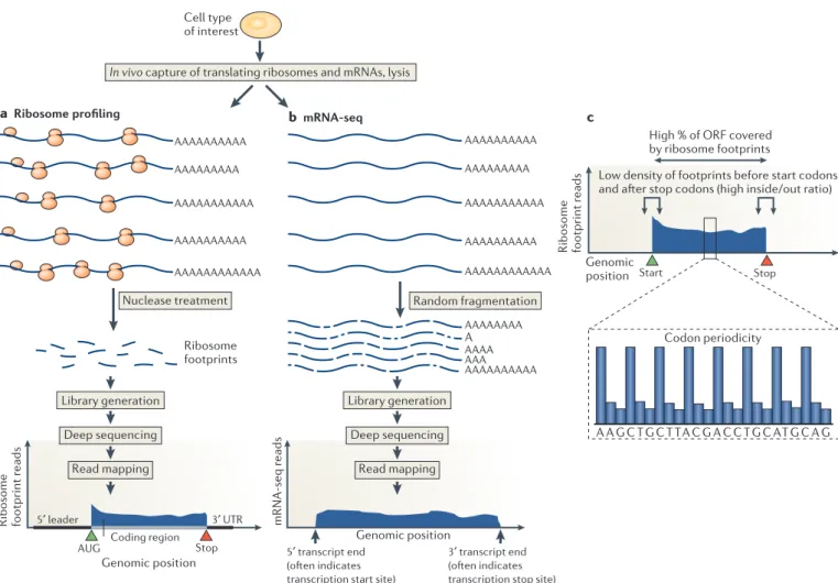
Figure 1 | An overview of ribosome profiling. a | Ribosome-bound mRNAs are isolated by size and treated with a nonspecific nuclease (typically RNase I or micrococcal nuclease), resulting in protected mRNA fragments termed ‘footprints’. These ribosome footprints are isolated and converted to a library for deep sequencing. The ribosome footprints typically show precise positioning between the start and the stop codon of a gene, which facilitates global and experimental identification of genomic coding regions. b | By comparison, mRNA sequencing (mRNA-seq) captures random fragments covering the entire mRNA transcript. The positional information determined by standard mRNA-seq
allows approximate determination of transcript boundaries, but it is less precise than that collected by ribosome profiling, owing to the loss of 5ʹ
and 3ʹ ends during the fragment generation method that is typically used. c | Translated open reading frames (ORFs) contain a stereotyped organization of ribosome footprints. Ribosome footprint density over ORFs begins sharply at the start codon, ends sharply at the stop codon and shows evidence of codon periodicity. True translated regions tend to show ribosome footprint coverage over the majority of the ORF and not typically in the regions before the putative start codon and after the putative stop codon. UTR, untranslated region.
the lack of genetic manipulation that is required for this approach make ribosome profiling highly adaptable to cells or tissues from essentially any organism, with mod-est modifications. Organisms that have been invmod-estigated thus far by ribosome profiling include a variety of bacte-ria, yeast, parasitic protozoa, zebrafish, flies, nematodes,
mice, rats, plants, viruses and human cells7,10–12,19,23–30.
Even mitochondrial translation within human cells has been effectively assayed by this method31, and a similar
approach has been applied to chloroplasts in plant cells32.
Many of these data sets have been compiled and made readily accessible for data mining and comparison33.
Nature Reviews |Molecular Cell Biology
AAAAAAAAAA AAAAAAAAAAA AAAAAAAAAAAA Ribosome footprint reads AAAAAAAAAAAA AAAAAAAAA AAAAAAAAAAA a b c AAAAAAAAAA AAAAAAAAAAAA mRNA-seq reads Gene annotations Ribosome footprints ORF mRNA ORF ORFs ORFs
Ribosome footprint counts
Protein synthesis rate (instantaneous)
mRNA counts
ORFs Transcript abundance
(steady state) Translation efficiency(relative)
uORF Translation pausing sORF Tr anslation efficiency =
Sample pool of mRNAs
Genome browser plot
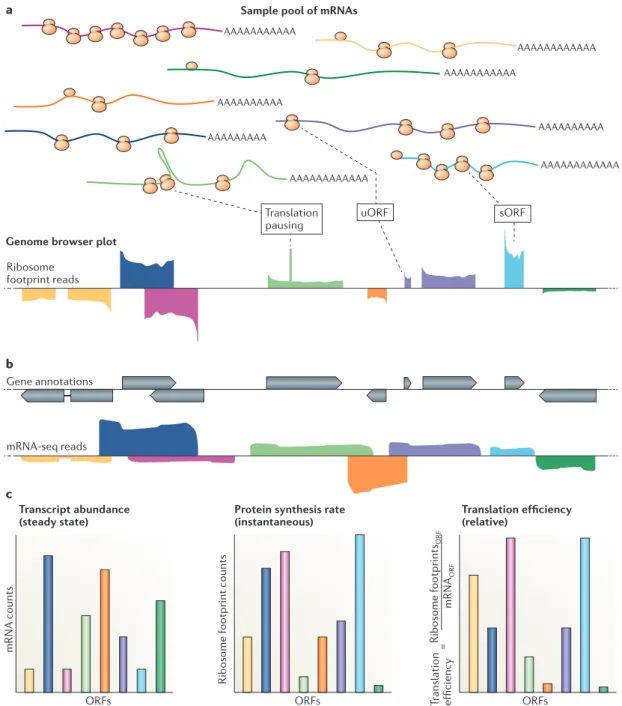
Figure 2 | Qualitative and quantitative data provided by ribosome profiling. a | A diverse sample pool of mRNAs, distinguished by colour, is shown, together with a corresponding representative genome browser plot of ribosome profiling data derived from this pool. Note that ribosome profiling facilitates experimental determination of translated regions, including short open reading frames (sORFs), which may be an important newly identified source of cellular peptides, and upstream ORFs (uORFs), which are thought to be largely regulatory. Pausing during translation elongation may result in peaks in ribosome footprint reads within ORFs. b | Overlaid gene annotations and mRNA sequencing (mRNA-seq) data for the examples in part a are shown. c | The graphs show examples of quantitative data derived from parts a and b. Note that transcript abundances may not correlate closely with the instantaneous protein synthesis rates. The collection of quantitative data for both transcript abundances and protein synthesis rates enables the relative translation efficiencies to be inferred. These can vary over several orders of magnitude within a given organism in a given state. The translation efficiencycan also change over time for a given mRNA, reflecting dynamic regulation at the level of translation.
Polysome gradients A method for fractionating ribosomes that are bound to mRNAs by velocity centrifugation of cell extract on sucrose gradients, allowing for the separation of mRNAs that are associated with one ribosome (monosome) from those being translated by multiple ribosomes (polysome). Sucrose gradient fractionation facilitates qualitative analysis of the translation status of cells. Ribosome P site The site within an actively translating ribosome that is usually associated with the tRNA attached to the growing peptide chain.
Codon periodicity The three-nucleotide pattern of ribosome occupancy, reflecting mRNA translocation in the ribosome by codon as translation occurs.
What are the strengths of ribosome profiling?
Despite its recent development, ribosome profiling has rapidly become a widely used tool for understand-ing diverse and complex biological issues. Three key features, which are outlined below, have facilitated the broad utility of this method.
Sensitivity and precision of quantification. Ribosome profiling provides a large dynamic range for the detec-tion and quantificadetec-tion of transladetec-tion in unperturbed cells. The sensitivity of the method, which results from the depth of sampling that is possible in sequencing studies, facilitates the measurement of even relatively rare translation events, with the range of detection generally limited only by the counting variability that is seen with very low numbers of sequencing reads. Complementary methods, including pulsed label-based mass spectrometry, analyses of transcript distributions on polysome gradients and 35S Met-based metabolic
label-ling, enable sensitive measurement of new protein syn-thesis; however, the highly parallel sequencing readout of all ribosome positions that is provided by ribosome profiling typically yields more quantitative and detailed information than is currently accessible by alternative methods.
Precision of positional information. In addition to its broad dynamic range of detection, ribosome profiling provides uniquely rich and precise positional informa-tion. The almost universal biophysical properties of ribo-somes across species yield a characteristic footprint size that allows prediction of the codon in the r ibosome P site
(that is, the position of peptide bond formation) and the detection of codon periodicity7(FIG. 1c). Analyses of
ribo-some footprint positions can be used to mechanistically probe aspects of translation, thus far identifying many novel examples of ribosomal frameshifting, stop codon readthrough, ribosome pausing, translation initiation at non-AUG codons and uORF translation7,10,12,34–37(FIG. 2a).
Furthermore, years after many genomes were originally annotated, the precise positional information obtained from ribosome profiling experiments has provided the first opportunity to experimentally define translated ORFs11–13,38,39(FIGS. 1,2), resulting in the identification of
new classes of coding regions in diverse organisms. Instantaneous measurements. A final valuable property of ribosome profiling is the instantaneous nature of the information that is collected, which reflects a snapshot of the dynamic process of translation. Although mRNA-seq and standard genome-scale mass spectrometry experi-ments are valuable in following gene expression globally, these widely used measurements report on steady-state levels of mRNA and protein, respectively. This informa-tion is important, but it may not reflect the rapid cellular decision making that accompanies developmental transi-tions and environmental responses. Ribosome profiling enables sensitive detection of changes in cellular protein expression as they occur7,12,40. The common, quantitative
output from ribosome profiling and mRNA-seq further allows for direct comparison of instantaneous protein
synthesis and steady-state transcript levels, providing an opportunity to quantify in vivo translation efficiencies in detail (FIG. 2c).
What are the limitations of the method?
We discuss below notable weaknesses and caveats of ribosome profiling that should be considered when using the method or interpreting data derived from its use.
Experimentally introduced distortions. The key techni-cal challenge of ribosome profiling is the need to rapidly inhibit translation to capture a snapshot of ribosomes in a particular physiological state. The reliability of this step is particularly important for any analyses of translation pausing, as the fast rate of translation elongation may result in signal blurring or the artificial accumulation of ribosomes at specific positions if inhibition is slow. The use of a translation elongation inhibitor (such as cycloheximide) can be valuable; however, it is clear that such inhibitors can alter the local distributions of ribo-somes on an mRNA, especially near translation start sites7,18,21,41. Although this does not seem to interfere with
the global measurements of the density of ribosomes on an mRNA that are used to determine rates of protein synthesis, it can cause spurious peaks of ribosome bind-ing at particular sites. Thus far, flash freezbind-ing has been the most robust approach in a wide range of diverse organisms and has enabled the physiological capture of local and global ribosome distributions21. In general,
each experimental step — from cell harvesting to nucle-ase digestion to library generation — has the potential to cause distortions in the data output. These distor-tions must be accounted for carefully, as the degree to which any given distortion might be problematic will depend strongly on the questions being addressed and the syste m being probed.
The need to infer protein synthesis rates. A caveat to con-sider when interpreting ribosome profiling data is that rates of synthesis are typically inferred from the aver-age ribosome density along the mRNA in question. The accuracy of this measure depends on the premise that all ribosomes finish translation and that, on average, the translation elongation rate is similar among different mRNAs in a cell. These assumptions can be tested and are appropriate for a wide range of conditions, but this will not always be the case. Known exceptions42–44 — including
the build-up of ribosomes at and immediately proximal to the start codon in a partially cycloheximide-dependent manner7 or regulated translation pausing and abortion
under starvation conditions45 — can be corrected for to
increase measurement accuracy, but there may be cases in which these and other, currently unknown, exceptions pose challenges for proper data analysis.
Contaminating footprint-sized fragments. Another important issue for ribosome profiling experiments is that footprints are inferred on the basis of their size and their association with assembled (80S) ribosomes. Contaminating RNA fragments, including those from
Fragment length
organization similarity score (FLOSS) analysis
A metric for determining the probability that ribosome footprints over a given region (or set of regions) result from translation. This analysis involves comparing size distributions of footprints over a query region and over validated coding regions and is based on the concept that the biophysical properties of translating ribosomes result in characteristic signatures in ribosome footprint sizes.
structured non-coding RNAs or large ribonucleopro-tein complexes that co-migrate in a sucrose gradient with the ribosome, may be processed with a ribosome profiling library and provide false readouts of trans-lation (see Supplementary information S1 (figure)). A recent approach, termed fragment length organization similarity score (FLOSS) analysis, aims to identify such
fragments and remove them post-experimentally (that is, in silic o)39. FLOSS analysis is based on the observation
that bona fide ribosome footprints have stereo typical distributions of footprint sizes (see Supplementary information S1 (figure, parts a and b)). The distribution of typical 80S footprint sizes used in FLOSS analysis is empirically measured for each experiment, by exam-ining the sizes of footprints in that same experiment from known protein-coding regions, and can be used to computationally identify contaminating fragments for removal. Nonetheless, there are examples in which genu-ine 80S mRNA footprints do not conform to the typical size pattern. Two recent cases that highlight interesting biology that was determined by analysis of alternatively sized ribosome footprints indicate effects that are due to both alternative ribosome conformations46 and
alter-native mRNA properties41 (see below). Nuclease
protec-tion assays can be a useful adjunct control for identifying the full range of ribosome footprint sizes in a new organ-ism or condition, thus informing the design of a ribo-some profiling experiment to best capture all translating ribosome s in a given system.
Ribosomal RNA (rRNA) fragments commonly result from the nuclease-treatment step of ribosome profiling and may substantially decrease the ribosome footprint sequencing space in a ribosome profiling experiment7,
particularly under conditions in which global translation levels are low. Whereas mRNA-seq often uses poly(A) selection as an effective method for the isolation of desired sequences, this approach is not possible with ribosome profiling. Selective subtraction of ribosomal fragments, however, is highly effective and is recom-mended, particularly for samples in which a small num-ber of specific footprint-sized rRNA fragments are seen as contaminants21.
Mapping ambiguous reads. A general challenge in the analysis of sequencing data is determining the correct alignment position for reads from repetitive or highly similar regions, such as gene families, or from alternative transcript variants. In the case of genome sequencing or mRNA-seq, longer reads or paired-end47 approaches can
help to resolve such ambiguities, but the inherently short size of ribosome footprints precludes these experimental approaches. However, computational methods that have been developed for mRNA-seq data to assign multiply mapping reads in a probabilistic manner on the basis of overall read distributions48 can be applied to ribosome
profiling data to mitigate this limitation.
Material quantities. Currently, the main limitation of ribosome profiling compared with mRNA-seq approaches is the requirement for relatively large sam-ples. In contrast to mRNA-seq49, ribosome profiling
cannot yet be applied to single cells. This limitation results from the extra processing step that is required to isolate ribosomes21, as well as the small proportion of
any given mRNA molecule that is being translated at any given instant and thus recoverable as footprints (FIG. 1a). It is likely that the types of technical advances that have greatly enhanced the sensitivity of RNA-seq approaches to small cell numbers will also be applicable in the future to ribosome profiling, although no such major effort has yet been undertaken.
Insights provided by ribosome profiling
With these advantages and disadvantages in mind, the application of ribosome profiling to specific biologi-cal questions has confirmed much of what we know about translation mechanism from decades of elegant structural, biochemical and genetic studies50. Ribosome
profiling has also made it possible to monitor transla-tion with unprecedented depth and precision, providing important — and at times surprising — insights. The application of this method to numerous organisms and cellular states has illuminated fundamental aspects of cell biology that were previously challenging to probe experimentally, providing measurements for how much of each protein is synthesized, how translation is regu-lated, where synthesis starts and stops and what is being synthesized.
How much? A quantitative view of protein synthesis. The simplest and broadest application of ribosome profiling is as a quantitative proteomics tool to monitor which proteins are being synthesized, and at what levels, thus providing rich molecular insight into a given cell state. Ribosome footprint density reflects the number of ribo-somes at a given position. Assuming that the average translation elongation rate is similar for different genes, ribosome profiling provides direct, global and quantita-tive measurements of rates of protein synthesis, thereby capturing information that has been largely invisible to gene expression measurements of mRNA levels alone. Mass spectrometry can, in principle, be used to measur e rates of protein synthesis; however, this is technically difficult, as it typically requires metabolic labelling and multiple measurements per sample. Analysis of the positions of mRNAs in polysome gradients provides valuable complementary information to that obtained with ribosome profiling, but again, this method is labo-rious and typically yields only a qualitative measure of protein synthesis.
In many cases, the ability to observe new protein syn-thesis globally and quantitatively provides insights that are not apparent from measurements of mRNA abun-dance. Bacterial operons provide a vivid example of the value of being able to directly measure rates of protein synthesis. As is the case for many protein complexes in bacteria, the eight different subunits of the FoF1-ATP
syn-thase are expressed from a single polycistronic mRNA, and thus measurements of mRNA levels would suggest that the subunits are all expressed at very similar levels. Ribosomal profiling, however, shows that the individual ORFs that encode the subunits of the FoF1-ATP synthase
operon are translated at a ratio of 1:1:1:1:2:3:3:10. Remarkably, these ratios precisely reflect the stoichiome-try of these components in the ATP synthase51,52(FIG. 3a).
This property of proportional synthesis, by which sub-units of multiprotein complexes are synthesized at rates that are proportional to their stoichiometry in the com-plex, turns out to be generally true for Escherichia coli and was also observed for some (but not all) complexes in budding yeast (Saccharomyces cerevisiae). Such meas-urements of instantaneous rates of protein synthesis may prove to be a general tool for exploring how proteins assemble and function together51.
Quantitative measurement of protein synthesis rates over multiple time points of a dynamic process can also provide information about specific gene function. For example, hierarchical clustering of patterns of new pro-tein synthesis for each gene over the dynamic process of meiosis in budding yeast resulted in an intricate map of gene expression that provided highly detailed functional
information12. In these data, the genes responsible for
the complex, conserved and meiosis-specific processes of homologous recombination and synaptonemal com-plex assembly emerged as a single cluster of 46 genes. This observation was surprising, because these pro-cesses are known to be regulated extensively at the post-translational level, and also because the cluster included almost every gene that had been found through decades of intensive genetic and cytological screening focused on these processes. This cluster also included several uncharacterized genes, two of which (GMC1 and GMC2) were subsequently shown to have roles in recombination and synaptonemal complex formation12,53.
Another striking recent example of this type of analysis used ribosome profiling to identify the factors that are responsible for initiation of the zygotic develop-mental programme in zebrafish54(FIG. 3b). The initiation
of zygotic development in vertebrates depends heavily on translational control, as maternal mRNAs provide the starting pool of material for translation. Zygotic activation then requires destruction of these mater-nal mRNAs and transfer of developmental control to the zygote itself. To determine the factors that medi-ate the first wave of zygotic transcription, ribosome profiling data were analysed for samples collected just before zygotic activation. This study identified Nanog, Sox19b and Pou5f1 as the three transcription factors that were most heavily translated, from the large pool of maternal mRNAs at this stage (FIG. 3b). Subsequent morpholino knockdown experiments showed that specifically blocking translation of these three factors resulted in a shutdown of the first wave of zygotic tran-scription and development, indicating that they are the key factors responsible for the initiation of the zygotic developmenta l programme54.
Other recent studies in disparate systems — from the Drosophila melanogaster oocyte-to-embryo transi-tion55 to the Trypanosome life cycle56 to the
mamma-lian cell cycle57 to plants under hypoxic conditions27
— have used ribosome profiling to identify specific proteins that drive these complex processes. Cases in which ribosome profiling data provide markedly dif-ferent information than can be obtained by traditional mRNA abundance measurements for gene expression tend to fall into two categories: systems in which tran-scriptional regulation is minimal26,54,55; and dynamic
cellular programmes11,12,27,35,57–59. The latter category
includes cellular differentiation, organismal develop-ment and dynamic responses to cellular stress, which are all cases in which the instantaneous and downstream gene expression measurements provided by ribosome profiling are particularly illuminating for understanding molecular control.
How? Insights into the mechanism of translational control. The basic mechanism by which the riboso-mal machinery reads codon information in mRNAs to create proteins is conserved, and many features of this process are well understood50. Nonetheless, there are
aspects of translational control that are not amenable to recapitulation in vitro and for which results from Nature Reviews |Molecular Cell Biology
a E. coli FoF1-ATP synthase operon
B E F H A G D C mRNA-seq reads 10 3 1 2 Ribosome footprint reads
b D. rerio zygotic development
Maternal mRNAs by rank
Ribosome footprint abundance (2 hpf)
5,000 10,000
Synthesis r
ate
(molecules per gener
ation)
Stoichiometry of protein in complex 0 0 4 8 10 104 6 2 BCGH AD F E D. rerio embryo Nanog (rank 25, TF rank 1) Sox19b (rank 101, TF rank 2) Pou5f1 (rank 185, TF rank 3)
Maternal translation only (2 hpf; 64-cell stage) Zygotic transcription (4 hpf; sphere stage) Primarily zygotic translation (6 hpf; shield stage) Nanog Sox19b Pou5f1
Figure 3 | Ribosome profiling facilitates quantitative proteomic discovery in diverse systems. a | Bacterial cells translate components of multi-member protein complexes in ratios that are proportional to their stoichiometry in these complexes. A notable example is the FoF1-ATP synthase, which is composed of eight different proteins (A to H), translated from a single operon. mRNA abundance for each gene is thus similar, but ribosome profiling reveals intricate translational control. b | Zebrafish zygotic development
requires the initiation of zygotic transcription 2 hours post-fertilization (hpf), although
the specific transcription factors responsible for this transcription have been unclear. Ribosome profiling of embryos at 2 hpf showed that the three most highly translated transcription factors (TFs) from maternal mRNAs were Nanog, Sox19b and Pou5f1, and subsequent experiments confirmed that these three proteins drive zygotic activation.
D. rerio, Danio rerio; E. coli, Escherichia coli; mRNA-seq, mRNA sequencing. Part a is
modified with permission from REF. 51, Elsevier.Part b is modified from REF. 54, Nature Publishing Group.
genetic approaches alone may be difficult to interpret, owing to complex secondary effects that result from cel-lular adaptation to chronic abnormal protein synthesis. Furthermore, ribosome profiling facilitates the iden-tification of translation mechanisms that vary across organisms, cellular state and individual transcripts, as well as the study of the roles of specific translation fac-tors. Several important examples of discovery in trans-lation mechanism have been highlighted in previous reviews22,60,61. Here, we focus on just two recent studies
in which ribosome profiling has illuminated important aspects of translation.
Dom34 (a homologue of eukaryotic release factor 1) has been shown to help to dissociate stalled ribosomes in vitro, but how and where it acted in vivo was unclear. Recent work explored the function of this protein through ribosome profiling of wild-type and dom34Δ budding yeast cells. The authors reasoned that if Dom34 was either dissociating ribosomes on truncated tran-scripts or causing multiple ribosomes to stack up owing to stalling, then the relevant footprints might be smaller or larger, respectively, in the absence of Dom34 (REF. 41) (FIG. 4). Indeed, in the case of the HAC1 (homologous to Atf/Creb1) transcript, which was previously shown to exist in a truncated form in the cytosol62, ribosome
profiling showed that dom34Δ budding yeast cells accu-mulated ribosomes with abnormal footprint sizes, indi-cating a defect in ribosome recycling at these sites41.
The largest effect that was revealed by ribosome pro-filing of dom34Δ cells — the presence of abundant ribo-some footprints in 3′ untranslated regions (3′ UTRs) on a subset of mRNAs — was unexpected. In contrast to ribosome footprints in coding regions, the footprints
that mapped to 3′ UTRs in the absence of Dom34 were not restricted to a single reading frame (FIGS. 1c, 4). This observation suggested that these footprints did not rep-resent canonical translating ribosomes but were instead likely to result from a population of ribosomes that had failed to be released from mRNAs following translation termination (FIG. 4). Together, these data indicate that ribosomes are not always automatically released follow-ing stop codon recognition, and that Dom34 has a role in freeing ribosomes from truncated transcripts and 3′ UTRs41.
Another important application of ribosome pro-filing has been the analysis of the mechanisms of drugs that target translation. Macrolides, for example, are a class of clinically important antibiotics that are known to bind in the nascent peptide exit channel of the ribo-some. Macrolide activity has long been thought to cause early translational inhibition by blocking the egress of nascent peptides from the ribosome. However, this view has been overturned by recent ribosome profiling stud-ies63–65, which found that macrolides function
primar-ily by selectively affecting the ability of the ribosome to form peptide bonds in specific sequence contexts. A key observation was that in bacteria that were treated with high doses of erythromycin or of telithromycin, a next-generation macrolide, not all protein synthesis was inhibited. In fact, telithromycin inhibited the translation of fewer proteins than erythromycin, despite being a more effective antibiotic64.
The application of ribosome profiling to bacterial cells treated with erythromycin or telithromycin also showed that, even in cases of inhibited translation for a given mRNA, the ribosome did not always stop trans-lating early in the transcript, as predicted by the classical model for macrolide action. Rather, ribosome footprint build-up, which is indicative of ribosome stalling, could be seen at various regions in the subset of mRNAs that were inhibited. The precise positional information that was obtained from these experiments made it possible to determine that these points of translation interruption were dependent on specific positively charged sequences ([R/K]X[R/K]) that were present in the peptidyl trans-fer centre of the ribosome. Macrolide-mediated inhi-bition of translation thus was not occurring primarily through obstruction of the peptide exit channel of the ribosome but instead was a result of ineffective peptide bond formation for certain amino acid sequences. This effect could be recapitulated precisely in vitro for some mRNAs, but poorly for others, suggesting that additional cellular factors might contribute to macrolide action63.
This improved understanding of macrolide mechanism has direct relevance to the development of newer, more effective antibiotics.
Where? Monitoring localized translation. A hallmark of eukaryotic cells is the presence of intricate subcellular structures that facilitate the compartmentalization of dif-ferent biological processes. Localized protein synthesis has a crucial role in creating these subcellular structures by allowing proteins to be produced at their sites of action and in response to local cellular need (see REF. 66 A A G C T G C T TA C G A A G C T G C T TA C G TAT G TATAT C C A G
Wild-type cell dom34Δ mutant cell
(subset of genes)
Stop Stop
AAAA
Nature Reviews |Molecular Cell Biology
Ribosome footprint reads
ORF
3′ UTR
Codon periodicity Codon periodicity No codon periodicity
Translating
ribosomes Translatingribosomes Ribosomes that fail to bereleased from mRNA after
translation termination Figure 4 | Dom34 facilitates the release of 80S ribosomes from a subset of 3ʹ
untranslated regions (UTRs). Ribosome footprints indicative of assembled 80S ribosomes are seen in a subset of 3′ UTRs in dom34Δ mutant cells. Unlike 80S ribosome footprints from open reading frames (ORFs), however, these do not show codon periodicity and represent ribosomes that have failed to properly release following translation termination.
Translocon
The proteinaceous tunnel through which nascent proteins cross the endoplasmic reticulum membrane.
for a review). As translation is an important amplifica-tion step, localizaamplifica-tion of a single mRNA molecule can allow for correctly localized synthesis of hundreds of protein molecules. In addition, such local synthesis pre-vents potentially toxic effects of proteins being present — even if only during transit time — in an inappropri-ate cellular compartment. Finally, localized translation allows for the regulation of protein synthesis on the basis of a proximal stimulus, such as that seen in dendrites in response to neuronal stimulation, which is thought to contribute to the learning process66.
Despite the broad importance of localized translation, few gene expression analysis tools are available that faith-fully preserve spatial information. Until recently, global approaches for studying subcellular control of protein
synthesis have been limited to bulk interrogations that cannot uniquely identify proteins or that require careful biochemical fractionation of the compartment of inter-est, which limits both the location and the resolution of analyses. Proximity-specific ribosome profiling now ena-bles in vivo measurement of localized translation within cells. The basis of proximity-specific ribosome profiling is selective biotinylation of ribosomes in a manner that depends on their subcellular location in intact, unper-turbed cells (FIG. 5). The use of in vivo labelling allows the recovery of ribosomes from defined locations, including those that cannot be purified by classical cell fractiona-tion techniques. Combining this purificafractiona-tion strategy with ribosome profiling provides a tool for the identi-fication of locally translated transcripts and sub-codon monitoring of translation at the site of interest.
So far, proximity-specific ribosome profiling has been used to probe two processes, translocation into mito-chondria and into the endoplasmic reticulum (ER), with both studies yielding unexpected results67,68. In the case
of mitochondria, the approach provided insight into a long-standing question: do mitochondrial proteins begin translocation co-translationally, or is the predominant route of mitochondrial translocation post-translational? Proximity-specific ribosome profiling showed that the majority of mitochondrial inner membrane proteins — but not proteins targeted to other mitochondrial sites — were co-translationally targeted67. These studies also
revealed exquisite specificity in protein trafficking, with the vast majority of translocated proteins that were iden-tified being targeted exclusively to either the ER or the mitochondria. A prominent exception was the fumarate reductase Osm1; follow-up studies showed that dual tar-geting of this protein resulted from the translation of alternative isoforms with distinct targeting signals67.
Monitoring of translation on the ER surface deter-mined several principles that are used by cells to coordi-nate translation and ER targeting68(FIG. 5). First, this work
showed that co-translational targeting to the ER is per-vasive and is principally determined by the location of the hydrophobic targeting sequence within the protein. The observation that co-translationally targeted mRNAs can be translated at the translocon immediately after or
even before translation of their targeting sequence sug-gested a crucial role for polysomes in retaining mRNAs at the ER. In addition, distinct translocon complexes engage nascent chains at different points during syn-thesis. ER-targeted nascent chains typically undergo a conformational rearrangement within the translocon that results in a ‘looped’ conformation of the nascent chains, with their amino termini facing the cytosol. However, proximity-specific ribosome profiling revealed that a subset of proteins, the targeting of which requires the translocon-associated factor secretory 66 (Sec66), engage the translocon only after 120 amino acids have been synthesized, which facilitates the direct adoption of the looped conformation. Finally, monitoring the fate of ER-associated ribosomes following translation termina-tion using pulsed biotinylatermina-tion experiments showed that any given ribosome can exchange readily between the ER and the cytosol, as ribosomes labelled on the ER are Figure 5 | Proximity-specific ribosome profiling at the endoplasmic reticulum
(ER). A ribosome subunit is fused to a biotin-acceptor (AVI) tag and BirA biotin ligase is fused to a localization element that spatially restricts its activity, for example, to the ER. Only ribosomes that orient AVI towards the ER surface, as seen during their close association with the ER membrane during protein translocation, are biotinylated when a controlled pulse of biotin is applied to cells. Cells are then frozen and ribosomes are collected. Ribosome profiling is carried out on all ribosomes and also specifically on ribosomes pulled down with streptavidin. The pulldown-enriched mRNA population (light blue) represents genes that are greatly enriched for translation at the ER.
The positional data from these analyses also reveals the point in the message at which a
translating ribosome is recruited to the ER. ORF, open reading frame. Modified from
Jan, C. H., Williams, C. C. & Weissman, J. S. Principles of ER cotranslational translocation
revealed by proximity-specific ribosome profiling. Science346, 1257521 (2014). Reprinted with permission from AAAS.
Nature Reviews |Molecular Cell Biology
Cytosol Ribosome AUG AVI AVI AVI AVI AVI ER lumen AAAA AAAA Translocon Localized biotin ligase Whole-cell ribosome
profiling Streptavidin-pulldownribosome profiling
+ + – – ER gene Cytosolic gene Gene-set enrichment Codon-r
esolved
enrichment
Biotinylation
Number of genes
Position across ORF mRNA
Nascent peptide chain
Recruitment of ribosome to the ER
Translating ribosome affinity capture
(TRAP). A method that allows identification of translated mRNAs on the basis of their
in vivo association with a tagged ribosomal subunit that is expressed in a cell type-specific manner. This method is a valuable tool for assaying tissue-specific translation in animal and plant systems. Nonsense-mediated decay mRNA degradation, which has traditionally been thought to result from stop codons that terminate translation more 5’ than is usual on an mRNA. Short ORFs
(sORFs). Open reading frames of fewer than 100 codons on mRNAs that are not known to encode a canonical (long) protein. sORFs are a class of ORF that have not traditionally been thought to be frequently translated, although ribosome profiling and other approaches have recently validated the translation of thousands of sORFs in a range of organisms. ORFs encoding alternative isoforms of known proteins Open reading frames (ORFs) that differ from another ORF at the same locus in either the start codon or the stop codon position but share the same reading frame. Translation of these ORFs may result in, for example, different subcellular targeting for a similar protein.
able to access the full pool of cytosolic mRNAs following at most a few rounds of translation at the ER68.
In principle, proximity-specific ribosome profiling could be applied to any subcellular location for which it is possible to target biotin ligase activity. It can also be combined with approaches that analyse different poly-some fractions55,69 or with the translating ribosome affinity
capture (TRAP)70–76 strategy. Together, these techniques
could make it possible to explore regulated localized translation in specific neuronal subtypes in response to learning programmes.
What is being made? Defining translation events. Perhaps the most surprising emergent area of discov-ery that has been facilitated by ribosome profiling results from the ability of the method to identify, in a systematic manner, the full set of ribosome-translated polypeptides in a cell. Algorithm-based analyses of the genomic sequence of an organism alone can direct iden-tification of probable coding sequences. Such strategies, however, are based on assumptions about what a coding region should look like, including start and stop codon identity, splice junction cues, conservation and the total codon length of an ORF. Such approaches for identify-ing protein-codidentify-ing genes could miss functional codidentify-ing sequences, particularly those that are short and/or spe-cies specific77. These approaches might also miss coding
regions that result from translational frame-shifting or stop codon read-through. Furthermore, translation and protein synthesis have effects beyond the production of stable proteins with discrete molecular functions. Polypeptide products from all cellular translation must be degraded, and non-canonical translation products yield unanticipated antigens that may have roles in viral detection or in autoimmunity39,78. Finally, the process
of translation can affect the stability of the template message by triggering co-translational decay pathways including nonsense-mediated decay79. Thus, knowing
which transcripts are translated has important implica-tions for the fate of the mRNA, the ribosome and the cell. Ribosome profiling provides a unique opportu-nity to experimentally address this question in a given biologica l system or cell state of interest.
Ribosome profiling data from many organisms have generally provided experimental evidence for the trans-lation of ORFs that had already been computationally predicted to encode proteins. These data have also sug-gested a diverse set of translated areas outside canonical coding regions (reviewed in REFS 60,80). These include, in some cases, ribosome footprints that are not clearly organized within ORFs, most commonly in 5′ leader regions and mammalian long non-coding RNAs. The importance of translation of these regions remains an open question, although the unusual patterns of ribo-some footprints that are often observed suggest that they may not reflect regions that are translated into canonical peptide products. In some cases, the translation that pro-duces these footprints may mediate translational regula-tion, as is the case for translation of regulatory uORFs. Alternatively, some such cases may reflect translation that is used to regulate mRNA stability81.
However, in diverse organisms and conditions, ribo-some footprints are seen that are organized within ORFs that were not previously known to encode proteins, in a manner that resembles those in canonical coding regions (as in FIG. 1c). This indicates that there is greater coding-region diversity and flexibility than had previously been recognized10–13. The translated ORFs that have been
defined by such ribosome footprints fall into two broad categories: translated short ORFs (sORFs) in predicted
intergenic regions, often on RNAs that had been pro-visionally characterized as non-coding; and translated
ORFs encoding alternative isoforms of known proteins.
Both categories could represent major emergent areas of biologica l importance.
How pervasive is sORF translation? Algorithms for pre-dicting protein-coding regions typically rely on assump-tions about translated ORF length. The minimum ORF length of 100 codons that is used by most computational annotation approaches was chosen both to minimize the number of false positive gene calls and to reflect the predicted biophysical folding stability of 100-amino-acid proteins relative to shorter amino acid strings. Recently, however, several short peptides have been shown to be translated and to have crucial intracellular and extra-cellular roles in metazoans14,82–84. Concomitant with
these findings, ribosome profiling data in several sys-tems, including mouse embryonic stem cells, meiotic yeast cells, hypoxic plants and virus-infected human fibroblasts, have identified many ribosome footprints that fall outside canonical coding regions but that cover short and discrete regions between an AUG and a stop codon10–12,16,27,85. These observations suggest that
canoni-cal protein-coding sequences may be only a subset of the sequences that are translated in cells.
There are, however, some features of the newly iden-tified translated sORFs that have led to doubts about their authenticity. First, some are present on RNAs that were thought to be non-coding10–12,82,83,86. In many cases,
these sORFs are not well conserved13,87,88. They also
sometimes seem to be translated in overlapping read-ing frames10–12,87,89, a feature that has been thought to be
unusual among typical eukaryotic genes (although ribo-some profiling data have recently been used to identify such cases among canonical genes, as well90). Finally,
translated sORF products are difficult to detect sys-tematically using mass spectrometry approaches. The validation or exclusion of these regions as examples of biologically relevant translation has been a major recent focus of interest.
Several analytical approaches to ribosome pro filing data allow rigorous testing of the degree to which ribo-some footprints over newly predicted translated sORFs match those that are seen for traditional protein-codin g sequences (TABLE 1). These analyses often examine whether ORFs that are predicted to be translated by ribosome profiling show footprint organization that is consistent with the canonical mechanism for transla-tion, such as sharp footprint-abundance transitions at known start codons and stop codons, and codon perio-dicity12,13,16,38,85,87,91(FIG. 1c;TABLE 1) (discussed in REF. 80).
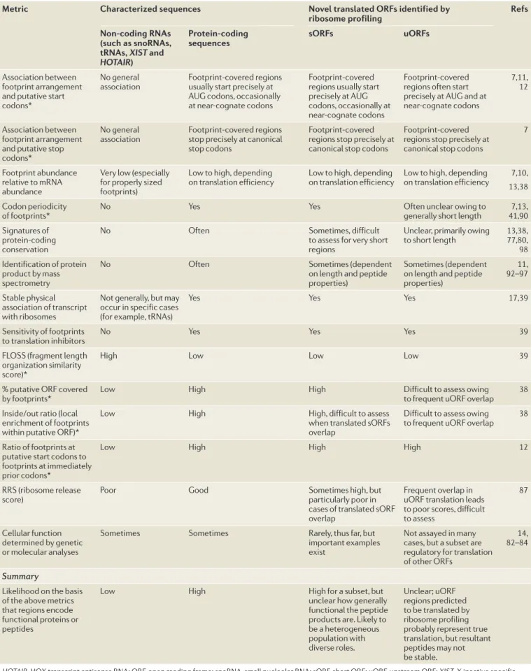
Table 1 | Novel translated ORFs compared with characterized translated ORFs by diverse metrics
Metric Characterized sequences Novel translated ORFs identified by
ribosome profiling Refs
Non-coding RNAs (such as snoRNAs, tRNAs, XIST and
HOTAIR)
Protein-coding
sequences sORFs uORFs
Association between footprint arrangement and putative start codons*
No general
association Footprint-covered regions usually start precisely at AUG codons, occasionally at near-cognate codons
Footprint-covered regions usually start precisely at AUG codons, occasionally at near-cognate codons
Footprint-covered regions often start precisely at AUG and at near-cognate codons
7,11, 12
Association between footprint arrangement and putative stop codons*
No general
association Footprint-covered regions stop precisely at canonical stop codons
Footprint-covered regions stop precisely at canonical stop codons
Footprint-covered regions stop precisely at canonical stop codons
7
Footprint abundance relative to mRNA abundance
Very low (especially for properly sized footprints)
Low to high, depending
on translation efficiency Low to high, depending on translation efficiency Low to high, depending on translation efficiency 13,387,10, Codon periodicity
of footprints* No Yes Yes Often unclear owing to generally short length 41,907,13,
Signatures of protein-coding conservation
No Often Sometimes, difficult
to assess for very short regions
Unclear, primarily owing
to short length 13,38, 77,80, 98 Identification of protein
product by mass spectrometry
No Often Sometimes (dependent
on length and peptide properties)
Sometimes (dependent on length and peptide properties) 11, 92–97 Stable physical association of transcript with ribosomes
Not generally, but may occur in specific cases (for example, tRNAs)
Yes Yes Yes 17,39
Sensitivity of footprints
to translation inhibitors No Yes Yes Yes 39
FLOSS (fragment length organization similarity score)*
High Low Low Low 39
% putative ORF covered
by footprints* Low High High Difficult to assess owing to frequent uORF overlap 38
Inside/out ratio (local enrichment of footprints within putative ORF)*
Low High High, difficult to assess
when translated sORFs overlap
Difficult to assess owing
to frequent uORF overlap 38 Ratio of footprints at
putative start codons to footprints at immediately prior codons*
Low High High High 12
RRS (ribosome release
score) Poor Good Sometimes high, but particularly poor in
cases of translated sORF overlap
Frequent overlap in uORF translation leads to poor scores, difficult to assess
87
Cellular function determined by genetic or molecular analyses
Sometimes Sometimes Rarely, thus far, but
important examples exist
Not assayed in many cases, but a subset are regulatory for translation of other ORFs
14, 82–84
Summary
Likelihood on the basis of the above metrics that regions encode functional proteins or peptides
Low High High for a subset, but
unclear how generally functional the peptide products are. Likely to be a heterogeneous population with diverse roles. Unclear; uORF regions predicted to be translated by ribosome profiling probably represent true translation, but resultant peptides may not
be stable.
HOTAIR, HOX transcript antisense RNA; ORF, open reading frame; snoRNA, small nucleolar RNA; sORF, short ORF; uORF, upstream ORF; XIST, X inactive specific transcript. *See the glossary terms, FIG. 1c, FIG. 2 and Supplementary information S1 (figure) for class definitions and examples.
Signatures of protein-coding conservation
Purifying evolutionary selection results in higher levels of synonymous than nonsynonymous substitutions, specifically among
homologous coding sequences. The pattern of nonsynonymous to synonymous differences among homologous regions compared in a phylogenetic group can be used to predict the likelihood that a genomic locus encodes a translated open reading frame (ORF).
Most of these approaches provide support for the pre-dicted widespread translation of short and alternative ORFs11–13,15,16,38,67,85,91(TABLE 1). Nevertheless, even with
ribosome profiling data, reliably identifying the full set of translated ORFs remains a challenge, especially in cases in which protein-coding sequences overlap.
Numerous complementary experimental approaches have aimed to further probe the degree to which newly predicted protein-coding sequences represent true cellular translation (TABLE 1). So far, these approaches generally confirm that the reads that are detected in regions predicted to be translated by ribosome profil-ing experiments represent translatprofil-ing 80S ribosomes. For example, ribosome footprints over putative trans-lated sORFs tend to respond to translation inhibitors in a manner comparable to benchmarked translating ribosomes39. Translated mRNA regions predicted from
mouse ribosome profiling data immunoprecipitate with tagged 60S ribosomal subunits in a specific man-ner, similar to that seen for characterized translated ORFs39. This finding suggests that true translating
somes produce the footprints that are detected by ribo-some profiling over ORFs not previously annotated as being translated, rather than these mRNA fragments being artefacts resulting from the protection of mRNA by scanning translation initiation complexes or alter-native RNA–protein complexes. An important open question is whether these translated regions produce stable peptides. Suggesting that they may, sORFs identi-fied as being translated by ribosome profiling that have been carboxy-terminally tagged in yeast and in human cytomegalovirus (HCMV)-infected cells can be seen to accumulate in a regulated manner that mirrors pre-dictions from ribosome profiling data11,39. Meanwhile,
specialized mass spectrometry approaches continue to identify a subset of peptides resulting from such sORFs in several systems11,92–97, suggesting that at least some of
these sORFs do encode abundant, stable peptides. Most convincingly, a few sORFs that were predicted to be translated from polysome association and ribo-some profiling data have now been shown to have bio-logical function14,39,83. In D. melanogaster, the peptides
encoded by two such translated sORFs contained in the sarcolamban locus have been shown to directly bind to a calcium transporter in heart cells and thus regu-late normal heart function83(FIG. 6a). In zebrafish, the
short protein Toddler was found to drive gastrulation by functioning as a secreted developmental signal14. In
mammals, a prominent example is the several translated sORFs, predicted on the basis of ribosome profiling of HCMV-infected human foreskin fibroblasts, that reside on the β2.7 RNA, which has traditionally been defined as non-coding11. Peptides resulting from the translation of
two of these sORFs have been shown by mass spectrom-etry to accumulate during HCMV infection. In addi-tion, analysis of serum samples from HCMV-positive and HCMV-negative blood bank donors showed a robust immune response to the peptides produced from several of these β2.7 sORFs, specifically in the HCMV-positive individuals39(FIG. 6b). This result
sug-gests that the ribosome-occupied sORFs are translated,
and that their products are processed and presented on MHC molecules as functional antigens in humans, thus expanding the range of epitopes displayed during viral infections. The condition-specific translation of many sORFs suggests that they could similarly be used to dis-tinguish cancer cells from normal cells, with important implications for immunomodulatory therapies.
The translation of some sORFs could also help to fuel the evolution of new proteins88. It is possible that
transcriptional noise, together with the propensity of the ribosome to translate capped cytosolic RNAs, may allow novel transcripts to engage the ribosome and allow trans-lational sampling of new, short motifs. Initially these sORFs may evolve under neutral selection. However, a subset could provide a small fitness advantage, resulting in positive selection and possible stabilization through lengthening over time, until they resemble canonical long protein-coding genes (FIG. 6c). Such regions would not necessarily be initially expected to show signatures of protein-coding conservation (as in REF. 98), and many might not produce a robust mutant phenotype when disrupted, making their study challenging.
How plastic is translation? Alternative isoforms abound. The results of ribosome profiling in yeast and in mam-mals have indicated that many genes may yield two or more protein variants independently of splicing, which indicates that there may be surprising flexibility in both where translation starts and where it stops in eukaryotes. Such alternative isoforms have been seen and charac-terized previously; in budding yeast, for example, both alanyl-tRNA synthetase 1 (ALA1) and glycyl-tRNA syn-thase 1 (GRS1) have been shown to exist in two isoforms, providing populations of the protein that are either cyto-solic or mitochondrial, depending on the presence or absence of an N-terminal in-frame extension99,100. These
examples are also detected by ribosome pro filing12 and
seem to be just a few of many10,12,67,101, supporting a
model in which diverse but targeted localization might be achieved for many proteins through sometimes small alterations in the site of translation initiation18,91,101.
Conversely, ribosome profiling of several yeast spe-cies, and of D. melanogaster embryos and cultured cells, revealed extensive heterogeneity in translation termina-tion sites15,102,103, resulting from regulated read-through
of hundreds of genes. As with the N-terminal-extension isoforms, many of these C-terminal extensions are pre-dicted to confer new subcellular localizations to the protei n products15,104.
Use of ribosome profiling has also facilitated the identification of interesting examples of regulated trun-cated protein isoforms10–12,89. In human cells, a recent
study identified a shortened alternative isoform of mito-chondrial antiviral signalling protein (MAVS), which is an important player in innate immune signalling89. The
alternative MAVS isoform results from translation ini-tiation downstream of the canonical start site to create an in-frame truncation, which the authors term ‘mini-MAVS’. Whereas full-length MAVS induces interferon production, miniMAVS antagonizes MAVS function by interfering with such production.
The large and diverse set of unconventional regions of translation suggested by ribosome profiling shows that there is considerably more to translational regulation and cellular content than was previously known. Some of these regions are likely to be translated into functional proteins, but it is likely that others will not produce sta-ble protein products that are similar to those from tra-ditional genes. Rather, subsets of these newly identified regions of translation may have regulatory, immune or currently neutral cellular roles. Unravelling the set of functions that are carried out by translated genomic regions poses a fascinating and daunting challenge.
Perspective
Protein synthesis consumes a large proportion of cellu-lar resources and is central to almost every function of a cell. Ribosome profiling allows, for the first time, in vivo and global measurement of translation, providing a pre-cise and quantitative account of what cells are translating, how this translation is regulated, and when and where translation happens. The rich and quantitative nature of ribosome profiling data provide an unprecedented oppor-tunity to explore and model complex cellular processes.
Although it has long been known that translational regulation has important roles in development, in cellu-lar responses to stimuli and in disease, the limited num-ber of well-studied examples of regulation at the level of protein synthesis have generally been identified in an ad hoc manner. When paired with RNA-seq measure-ments of mRNA levels, ribosome profiling now allows
instantaneous measurement of all translational control in a given system, providing a tool for broad discovery of the underlying biology of a cellular process or state of choice. Furthermore, the detailed information that is yielded by this method provides valuable insight into fundamental aspects of how translation works. Despite the conserved nature of much of the translation machinery, important open questions about the mechanism of protein syn-thesis remain, including the basis for most specificity of translation among different mRNAs and the connections between translation and nascent protein folding.
Finally, owing to the precise genomic positional information provided by ribosome profiling, the pro-tein-coding capacity of genomes can now be defined experimentally. This has led to the identification of a broad range of non-canonical translation events, includ-ing the translation of novel sORFs and alternative forms of previously annotated proteins, thereby challenging traditional views of protein-coding regions and gene diversity. Analytical advances that facilitate more com-prehensive identification of other non-canonical transla-tion events, such as those resulting from frame-shifting and stop codon read-through will continue to expand our understanding of the protein-coding capacity of complex genomes. The functions of the many novel short and alternatively translated regions that have been identified so far by ribosome profiling remain an intriguing and largely open question, the answer to which could funda-mentally change the way we think about the encoding of information in genomes. Newly available CRISPR-based Nature Reviews |Molecular Cell Biology
Ca-P60A SERCA cardiac calcium transporter Sarcolamban peptide A or B Time Cardiac kymograph Voltage Time Voltage – sarcolamban + sarcolamban
a Regulation of larger protein activity b Antigens
MHC class II
T cell
Peptide from sORF on HCMV β2.7 RNA c An evolutionary pool for new protein function
Translation sORF Function of protein in cell None or minimal, nonspecific sORF sORF or ORF ORF Weak, specific Strong, specific Strong, specific
Stop codon mutation and selection over time
Antigen-presenting cell
Figure 6 | Proposed cellular roles for the peptide products of translated short open reading frames (sORFs) identified by ribosome profiling. a | The two sarcolamban peptides are 28 and 29 amino acids in length, are conserved from fruit flies to human and regulate normal heart function in flies through direct binding to the sarcoendoplasmic reticulum Ca2+ ATPase (SERCA) calcium transporter Ca-P60A in cardiac tissue. b | Sera from human cytomegalovirus
(HCMV)-positive blood donors were used to identify a specific and robust immune response against multiple short peptides translated from the β2.7 RNA, which was previously thought to be a non-coding RNA. c | Spurious translation of short regions may produce a pool of peptides with weak or no cellular function. New protein domains may evolve through selection for maintenance of peptides with weak cellular function, followed by stop codon mutation and further selection for increasingly specific and important cellular function over time. Part a from Magny, E. G. et al. Conserved regulation of cardiac calcium uptake by peptides encoded in small open reading frames. Science341, 1116–1120 (2013). Redrawn with permission from AAAS.