warwick.ac.uk/lib-publications
Manuscript version: Author’s Accepted Manuscript
The version presented in WRAP is the author’s accepted manuscript and may differ from the
published version or Version of Record.
Persistent WRAP URL:
http://wrap.warwick.ac.uk/123676
How to cite:
Please refer to published version for the most recent bibliographic citation information.
If a published version is known of, the repository item page linked to above, will contain
details on accessing it.
Copyright and reuse:
The Warwick Research Archive Portal (WRAP) makes this work by researchers of the
University of Warwick available open access under the following conditions.
Copyright © and all moral rights to the version of the paper presented here belong to the
individual author(s) and/or other copyright owners. To the extent reasonable and
practicable the material made available in WRAP has been checked for eligibility before
being made available.
Copies of full items can be used for personal research or study, educational, or not-for-profit
purposes without prior permission or charge. Provided that the authors, title and full
bibliographic details are credited, a hyperlink and/or URL is given for the original metadata
page and the content is not changed in any way.
Publisher’s statement:
Please refer to the repository item page, publisher’s statement section, for further
information.
via Attentional Representation Learning
Kai Wu1, Bowen Du2, Man Luo2, Hongkai Wen2?, Yiran Shen3, and Jianfeng
Feng1,2
1 Fudan University, China 2
University of Warwick, UK 3 Data61, CSIRO, Australia
Abstract. In this paper, we propose a new weakly supervised 3D brain lesion segmentation approach using attentional representation learning. Our approach only requires image-level labels, and is able to produce accurate segmentation of the 3D lesion volumes. To achieve that, we de-sign a novel dimensional independent attention mechanism on top of the Class Activation Maps (CAMs), which refines the 3D CAMs to obtain better estimates of the lesion volumes, without introducing significantly more trainable variables. The generated attentional CAMs are then used as a source of weak supervision signals to learn a representation model, which can reliably separate the voxels belong to the lesion volumes from those of the normal tissues. The proposed approach has been evaluated on the publicly available BraTS and ISLES datasets. We show with com-prehensive experiments that our approach significantly outperforms the competing weakly-supervised methods in both initial lesion localization and the final segmentation, and is able to achieve comparable Dice scores in segmentation comparing to the fully supervised baselines.
Keywords: Weakly Supervised·Lesion Segmentation·Representation Learning.
1
Introduction
Magnetic Resonance Imaging (MRI) has become one of the key diagnostic tech-niques for brain lesion analysis, offering a non-intrusive and radiation-free modal-ity to detect abnormalities. For many diseases such as brain tumour, the precise identification and segmentation of the lesion volumes is a fundamental prerequi-site for further treatments including surgery or radiation therapy. Traditionally, this process is performed by the experienced medical experts, which is expensive, tedious and more importantly prune to the inter-rater/test-retest variability [11]. Recently, there has been a solid body of work on automatic lesion segmenta-tion [5, 8, 13], offering a faster and more objective alternative to human labor. In particular, the deep learning based methods have demonstrated impressive performance in various segmentation tasks, and are robust enough to process
?
2 K. Wu et al.
lesion volumes with irregular shapes or challenging sizes [8, 13]. However, most of the existing deep learning methods aresupervised, whose success depends on substantial amount of training data with accurate labels. Particularly for the task of brain lesion segmentation, such labels have to be the accurate lesion boundaries in the 3D space, which are often annotated by the experts manually. They are extremely expensive and time-consuming to obtain, which significantly limits the application of those approaches in clinical practice.
On the other hand, weakly supervised segmentation approaches which only require the coarse-grained (e.g. image-level) labels point to a promising direc-tion [4, 3, 6, 7, 10]. These approaches aim to exploit the minimum level of expert annotations, while being able to generate the fine-grained segmentation automat-ically. Many of these weakly supervised approaches leverage the Class Activation Maps (CAMs) [14] as a key step for segmentation. For example, the work in [7] uses multi-layer CAMs to detect histological features of glioma in CLE images, while [3] proposes an iterative mining pipeline to localise lesion in different areas. However, a major drawback of CAMs is that they can be very noisy in practice, and thus it is often challenging to derive good initial segmentation proposals from them, especially for 3D images.
In this paper, we propose a novel weakly supervised segmentation approach for brain lesion in MRI images, which only requires image-level binary labels in-dicating whether the lesion is present or not. Such weak labels are much easier to obtain in practice, and thus our approach can generate high quality segmentation proposals on the vast unlabeled data with minimum human input, which can then be refined by experts to produce accurate voxel-level labels in 3D. In partic-ular, we develop a new dimensional independent attention mechanism on top of the standard CAMs, which can significantly improve lesion localization without introducing excessive trainable variables to the network. This attentional CAM provides high quality estimates of the lesion regions as well as normal tissues, which are then used to train a representation model. The learned model can reliably distinguish the voxels of lesion volumes from those belong to the nor-mal tissues, and thus generate the accurate lesion segmentation. We evaluate the proposed approach on the public BraTS [2] and ISLES [9] datasets. Extensive ex-periments show that with the proposed attentional CAMs, our approach is able to acquire much better initial estimates of lesion regions comparing to the com-peting methods, and the proposed representation learning technique is evidently beneficial in improving segmentation performance, significantly outperforming the state-of-the-art while offering comparable accuracy with the fully supervised baselines.
2
Method
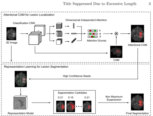
The proposed weakly supervised 3D lesion segmentation approach consists of three steps: i) obtaining the initial lesion regions with image-level labels; ii) learning a representation model to distinguish the voxels belong to normal and lesion tissues; and iii) using the learned model to obtain the fine-grained
voxel-Attentional CAM 3D Image
Final Segmentation Representation Model
Dimensional Independent Attention
h w d CAM Non Maximum Suppression Segmentation Cadidates 0.15 0.31 0.21 Classification CNN
Attentional CAM for Lesion Localization
Representation Learning for Lesion Segmentation
High Confidence Seeds
Attention Scores
Fig. 1.The overview of the proposed weakly supervised lesion segmentation approach.
level segmentation of lesion volume. Fig.1 shows the overview of the proposed approach. In particular, our approach is based on two deep neural networks: a) a classification network which is used to compute the 3D Class Activation Maps (CAMs); and b) another network which learns the voxel representation model and performs the segmentation. The remainder of this section describes the design of the two networks and training techniques for them in more details.
2.1 Lesion Localization with Attentional CAM
Generating CAMs with Image-level Labels: As in many state of the art weakly supervised approaches, the Class Activation Maps (CAMs)[14] are often considered as the seeds for later segmentation. In the proposed approach, we also consider CAMs as the initial segmentation proposals, which can roughly localize the lesion volumes within the images. In particular, given the image-level labels i.e. binary labels indicating if lesion tissues exist, the standard technique [15] to obtain the CAM is to train a classification network with global average pooling (GAP) or global mean pooling (GMP) followed by a fully connected layer. The CAM of a positive sample (image with lesion) is computed by:
C(h, w, d) =wCAMT F(h, w, d) (1) where w is the weights associated to the positive class, F is the feature map extracted by the last convolutional layer before the GAP/GMP operation, and
4 K. Wu et al.
F(h, w, d) is the feature vector at location (h,w,d). In practice, C is typically normalized to make the maximum activation equals to one, and a threshold is often used to extract the region of target objects, which can be used as an initial proposal for segmentation.
Refining CAMs with Attention: However for 3D medical images, in some cases the lesion tissues may only occupy a small fraction of the entire volume, e.g., in our experiments with the ISLES [9] and BraTS datasets [2], the tumor or stroke volumes can be just 0.1% of the whole brain. For those samples, the CAMs tend to be very noisy, making it challenging to select the appropriate threshold to obtain the desired lesion regions. In practice, if GAP is used in the network then we typically need to consider larger activation values which make training more difficult to converge. On the other hand, if GMP is considered, the network may focus too much on small areas with stronger responses, and generate incomplete regions that won’t be able to cover the whole lesion volumes. To address this problem, we proposed to use the attention mechanism to refine the generated CAMs. Conceptually this can be achieved by directly ap-plying the learned attention sores to the 3D CAMs. However, in practice it is prohibitively expensive for 3D images since the amount of trainable parameters are significantly larger than 2D, which can easily lead to over-fitting. Therefore, in this paper, we consider an approximation approach, which enforces atten-tion along individual dimensions and fuses the re-weighted features afterwards. Concretely, let F ∈ RH×W×D×K be the activation map extracted by the last convolutional layer. F can be viewed as K 3D feature cubes Fk ∈ RH×W×D. For each cube Fk, we first apply average pooling to its slices along the three different dimensions. For example along the dimension of sizeD, we do average for theD feature slices, each of which is aH×W feature, and obtain a vector
FD ∈ RD×K across the K cubes. We then consider a self-attention over this featureFD, where the attention scoressd (1≤d≤D) is calculated as:
sd= exp(gd) PD d=1exp(gd) , wheregd=wTattF D(d) (2)
Here FD(d) is the d-th feature in FD, and w
att are the weights learned via
training. The attention scoressdare then used to re-weight the generated CAM along the dimension of sizeD, i.e. multiplied to the 2D slices of the activation values (with size H ×W) in the CAM: CD0 (d) = sd·C(d). Similarly, we can learn the attention scores along the other two dimensions (with sizeH andW), which are used to obtain the re-weighted CAMs C0
H and CW0 . Then the final attentional CAM (ACAM)C0 is computed by fusing the threeC
D,CHandCW, which is also normalized to [0,1].
Intuitively, the learned attention scores specify how important the feature slices are with respect to the final classification results (lesion or not) along a certain dimension, which is exploited to refine the standard CAM. It is also worth pointing out that here we implicitly assume that the attention scores along different dimensions are independent, and thus they are actually approximations to the full 3D self-attention, which however would require much more training
effort. In our experiment, we show that the proposed attentional CAM (ACAM) can achieve much better performance than the standard CAMs (using GMP or GAP), while won’t incur significantly expensive training cost.
2.2 Weakly Supervised Representation Learning for Segmentation
As discussed above, the ACAMs generated by the classification network provide rough estimates of the 3D lesion volumes. To obtain the accurate voxel-level segmentation, in this paper we use another network, which exploits the ACAMs as a source of weak supervision. The key idea is that although ACAMs are not globally accurate segmentation, in regions with high confidence they can still pro-vide valuable information on features of the foreground (lesion) and background (normal tissues). We could then leverage that to learn a universal representation model, with which voxels from the foreground can be effectively separated from those from the background, and thus obtain the voxel-level segmentation.
Learning the Representation Model: To learn the representation model, we first randomly select the voxels with high confidence from both the fore-ground (lesion) and backfore-ground (normal tissues) according to the ACAMs. In particular, we select a foreground voxel if the ACAMC0(h, w, d)≥θ
F G , while the background ifC0(h, w, d)≤θBG. In our experiments, we setθF G to 0.1 and
θBG to 0.01 respectively.
With the selected set of high confidence voxelsV, we aim to learn a repre-sentation model fR in the form of a neural network, which maps an arbitrary voxels pto its embedding vectorfR(p). In this case, the distance between two voxels pand q can be defined as D(p, q) = (1 + exp{−kfR(p)−fR(q)k2
2})−1,
wherek · k2 is theL2norm. We trainfR using a cross-entropy loss:
L=− 1 |V| X p,q∈V log[D(p, q)]1(p,q)+ log[1−D(p, q)]1−1(p,q) (3)
where the indicator function1(p, q) returns 1 if the voxels pandq both belong to the foreground or background, while 0 if pandqare different.
Segmentation based on Voxel Affinity: Letpbe a foreground voxel inV. With the learned representation modelfR, we could obtain a lesion segmentation candidateSp by selecting all voxelsq that are close enough (with high affinity) to p, i.e.Sp={q|D(p, q)≥λ}, where the rest is considered as the background (normal tissues). In practice, the quality of the segmentationSpdepends on the selection of the seed voxel p, which can lead to noisy results in some cases. To mitigate that, in this paper we firstly use multiple foreground seed voxels in
V to generate an array of segmentation candidates S. Then for each candidate
Sp ∈S, we assign a confidence scorecp, which is computed as the mean of the voxel ACAM values, i.e. the average confidence that the voxels inSpare belong to the foreground. Finally we combine the segmentation candidates S by non-maximum suppression according to the confidence scores, and obtain the final segmentation of the lesion volume.
6 K. Wu et al.
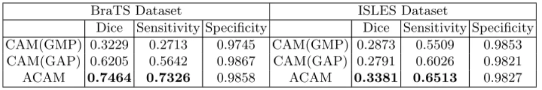
BraTS Dataset ISLES Dataset
Dice Sensitivity Specificity Dice Sensitivity Specificity CAM(GMP) 0.3229 0.2713 0.9745 CAM(GMP) 0.2873 0.5509 0.9853 CAM(GAP) 0.6205 0.5642 0.9867 CAM(GAP) 0.2791 0.6026 0.9821
ACAM 0.7464 0.7326 0.9858 ACAM 0.3381 0.6513 0.9827
Table 1.Lesion localisation performance of the competing approaches.
3
Evaluation
3.1 Experimental Setup
Datasets: We consider two datasets in our experiments: i) the Multimodal Brain Tumor Segmentation dataset (BraTS 2017) [2] and ii) the Ischemic Stroke Lesion Segmentation (ISLES 2017) dataset [9]. The BraTS dataset contains mul-timodal MRI images including T1, T1c, T2 and Flair, where the training set has 210 samples and the test set contains 47 samples. The ground truth segmenta-tion includes four tumor tissue classes, but in this paper we only consider those for the whole tumors. The ISLES dataset also contains multi-spectral MRI data of 43 stroke patients, with ground truth segmentation provided.
Competing Approaches: For initial lesion localization, we compare the pro-posed attentional CAM with CAM(GMP) and CAM(GAP), which are the stan-dard approaches of generating CAMs using Global Maximum Pooling(GMP) and Global Average Pooling(GAP) respectively. For lesion segmentation we compare the proposed weakly supervised representation learning approach (ACAM+WSR) with: i) a fully supervised UNet [12] (S-UNet) trained with the ground truth; and ii) a weakly supervised UNet (WS-Unet), which uses the results of our ACAM as the weak supervision signals to train the UNet.
Metrics: We consider the Dice, Sensitivity and Specificity as the evaluation metrics, which are computed as: Dice = 2TP/(2TP+FP+FN); Sensitivity = TP/(TP+FN); and Specificity = TN/(TN+FP) respectively. Here TP, FP, TN and FN are true positive, false positive, true negative and false negative.
Implementation: Our networks are implemented with Tensorflow [1], and trained on a single Titan X GPU with the Adam optimiser of learning rate 0.001. The network to generate ACAM is a VGG-like 3D convolutional network, where at the end of the conv layer we incorporate the proposed attention mechanism. The network for representation learning shares the similar structure with 3D UNet. We train the networks with only binary labels, i.e. if this image contains lesion or not, and report the segmentation performance with respect to the ground truth segmentation labels. For BraTS dataset, we report results returned from the official evaluation server, while for the ISLES dataset, we randomly divide training and testing sets (3:1) and report results on the testing set.
3.2 Results
Lesion Localization: Table 1 shows the lesion localisation performance of the competing approaches. We can see that on the BraTS dataset, the standard
BraTS Dataset ISLES Dataset
Dice Sensitivity Specificity Dice Sensitivity Specificity S-UNet 0.8811 0.8691 0.9950 S-UNet 0.4459 0.3753 0.9984 WS-UNet 0.7682 0.7727 0.9874 WS-UNet 0.3374 0.7348 0.9795 ACAM+WSR0.7997 0.8973 0.9785 ACAM+WSR0.3827 0.8306 0.9791
Table 2.Lesion segmentation performance of the competing approaches.
CAM with GMP has the much lower Dice and Specificity scores (around 0.32 and 0.27), which won’t be able to localise the lesion volume. The CAM(GAP) works better than CAM(GMP), but it is the proposed ACAM that achieves the best results overall (up to 42% improvement in Dice and 46% in Sensitivity). It confirms that the attention mechanism can help the network to focus more on the lesion volumes, and significantly improve the localization performance (see Fig. 2(b) and (c)). On the other hand, we see that on ISLES dataset the gap between CAM(GMP) and CAM(GAP) is smaller, where CAM(GMP) even has slightly better Dice score. This is because unlike in the BraTS dataset, the lesion volumes in ISLES dataset tend to be smaller, where the max pooling used in CAM(GMP) is more suitable to detect such small regions. Again we see the proposed ACAM achieves the best results, offering up to 6% improvement in Dice and 10% better Sensitivity scores.
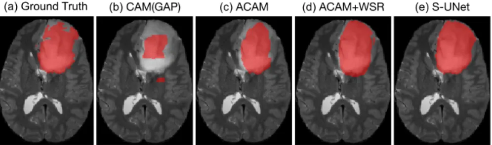
Lesion Segmentation: We are now in a position to evaluate the performance of lesion segmentation of the competing approaches. Table 2 shows the results on both BraTS and ISLES datasets. Note that here the S-UNet is a fully supervised approach, which has access to the ground truth segmentation. On the other hand, both WS-UNet and the proposed ACAM+WSR only use the image level labels. We see that the proposed ACAM+WSR generally outperforms the weakly supervised WS-UNet, e.g., in the ISLES dataset it enjoys roughly 5% better dice and 10% better sensitivity scores, while in the BraTS the improvements are 3% and 12.5% respectively. This means the representation learning does help to effectively extract lesion volumes from the normal tissues, leading to more accurate segmentation. On the other hand, we see that the gap between the proposed ACAM+WSR and the fully supervised S-UNet is only about 6% in Dice (note that for ACAM+UNet the gap is 11%), which means even with image-level labels, our weakly supervised approach could achieve comparable accuracy with fully supervised baseline (see Fig. 2(d) and (e) for an example).
4
Conclusion
In this paper, we present a new weakly supervised brain lesion segmentation ap-proach that only requires image-level labels to generate the accurate voxel-level 3D boundaries of the lesion volumes. We develop a novel attentional CAM tech-nique to better localize the lesion regions, and exploit representation learning to further improve lesion segmentation. The proposed approach has been validated on two public datasets, the BraTS and ISLES, where experimental results show
8 K. Wu et al.
(a) Ground Truth (b) CAM(GAP) (c) ACAM (d) ACAM+WSR (e) S-UNet
Fig. 2. Qualitative results on BraTS dataset showing extracted lesion volumes by (a) ground truth, (b) CAM(GAP), (c) the proposed ACAM, (d) the proposed ACAM+WSR and (e) fully supervised S-UNet.
that our approach offers superior performance to the state-of-the-art, and can achieve comparable segmentation accuracy with the fully supervised methods. For future work, we would like to explore approaches that can detect multiple classes of the lesion structure with similar weak supervision signals and consider more types of lesion data.
References
1. Tensorflow.https://www.tensorflow.org, accessed: 2019-04-01
2. Bakas, S., Akbari, H., Sotiras, A., Bilello, M., Rozycki, M., Kirby, J.S., Freymann, J.B., Farahani, K., Davatzikos, C.: Advancing the cancer genome atlas glioma mri collections with expert segmentation labels and radiomic features. Scientific data
4, 170117 (2017)
3. Cai, J., Lu, L., Harrison, A.P., Shi, X., Chen, P., Yang, L.: Iterative attention mining for weakly supervised thoracic disease pattern localization in chest x-rays. In: International Conference on Medical Image Computing and Computer-Assisted Intervention. pp. 589–598. Springer (2018)
4. Cai, J., Tang, Y., Lu, L., Harrison, A.P., Yan, K., Xiao, J., Yang, L., Summers, R.M.: Accurate weakly-supervised deep lesion segmentation using large-scale clin-ical annotations: Slice-propagated 3d mask generation from 2d recist. In: Interna-tional Conference on Medical Image Computing and Computer-Assisted Interven-tion. pp. 396–404. Springer (2018)
5. Despotovi´c, I., Goossens, B., Philips, W.: Mri segmentation of the human brain: challenges, methods, and applications. Computational and mathematical methods in medicine2015(2015)
6. Hwang, S., Kim, H.E.: Self-transfer learning for weakly supervised lesion localiza-tion. In: International Conference on Medical Image Computing and Computer-Assisted Intervention. pp. 239–246. Springer (2016)
7. Izadyyazdanabadi, M., Belykh, E., Cavallo, C., Zhao, X., Gandhi, S., Moreira, L.B., Eschbacher, J., Nakaji, P., Preul, M.C., Yang, Y.: Weakly-supervised learning-based feature localization for confocal laser endomicroscopy glioma images. In: International Conference on Medical Image Computing and Computer-Assisted Intervention. pp. 300–308. Springer (2018)
8. Kamnitsas, K., Ledig, C., Newcombe, V.F., Simpson, J.P., Kane, A.D., Menon, D.K., Rueckert, D., Glocker, B.: Efficient multi-scale 3d cnn with fully connected crf for accurate brain lesion segmentation. Medical image analysis36, 61–78 (2017) 9. Maier, O., Menze, B.H., von der Gablentz, J., H¨ani, L., Heinrich, M.P., Liebrand, M., Winzeck, S., Basit, A., Bentley, P., Chen, L., et al.: Isles 2015-a public eval-uation benchmark for ischemic stroke lesion segmentation from multispectral mri. Medical image analysis35, 250–269 (2017)
10. Mlynarski, P., Delingette, H., Criminisi, A., Ayache, N.: Deep learning with mixed supervision for brain tumor segmentation. arXiv preprint arXiv:1812.04571 (2018) 11. Porz, N., Bauer, S., Pica, A., Schucht, P., Beck, J., Verma, R.K., Slotboom, J., Reyes, M., Wiest, R.: Multi-modal glioblastoma segmentation: man versus ma-chine. PloS one9(5), e96873 (2014)
12. Ronneberger, O., Fischer, P., Brox, T.: U-net: Convolutional networks for biomedi-cal image segmentation. In: International Conference on Medibiomedi-cal image computing and computer-assisted intervention. pp. 234–241. Springer (2015)
13. Tseng, K.L., Lin, Y.L., Hsu, W., Huang, C.Y.: Joint sequence learning and cross-modality convolution for 3d biomedical segmentation. In: Proceedings of the IEEE Conference on Computer Vision and Pattern Recognition. pp. 6393–6400 (2017) 14. Zhou, B., Khosla, A., Lapedriza, A., Oliva, A., Torralba, A.: Learning deep features
for discriminative localization. In: Proceedings of the IEEE conference on computer vision and pattern recognition. pp. 2921–2929 (2016)
15. Zhou, B., Khosla, A., Lapedriza, A., Oliva, A., Torralba, A.: Learning deep features for discriminative localization. In: CVPR. pp. 2921–2929 (2016)