ORIGINAL ARTICLE
Glycan-binding specificities of
Streptococcus mutans
and
Streptococcus sobrinus
lectin-like adhesins
Viviane Schüler&Adrian Lussi&Andreas Kage&
Rainer Seemann
Received: 6 December 2010 / Accepted: 13 May 2011 / Published online: 17 June 2011 #Springer-Verlag 2011
Abstract Since the adhesion of bacteria to the tooth surface is a prerequisite for dental plaque and subsequent caries development, a promising caries preventive strategy could be to block the lectin–glycan-mediated adherence of cariogenic bacteria. The aim of the study was to evaluate potential differences in glycan-binding specificities of two Streptococcus mutansstrains (DSM 20523 and DSM 6178) and Streptococcus sobrinus (DSM 20381). A competitive enzyme-linked lectin-binding assay was used to identify the binding specificities of isolated bacterial surface lectins. Blotting of the microbial proteins on neoglycoprotein-coated PVP membranes enabled a qualitative protein analysis of all specific bacterial lectins. Different glycan-binding sites could be identified for theS. mutans strains in comparison to S. sobrinus. An earlier reported glycan-binding specificity for terminal galactose residues could be confirmed for theS. mutansstrains. For theS. sobrinusstrain, more than one glycan-binding specificity could be found (oligomannose and terminal sialyl residues). Each of the tested strains showed more than one surface lectin responsible for the specific lectin-binding with varying molecular weight
(S. mutans, 90/155 kDa and S. sobrinus, 35/45 kDa). The established experimental setup could be used as future standard procedure for the identification of bacterial lectin-derived binding specificities. The findings from this study might serve as basis for the design of an individual‘glycan cocktail’ for the competitive inhibition of lectin-mediated adhesion of mutans streptococci to oral surfaces.
Keywords Dental caries .Streptococcus mutans. Streptococcus sobrinus. Lectin-like adhesins . Glycan pattern
Introduction
Several microorganisms have been identified from carious lesions of which Streptococcus mutans and Streptococcus sobrinus are the most prevalent pathogenic species in humans involved in the initiation and development of dental caries [1–4]. The high cariogenic potential especially of S. mutans derives from the ability to produce high amounts of organic acid and extracellular glycans in the presence of low molecular sugars [3,5,6].
Consequently, established preventive strategies such as dietary control, topical or systemic use of fluorides and fissure sealants have led to a significant caries decline [7, 8]. However, caries is still causing high costs in health systems. Due to the current stagnation within caries prevention [9], continuing development of innovative preventive strategies and techniques seems to be indispensable to minimize further expansion of this widespread disease.
Since the adhesion of bacteria to the tooth surface represents the initial step in the pathogenesis of dental plaque formation and subsequent caries development [10], an anti-adhesive therapy gains significant importance by a
V. Schüler
:
R. SeemannDepartment of Operative and Preventive Dentistry, Charité, Humboldt University,
Berlin, Germany
A. Lussi
:
R. Seemann (*)Department of Preventive, Restorative and Pediatric Dentistry, University of Bern,
Freiburgstrasse 7, 3010 Bern, Switzerland
e-mail: rainer.seemann@zmk.unibe.ch A. Kage
Technical University Berlin, Berlin, Germany
possible prevention of specific lectin-mediated adherence mechanisms [11–13]. Studies concentrating on salivary glycans have indicated that specific soluble glycoconju-gates in sufficient concentrations have the ability to competitively inhibit the adherence of bacterial lectins of caries-inducing microorganisms to immobilized glycocon-jugates on oral surfaces [14,15]. Thus, the aim of this study was to evaluate potential differences in glycan-binding specificities of twoS. mutansstrains andS. sobrinus.
Materials and methods
BothS. mutansstrains (DSM 20523 and DSM 6178) andS. sobrinus(DSM 20381) were obtained in lyophilisated form by the DSMZ (Deutsche Sammlung von Mikroorganismen und Zellkulturen, Braunschweig, Germany). Individual colonies of each strain were cultivated in universal broth agar (CASO-Bouillon, heipha,D-Eppelheim) within 1 week.
The resulting bacterial suspensions were each biotinylated (0.5 mg Sulfo-NHS-LC-Biotin per millilitre reaction volume, solved in 50 mM Na2CO3buffer, pH 8.0), lysed (five times,
50 ml lysing buffer, pH 8.0, 10 min, 5,000 rpm) and finally filtrated (PES filter 0.2μm/10 kDa, Vivaflow 50, Vivascience, Göttingen, Germany/10 kDa palfiltrons, Amicon® Ultra-4, Millipore, Billerica, MA, USA) in order to obtain bacterial homogenates >10 kDa, <0.2μl, including their biotinylated surface membrane proteins of lectin type. The BCA™Protein Assay-Kit (Pierce, Rockford, IL, USA) was used to adjust all bacterial homogenates to the same total protein concentration.
A competitive enzyme-linked lectin-binding inhibition assay was used to determine the binding specificity of bacterial surface lectin-like adhesins towards lectin-specific glycan patterns, as well as the potential binding inhibition by soluble complementary glycans in dependence on their concentration (Fig. 1). Based on the principle of the competitive lectin-binding inhibition assay described by Kage et al. [16], several microtitre plates were pre-coated with immobilized neoglycoproteins (1:1,000, solved in 0.1 M Na2CO3buffer, pH 8.5, 100μl), with corresponding
binding specificities towards well-known lectins. The fol-lowing purified plant and animal lectins with known specificity for different glycan patterns were used in this study: peanut agglutinin (PNA, specific for β-galacto-1,3-N-acetyl-galactosamine, Sigma-Aldrich, Steinheim, Germany), Galanthus nivalis agglutinin (GNA, specific for terminal mannose residues, EY Laboratories Inc., San Mateo, CA, USA), Concanavalin A (ConA, specific for trimannosyl structures in high mannose type N-glycans, Sigma-Aldrich, Steinheim, Germany), Sambucus nigra agglutinin (SNA, specific for α-2,6-sialyl residues, EY Laboratories Inc., San Mateo, CA, USA) and Anguilla anguilla agglutinin (AAA, specific for α-1,6-fucose, EY Laboratories Inc., San Mateo, CA, USA) [17]. The following neoglycoproteins were used for pre-coating of the microtitre plates: BSA-galactose-1,3-N-acetyl-galactosamine (specific for PNA, Dextra Laboratories, Reading, UK), BSA-α-mannopyranoside (specific for GNA and ConA, Sigma-Aldrich, Steinheim, Germany), BSA-fucosylamide (specific for AAA, Sigma-Aldrich, Steinheim, Germany) and fetuin
Fig. 1 Scheme of the competi-tive lectin-binding inhibition assay used to determine the binding specificity of bacterial surface lectin-like adhesins towards lectin-specific glycan patterns as well as the potential binding inhibition by soluble complementary glycans in dependence on their concentration
(specific for SNA, Sigma-Aldrich, Steinheim, Germany). Unbound neoglycoproteins were removed during washing (three times, 200 μl, 50 mM tris(hydroxymethyl)-amino-methane, 0.1% Tween 20, pH 7.5) after overnight incubation at 4°C. Subsequently, double rows on each microtitre plate were incubated with 75 μl of the specific standard lectin (1 mg/ml, 1:20,000 in probing buffer: 50 mM Tris–HCl, 150 mM NaCl, 0.1 mM MnCl2, 0.5 mM CaCl2, 2.0 mM
MgCl2, 0.025 mM ZnCl2, pH 7.5) and analogous all
bacterial homogenates (approximately 730 μg/ml, 1:1,500 in probing buffer, pH 7.5), each together with 25μl soluble complementary glycans (methyl-D-galactopyranoside (specific for PNA, Sigma-Aldrich, Steinheim, Germany), mannan (specific for GNA, Sigma-Aldrich, Steinheim, Germany), α-L-(−)-fucose (specific for AAA, Sigma-Aldrich, Steinheim, Germany), methyl-α-mannopyranoside (specific for ConA, Sigma-Aldrich, Steinheim, Germany) andN -acetyl-neuramin-lactose (specific for SNA, Dextra Laboratories, Reading, UK) in different concentrations following a geometrically dilution (in probing buffer, pH 7.5). Additionally, each double row included a blank value to evaluate possible unspecific bindings between buffer and immobilized neo-glycoproteins, as well as the maximal lectin binding without the addition of complementary soluble glycan. Finally, all plates were incubated shaking for 30 min at room temperature, then overnight at 4°C. Washing (three times, 200μl) allowed the elimination of all unbound or by soluble complementary glycans bound lectins. Only lectin molecules bound to the immobilized glycoproteins were detected by the neutravidin peroxidase detection system (100 μl, 1 mg/ml, 1:20,000 in probing buffer) during a 1-h incubation at room temperature (followed by a washing procedure, four times, 200μl). The colour change induced by tetramethylbenzidin substrate (1:100 solved in galatti buffer, stopped with 50 μl 4 N H2SO4) allowed finally the colorimetrically determination in
a spectral photometer (450 to 492 nm, Ultra 384, Tecan, Crailsheim, Germany). Non-carbohydrate–lectin-specific bindings between the lectins and the immobilized neo-glycoproteins could be further measured by incubation with the lectins in the presence of deglycosylated, immobilized neoglycoproteins in the same experimental setup. In the attempt to identify only the bacterial surface proteins responsible for the lectin-mediated binding specificities, deglycosylated glycoconjugates were obtained by using oxidative separation of their terminal glycan structures.
Sodium dodecyl sulphate-polyacrylamide gel electro-phoresis (SDS-PAGE) of the bacterial homogenates was performed with a 10% polyacrylamide gel (CleanGel, 52S, ETC, Kirchtellinsfurt, Germany) under reducing conditions (10μl 2.5 M DTT to 1 ml buffer). The gels were calibrated with the following molecular weight standards: Precision Plus (10–250 kDa, Bio-Rad, Hercules, CA, USA), pre-stained Multi Mark (4–250 kDa, Invitrogen, Karlsruhe, Germany)
and biotinylated SDS Molecular Weight Standard (6.5– 180 kDa, Sigma-Aldrich, Steinheim, Germany). Silver stain-ing (GE Healthcare Biosciences, Freiburg, Germany) enabled to uncover all proteins of each bacterial homogenate separated by their molecular weight. Based on the Western Blot technique, a contact blot was newly developed to transfer the SDS-PAGE separated proteins by diffusion to glycan-specific polyvinyl difluoride (PVP) membranes (Fig.2). The glycan-specific PVP membranes were obtained by incuba-tion (overnight at 4°C after activaincuba-tion with 100% methanol for 10 min) with the same complementary glycoconjugates used in the lectin assay described above. Both the PVP membranes and the SDS gels were equilibrated in transfer buffer (12 mM tris(hydroxymethyl)-aminomethane, 96 mM glycine, 20% methanol) for 15 min. The PVP membranes were then blotted under the weight of 5 kg in a blot sandwich (lower glass plate–SDS gel–PVP-membrane–5× filter paper–upper glass plate) for 1.5 h with the by electrophoresis-separated microbial proteins. This technique enabled a qualitative protein analysis of only the bacterial lectins responsible for the specific binding to a specific neoglycoprotein. After blocking remaining binding sites (60 min at room temperature, 20 mM tris(hydroxymethyl)-aminomethane, 137 mM NaCl, 2.0% Tween 20, pH 7.6) and incubation with the neutravidin peroxidase detection system (60 min, 1 mg/ml, 1:10,000 in probing buffer) followed by
Fig. 2 Scheme of the contact blot. SDS-PAGE-separated bacterial proteins were blotted on glycan-specific polyvinyl difluoride (PVP)-membranes enabling a qualitative protein analysis of only the bacterial lectins responsible for a specific glycan binding. The following glycans were used: methyl-D-galactopyranoside (specific for PNA), mannan (specific for GNA), α-L-(−)-fucose (specific for AAA), methyl-α-pyranoside (specific for ConA) and NAC-neuraminlactose (specific for SNA). Using the neutravidin peroxidase detection system, only the specific microbial biotinylated surface proteins could be visualized by Enhanced Chemo-luminescence
several washing steps (1×15 and 3×5 min, shaking in washing buffer), the visualization of these specific proteins was achieved by Enhanced Chemo-luminescence (solution 1 +2 mixed 1:1, 0.125 ml/cm2, Pharmacia Biotech, Piscat-away, NJ, USA) onto a sensitive film (exposure time from 0.25 to 5 min, High Performance Chemiluminescence Film, Amersham Biosciences). The exclusion of all non-carbohydrate–lectin-based binding mechanisms was again obtained by applying deglycosylated glycan patterns to the PVP membranes in the same experimental setup using the purified plant and animal lectins (PNA, GNA, ConA, SNA and AAA) with the corresponding specificities as control. Accordingly, to the competitive enzyme-linked lectin-binding inhibition assay, all lectin-like adhesins with lectin-binding specificities to the deglycosylated PVP membranes were excluded from further consideration.
Results
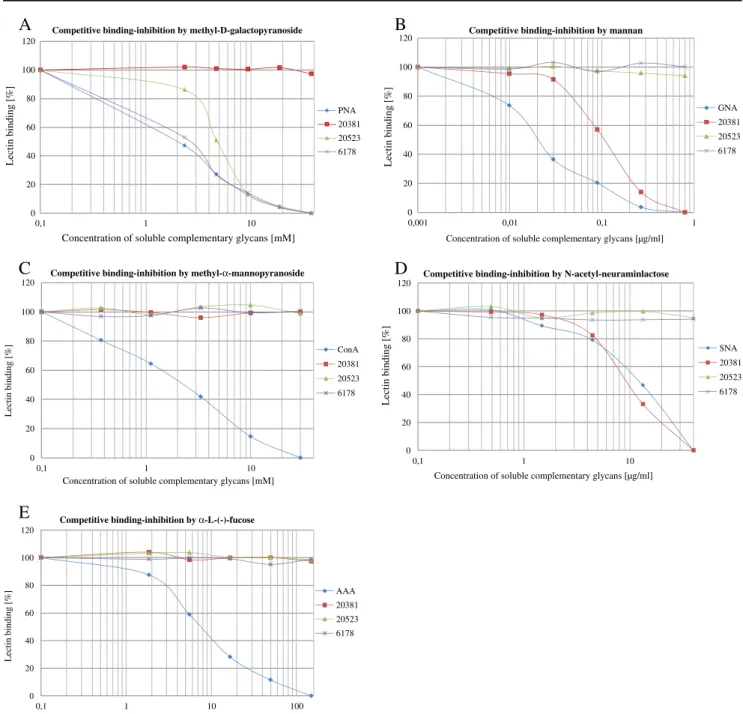
All bacterial lectins showed a reproducible individual binding behaviour with regard to the glycans tested. Figure 3 illustrates the competitive binding inhibition by different soluble complementary glycans towards the tested bacterial strainsS. mutansDSM 20523 and DSM 6178, as well as S. sobrinus DSM 20381 in comparison to the corresponding standard lectin with known specificity for the tested glycans. Each purified standard lectin (PNA (peanut agglutinin), GNA (G. nivalis agglutinin), ConA (Concanavalin A), SNA (S. nigraagglutinin) and AAA (A. anguilla agglutinin)) presents the typical graph of a sigmoidal curve with a clear decrease of the lectin binding (percent) to a rising concentration of the soluble comple-mentary glycan. By using deglycosylated neoglycoproteins, all identified non-lectin bindings between the bacterial homogenates and the immobilized neoglycoproteins could be excluded from further regards.
Earlier reported glycan-binding specificity for terminal galactose residues could be confirmed for the twoS. mutans strains tested. Although minimal concentrations of the soluble complementary glycan seem to be enough to inhibit the lectin binding, differences appear in the degree of the 50% binding inhibition. While approximately 2.5 mM of methyl-D-galactopyranoside was sufficient to reduce S.
mutansDSM 6178, approximately 5.0 mM was necessary to evoke the 50% binding inhibition for S. mutans DSM 20523 (Fig. 3a). Additionally, the finding of a significant mannose-binding specificity in the form of an oligomannose of the S. sobrinus strain could be made. Figure 3b demonstrates that only 0.1 μg/ml appears to be enough to cause the 50% binding inhibition of the bacterial lectin binding. Contrarily, branched mannose structures without terminal mannose residues had no influence on the binding
specificity on none of the tested strains (Fig.3c). Terminal sialyl residues were shown to inhibit S. sobrinusas well. A comparatively high amount of N-acetyl-galactosamine (10 mg/ml) was needed for the 50% inhibition (Fig. 3d). The specificity for terminal fucose could be excluded for all tested mutans streptococci (Fig.3e).
Blotting of the bacterial proteins separated by their molecular weight onto glycan-specific PVP membranes revealed more than one surface lectin responsible for the specific lectin binding for each of the tested strains (Table1
and Fig.4).S. mutansDSM 20523 and DSM 6178 showed PNA-specific surface lectins with varied molecular weights of 90 and 155 kDa.S. sobrinusDSM 20381 showed GNA-specific surface lectins of 35 and 45 kDa. Regarding the SNA specificity of S. sobrinus DMS 20381, no clear information can be given according to the molecular weight of the corresponding surface lectin because of only slight differences between the glycosylated and deglycosylated PVP membranes (Fig.4). A molecular weight around 25 to 35 kDa can be assumed; however, further verification is needed.
Discussion
Long-term survival of bacteria in the oral cavity requires that the microorganisms have the ability to adhere to a tissue surface or colonize a suitable niche in the complex biofilm. One of the many molecular mechanisms involved in initial adherence of bacteria to the acquired pellicle and subsequent development of mixed species oral biofilms is the specific lectin–carbohydrate interaction between the microorganisms and their host [12,13].
Individual host defence factors of the saliva and crevicular fluid protect the human organism to a certain degree of accumulating or invading microorganisms. Besides the systemic immune system, available soluble glycoconjugates in saliva play an important role as a constitutional host defence factor in terms of afirst line of defence[14]. While promoting the bacterial adhesion on the one hand, they can specifically prevent the binding of bacteria by competitively blocking their adhesion on the other hand [12,18] depending on their concentration available.
Whereas Zehetbauer et al. could not detect any significant differences in the salivary protein profiles between children with early childhood caries and caries-free children [19], Seemann et al. found that salivary glycoconjugates of caries-resistant children from a representative sample showed a higher binding inhibition against the lectin PNA (specific for terminal galactosyl residues) compared to saliva of caries-susceptible children [15]. Studies concentrating on patients with periodontal disease revealed a lack of terminalN -acetyl-galactosamine residues, specific for Vicia villosa agglutinin
[16]. Further research demonstrated a significant increase of specific salivary glycoconjugates during a progressive bacterial contamination in the oral cavity [20]. These data indicate that a reduced availability of distinct glycosylation patterns may act as an additional host-derived factor for an increased occurrence of specific microorganisms within a certain disease.
Anti-adhesion therapy aims the inhibition or complete blocking of lectin-mediated adhesion with the intention to reduce the initial adhesion of pathogenic microorganisms
on host tissues without affecting their viability to prevent selective pressure and overgrowth of resistant bacteria. The infection of the urothelium byEscherichia colifor instance could be effectively inhibited after applying lectin-specific methyl-α-D-mannopyranoside [21]. Consequently, this
model can be applied to the colonization of oral pathogens such as cariogenic S. mutans and S. sobrinus in order to prevent initial adhesion as a first step in the multi-factorial caries aetiology. Although the lectin-mediated binding specificities of caries-inducing microorganisms are not very
A
B
C
D
E
0 20 40 60 80 100 120 0,1 1 10 Lectin binding [%]Concentration of soluble complementary glycans [mM] Competitive binding-inhibition by methyl-D-galactopyranoside
PNA 20381 20523 6178 0 20 40 60 80 100 120 0,001 0,01 0,1 1 Lectin binding [%]
Concentration of soluble complementary glycans [µg/ml] Competitive binding-inhibition by mannan
GNA 20381 20523 6178 0 20 40 60 80 100 120 0,1 1 10 Lectin binding [%]
Concentration of soluble complementary glycans [mM] Competitive binding-inhibition by methyl-α-mannopyranoside
ConA 20381 20523 6178 0 20 40 60 80 100 120 0,1 1 10 Lectin binding [%]
Concentration of soluble complementary glycans [µg/ml] Competitive binding-inhibition by N-acetyl-neuraminlactose
SNA 20381 20523 6178 0 20 40 60 80 100 120 0,1 1 10 100 Lectin binding [%]
Concentration of soluble complementary glycans [mM] Competitive binding-inhibition by α-L-(-)-fucose
AAA 20381 20523 6178
Fig. 3 Competitive binding inhibition of the bacterial homogenates fromStreptococcus mutans(DSM 20523),S. mutans(DSM 6178) and
Streptococcus sobrinus (DSM 20381) in the presence of soluble glycans with PNA-specific glycan patterns (a), GNA-specific glycan
patterns (b), ConA-specific glycan patterns (c), SNA-specific glycan patterns (d) and AAA-specific glycan patterns. (e) PNA peanut agglutinin, GNA Galanthus nivalis agglutinin, ConA Concanavalin A,SNA Sambucus nigraagglutinin,AAA Anguilla anguillaagglutinin
well characterized yet, studies revealed that a high molecular mass cranberry constituent was capable of reducing mutans streptococci level in saliva and effectively inhibiting in vitro adhesion to hydroxyapatite [22, 23]. Furthermore, studies in an artificial mouth model showed a caries-preventive effect of an experimental glycan solution prepared by hydrolysis of porcine gastric mucine on natural teeth [24]. Early adhesion experiments by Gibbons and Qureshi revealed that the adhesion of different S. mutans serotypes to saliva-coated hydroxyapatite could be inhibited by galactose and melibiose (6-α-D-galactosyl-glucose) but not
by lactose [25]. Further experiments by other investigators supported the assumption that galactose residues seem to have a significant meaning within the lectin-mediated adhesion of S. mutans [26–28]. In the present study, these findings could be confirmed for the two S. mutans strains (DSM 20523 and DSM 6178) tested in the present study.
Nevertheless, terminal galactose residues had no influence on the adherence ofS. sobrinusDSM 20381, which supports early observations that S. mutans and S. sobrinus attach to different receptors in experimental pellicles [29]. Instead,S. sobrinusshowed a significant mannose-binding specificity in
Fig. 4 Overview of the different binding specificities of Streptococ-cus mutans DSM 20523, S. mutans DSM 6178 and Streptococcus sobrinusDSM 20381 regarding the molecular weights of their specific bacterial surface lectins. For each tested strain, the proteins are displayed in four columns, always in comparison to the Precision Plus molecular weight standard (left column). While the total protein identifies all verifiable proteins within the bacterial homogenate of each strain (both from the surface and the bacterial inside), the biotinylated column shows only the proteins of the bacterial surface membrane with possible lectin specificity. Using glycosylated mem-branes, only the bacterial surface proteins responsible for a specific binding to a certain neoglycoprotein could be identified (PNA-specific
binding ofS. mutansDSM 20523 andS. mutansDSM 6178, as well as the GNA-/ConA-specific and SNA-specific binding ofS. sobrinus
DSM 20381 could be found). By using deglycosylated membranes, the bacterial surface proteins responsible for the lectin-mediated binding specificities could be identified. Only the proteins marked by the arrows bind on carbohydrate–lectin-mediated binding mech-anisms (binding reduction because of the separation of the terminal glycan structures of the neoglycoproteins). For each specific lectin-binding of the tested strains, more than one surface lectin with varied molecular weight could be identified (PNA-specific 90/155 kDa ofS. mutansDSM 20523 andS. mutansDSM 6178; GNA-/ConA-specific 35/45 kDa ofS. sobrinusDSM 20381)
Table 1 Molecular weights of the specific bacterial surface lectins depending on their binding specificities
Binding specificities of the bacterial surface proteins (lectins) DSM 20523 DSM 6178 DSM 20381
β-galacto-1,3-N-acetyl-galactosamine (PNA-specific) 90 kDa 90 kDa –
155 kDa 155 kDa
Terminal mannose residues (GNA-specific) – – 35 kDa
45 kDa
Branched mannose residues (ConA-specific) – – –
α-2,6-Sialyl residues (SNA-specific) – – Further verification needed
form of an oligomannose (GNA specific). Interestingly, a branched monomannose structure without terminal mannose residues (ConA specific) had no influence on the binding specificity on the tested strains. The present results identified another novel aspect of bacterial lectins: terminal sialyl residues were also shown to inhibitS. sobrinus.
The comparable less distinctive binding inhibition of S. sobrinus by sialyl residues may be explained by a glycosylation pattern, which is very similar but does not absolutely correspond to the lectin-binding specificity. Respectively, it can be assumed that there are further surface lectin-binding specifities available forS. mutansas well.
In all bacterial strains, we identified more than one surface lectin (Fig.4) capable of the specific lectin binding with varied molecular weight (S. mutans, 90/155 kDa and S. sobrinus, 35/45 kDa), which supports speculations that epitopes necessary for a specific adherence can be found on multiple proteins [30]. For SNA-specific binding of S. sobrinus, a molecular weight of around 25 to 35 kDa can be speculated, but further investigation is needed (Fig. 4 and Table1). Because of the heterogeneous binding behaviour of caries-relevant microorganisms, anti-adhesive agents should be adapted to the individual adhesion specificity. Further studies concentrating on the revelation of the bacterial surface lectin-binding specificities will elucidate the effec-tiveness of glycoconjugates as anti-adherence agents.
Regarding the established experimental concept and technique, this present in vitro study might be used as a future standard procedure for the identification of the individual binding specificity of predominant caries-inducing microorganisms.
Conclusions
In conclusion, these findings suggest that mutans streptococci possess surface lectins with heterogeneous glycan-binding sites in order to adhere to oral surfaces. Different glycan-binding sites could be identified for theS. mutansstrains in comparison toS. sobrinus. For theS. sobrinus strain, more than one glycan-binding specificity could be detected. An earlier reported glycan-binding specificity for terminal galac-tose residues could be confirmed for theS. mutansstrains. Conflicts of interest The authors declare that they have no conflicts of interest.
References
1. Nurelhuda NM, Al-Haroni M, Trovik TA, Bakken V (2010) Caries experience and quantification ofStreptococcus mutansand
Streptococcus sobrinus in saliva of Sudanese schoolchildren. Caries Res 44:402–407
2. Choi EJ, Lee SH, Kim YJ (2009) Quantitative real-time polymerase chain reaction forStreptococcus mutansand Strepto-coccus sobrinusin dental plaque samples and its association with early childhood caries. Int J Paediatr Dent 19:141–147
3. Loesche WJ (1986) Role of Streptococcus mutans in human dental decay. Microbiol Rev 50:353–380
4. Fitzgerald RJ, Keyes PH (1960) Demonstration of the etiologic role of streptococci in experimental caries in the hamster. J Am Dental Assoc 61:9–19
5. Matsumura M, Izumi T, Matsumoto M, Tsuji M, Fujiwara T, Ooshima T (2003) The role of glucan-binding proteins in the cariogenicity of
Streptococcus mutans. Microbiol Immunol 47:213–215
6. Koo H, Xiao J, Klein MI (2009) Extracellular polysaccharide matrix—an often forgotten virulence factor in oral biofilm research. Int J Oral Sci 1:229–234
7. Ericsson Y (1978) Progress in caries prevention. Caries Res 12 (Suppl):1–112
8. Micheelis W, Schiffner U (2006) Vierte Deutsche Mundgesundheits-studie (DMS IV). Neue Ergebnisse zu oralen Erkrankungsprävalen-zen, Risikogruppen und zum zahnärztlichen Versorgungsgrad in Deutschland 2005, vol. 31. Deutscher Zahnärzte Verlag 9. Marthaler TM (2004) Changes in dental caries 1953–2003. Caries
Res 38:173–181
10. Gibbons RJ (1989) Bacterial adhesion to oral tissues: a model for infectious diseases. J Dental Res 68:750–760
11. Kelly CG, Younson JS (2000) Anti-adhesive strategies in the prevention of infectious disease at mucosal surfaces. Expert Opin Invest Drugs 9:1711–1721
12. Ofek I, Hasty DL, Sharon N (2003) Anti-adhesion therapy of bacterial diseases: prospects and problems. FEMS Immunol Med Microbiol 38:181–191
13. Sharon N (2006) Carbohydrates as future anti-adhesion drugs for infectious diseases. Biochim Biophys Acta 1760:527–537 14. Kage A, Fimmel S, Bernimoulin JP, Hagewald S, Nitschke I, Kage R,
Kottgen E (1995) Oligosaccharides in mucosal host defense: model, method, and first data. Adv Exp Med Biol 371B:1177–1182 15. Seemann R, Zimmer S, Bizhang M, Kage A (2001) Differences in
the salivary glycan pattern between children with high and low caries susceptibility. Caries Res 35:156–161
16. Kage A, Weitzel D, Köttgen E (1989) Novel competitive lectin-binding inhibition assay for quantitative characterization of glycoconjugates using different peroxidase labeled lectins. J Clin Chem Clin Biochem 27:701–702
17. Wu AM, Song SC, Tsai MS, Herp A (2001) A guide to the carbohydrate specificities of applied lectins-2 (updated in 2000). Adv Exper Med Biol 491:551–585
18. Levine MJ, Tabak LA, Reddy MS, Mandel ID (1985) Nature of salivary pellicles in microbial adherence: role of salivary mucins. In: Mergenhagen SE, Rosan B (eds) Molecular basis of oral microbiol adhesion. American Society for Microbiology, Wash-ington, pp 125–130
19. Zehetbauer S, Wojahn T, Hiller KA, Schmalz G, Ruhl S (2009) Resemblance of salivary protein profiles between children with early childhood caries and caries-free controls. Eur J Oral Sci 117:369–373
20. Drews J (2005) Konzentration Lektin-spezifischer Speichelglykane im Verlauf einer experimentellen Gingivits. Zahnmedizinische Dissertation, Zentrum für Zahnmedizin Campus Virchow-Klinikum der Medizinischen Fakultät der Charité, Berlin
21. Aronson M, Medalia O, Schori L, Mirelman D, Sharon N, Ofek I (1979) Prevention of colonization of the urinary tract of mice with
Escherichia coliby blocking of bacterial adherence with methyl alpha-D-mannopyranoside. J Infect Dis 139:329–332
22. Steinberg D, Feldman M, Ofek I, Weiss EI (2004) Effect of a high-molecular-weight component of cranberry on constituents of dental biofilm. J Antimicrob Chemother 54:86–89
23. Weiss EI, Kozlovsky A, Steinberg D, Lev-Dor R, Bar Ness Greenstein R, Feldman M, Sharon N, Ofek I (2004) A high molecular mass cranberry constituent reduces mutans streptococci level in saliva and inhibits in vitro adhesion to hydroxyapatite. FEMS Microbiol Lett 232:89–92
24. Seemann R, Kluck I, Kage A (2006) An in vitro microbial-based model for studying caries-preventive agents. Acta Odontol Scand 64:27–30
25. Gibbons RJ, Qureshi JV (1979) Inhibition of adsorption of
Streptococcus mutansstrains to saliva-treated hydroxyapatite by galactose and certain amines. Infect Immun 26:1214–1217 26. Nagata K, Shibata S, Inoshita E, Tamagawa H, Shizukuishi E,
Tsunemitsu A (1982) The effect of daily mouth rinsing with galactose on dental plaque formation. J Dent Health 32:104– 107
27. Levine MJ, Herzberg MC, Levine MS, Ellison SA, Stinson MW, Li HC, van Dyke T (1978) Specificity of salivary-bacterial interactions: role of terminal sialic acid residues in the interaction of salivary glycoproteins withStreptococcus sanguisand Strepto-coccus mutans. Infect Immun 19:107–115
28. Ligtenberg AJ, Veerman EC, de Graaff J, Nieuw Amerongen AV (1990) Saliva-induced aggregation of oral streptococci and the influence of blood group reactive substances. Arch Oral Biol 35:141S–143S
29. Gibbons RJ, Cohen L, Hay DI (1986) Strains of Streptococcus mutans and Streptococcus sobrinus attach to different pellicle receptors. Infect Immun 52:555–561
30. Murray PA, Prakobphol A, Lee T, Hoover CI, Fisher SJ (1992) Adherence of oral streptococci to salivary glycoproteins. Infect Immun 60:31–38