precursor acute lymphoblastic leukemia
Lay-out: Isabel Sophie Jerchel
Cover design: Henning Jerchel, Isabel S. Jerchel, and Jessica Dobrig
Cover- layout, print, and publishing by: ProefschriftMaken || www.proefschriftmaken.nl © 2017 Isabel Sophie Jerchel. No part of the material protected by this copyright may be reproduced, stored in a retrieval system, or transmitted in any form or by any means without prior written permission from the author, or when appropriate, from the publisher of the publications.
The work described in this thesis was performed at the Department of Pediatric Oncology/ Hematology of the Erasmus Medical Center Sophia Children’s Hospital Rotterdam, the Netherlands and was funded by the Netherlands Organisation for Scientific Research (Nederlandse Organisatie voor Wetenschapplijk Onderzoek, NWO).
Signaling Pathways as Therapeutic Targets in
Pediatric B-cell Precursor Acute Lymphoblastic
Leukemia
Signalwegen als therapeutische doelwitten in pediatrische acute lymfatische leukemie
Proefschrift
ter verkrijging van den graad van doctor aan de Erasmus Universiteit Rotterdam
op gezag van de rector magnificus Prof. dr. H.A.P. Pols
en volgens besluit van het College van Promoties op 14 Maart 2018 om 9:30
door
Isabel Sophie Jerchel
Prof. dr. Rob Pieters Overige leden: Prof. dr. C. Michel Zwaan
Prof. dr. Ivo P. Touw
No Man Is An Island No man is an island Entire of itself Every man is a piece of the continent A part of the main If a clod be washed away by the sea Europe is the less As well as if a promontory were As well as if a manor of thy friend’s Or of thine own were Any man’s death diminishes me Because I am involved in mankind And therefore never send to know for whom the bell tolls It tolls for thee.
Table of Contents
Chapter 1
General Introduction
9
Chapter 2
JAK2 aberrations in childhood B-cell precursor acute
lymphoblastic leukemia
23
Chapter 3
RAS pathway mutations as predictive biomarker for treatment
adaptation in pediatric B-cell precursor acute lymphoblastic
leukemia 61
Chapter 4
High PDGFRA expression does not serve as effective therapeutic
target in ERG-deleted B-cell precursor acute lymphoblastic
leukemia 103
Chapter 5
Fibroblast growth factor receptor signaling in pediatric B-cell
precursor acute lymphoblastic leukemia
123
Chapter 6
EMP1, a novel poor prognostic factor in pediatric leukemia
regulates prednisolone resistance, cell proliferation, migration
and adhesion
135
Chapter 7
General Discussion and Perspective
161
Chapter 8
Summary 175
Chapter 9
About the Author
183
Dankwoord 191
Appendix 197
Chapter 1
Ch
ap
ter 1
Leukemia, or blood cancer, occurs in children and adults, and is divided into subgroups based on cell type and presentation of the disease. It is therefore important to consider how normal blood is formed, which types of leukemia exist, and which mechanisms can cause blood cells to become malignant. This information will help developing treatment strategies.
The hematopoietic system
Blood transports nutrients, waste products, hormones, and cells throughout the body. Of the cellular components red blood cells transport oxygen, platelets form blood clots and mediate wound healing, and the immune system defends the body against pathogens with innate and adaptive immune cells. All blood cells are derived from hematopoietic stem cells (HSC), which reside in the bone marrow and ensure life-long reconstitution of the tissue (Figure 1). The progeny of HSC, so-called progenitor cells, are not yet fully mature (differentiated) and undergo further divisions and differentiation steps. They are divided into two lineages, the myeloid and the lymphoid lineage. The myeloid lineage gives rise to most cells of the innate immune system, platelet-producing megakaryoblasts, and erythrocytes. The lymphoid lineage consists of B- and T-lymphocytes, also called B- and T-cells. These constitute the adaptive immune system.
Clinical presentation of acute leukemia, risk factors, and
chemotherapy
Leukemia is caused by the malignant proliferation of a white blood cell progenitor. It originates from the bone marrow, but at the time of diagnosis cells have usually spread systemically. Depending on the growth speed and whether a uniform population or a mixture of maturation stages is present, clinicians distinguish between acute and chronic leukemia. In addition, the leukemia can bear lineage markers of myeloid or lymphoid cells. These groups define the four major categories chronic or acute myeloid leukemia (CML or AML), and chronic or acute lymphoid leukemia (CLL and ALL). Very rare forms of leukemia present markers of both blood cell lineages (mixed-lineage leukemia), or are myeloid but can be neither defined as acute nor chronic (juvenile myeolomonocytic leukemia, JMML).
Acute lymphoblastic leukemia usually presents with the immunophenotype of B- or T-cell progenitors, and is therefore denominated B-cell precursor ALL (BCP-ALL) and T-cell ALL (T-ALL). Some patients present as mixed-lineage or bilineage leukemia, a more aggressive type that bears markers of both lymphoid and myeloid lineages. These can be further classified according to the genetic changes that initiate the leukemia, and provide a biologically more homogeneous classification (discussed below).
Children rarely present with chronic leukemia, while it is more common in adults. In Europe, approximately 5000 childhood leukemia cases are diagnosed every year, of which three quarters are ALL. BCP-ALL accounts for about 80 to 85% of ALL cases, and is therefore the most common leukemia in children.1 They present with symptoms such as
fatigue, dizziness, anemia, and bone or joint pain. More severe consequences of leukemia are infections due to reduced immunity, and bleeding due to deficient coagulation. The definitive diagnosis is made after a bone marrow puncture.
that one of the hematopoietic stem or progenitor cells transforms into a leukemic cell increases with age. This is largely explained by the fact that exposure to environmental influences (e.g. radiation and carcinogens) increases the frequency at which leukemia-causing mutations accumulate in hematopoietic stem and progenitor cells. In children, the incidence of leukemia peaks around the age of 3 to 5 years, and then declines again. While direct causes remain unknown, environmental toxins and genetic predisposition are thought to play a minor role. Instead, many studies support the hypothesis of a two-step model involving common infections: In addition to a genetic lesions in a progenitor cell, either the aberrant response to, or active infection with one or more infectious agents is thought to promote the development of ALL.2,3
The therapy of leukemia mainly consists of a combination of cytostatic and cytotoxic drugs. Depending on the specific treatment protocols, the exact schedule and therapy can differ. However, the backbone of all ALL treatment protocols relies on five major components: Prednisolone, a glucocorticoid which is toxic to lymphoid cells at high concentrations, vincristine, a spindle-poison which inhibits cell division, and L-asparaginase which degrades the amino acid asparagine, and the antimetabolites methotrexate and 6-mercaptopurine.1 In the most recent Dutch treatment protocol
(DCOG-ALL11), children receive up to 14 different drugs over a period of two years. In cases with high risk of relapse, the therapy is concluded by a hematopoietic stem cell transplantation.
The risks associated with intense chemotherapy and stem cell transplantation are high, and a considerable number of patients die from treatment-related toxicities. Short-term
Ch
ap
ter 1
side effects include nausea, diarrhea, fatigue, moodiness, sleeplessness, allergic reactions, and severe infections. Possible late and long-term side effects are bone necrosis and growth retardation, heart problems, learning disabilities, infertility, and an increased chance of second cancers.
Finding cures for all children, especially those with poor response to current therapy, is a main goal for childhood cancer research.4 However, because the current therapy achieves
cure 75% of cases, a second important goal is minimizing short- and long-term side effects. Therapy is therefore adjusted depending on individual risk factors.
Many risk factors have been identified, including white blood cell count at diagnosis, age above 10 years, male sex, trisomy 21, certain cytogenetic aberrations (e.g IKZF1-deletions), T-ALL, central-nervous system involvement, and poor response to prednisone. Today, the most robust and accurate factor is the measurement of minimal residual disease (MRD).5 MRD is determined in bone marrow aspirates during the first months
of treatment, using leukemia-specific PCR or flow cytometry. Good responses with fast reduction in disease load indicate that further treatment may be safely reduced, but slow reductions are indicative for treatment failure and require treatment intensification.5,6
The identification of risk factors is only a first step, and effective treatment alternatives are warranted to successfully treat high risk cases. Intensified chemotherapy for patients with poor treatment response largely improved the prognosis in recent protocols, but has likely reached the maximum achievable effect.6
In cases of relapse, treatment options are equally limited. The drugs patients receive are usually similar to those that have been given in initial therapy. However, it has been shown that leukemic cells at relapse are more drug resistant, likely due to selection during therapy.7 In addition, relapse therapy often entails stem cell transplantation, which is
associated with high rates of treatment-related death. Novel relapse therapies are urgently needed, and identification of relapse-driving aberrations for targeted therapy and immunotherapy are currently investigated in children with hematological malignancies.8
Pathobiology and genetic aberrations of acute lymphoblastic
leukemia
Leukemogenesis is a multi-step process (Figure 2). In the case of BCP-ALL, it is initiated when a hematopoietic progenitor with B-cell capacities acquires a genetic mutation that disrupts differentiation. Initiating lesions largely determine the biology of leukemic cells, and are therefore used to distinguish the major cytogenetic subtypes. Many are chromosomal translocations that create fusion genes.
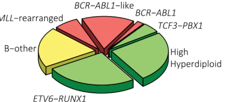
The most frequently detected fusion gene in pediatric BCP-ALL is translocation t(12;21), creating the ETV6-RUNX1 fusion gene (~26% of cases). Another very frequent aberration is the acquisition of additional chromosomes causing high hyperdiploidy (51-67 chromosomes), which is found in approximately 24% of patients. Hypodiploidy (<45 chromosomes) and translocations causing MLL-, TCF3-, or BCR-ABL1-fusions are rare (1%, 7%, 5%, and 2%, respectively).9 The remaining “B-other” group, accounting for
one in every four patients, had previously included all cases lacking the aforementioned aberrations. Recent advances in genomics and transcriptomics have identified several distinct entities in this clinically and biologically heterogeneous group.
The largest group are so-called BCR-ABL1-like cases, which are defined by a gene expression signature similar to that of BCR-ABL1-positive cases.10,11 Although definitions
vary, this poor prognostic group constitutes roughly half of all B-other cases and about 15% of all BCP-ALL cases.12,13BCR-ABL1-like leukemia is, just as BCR-ABL1-positive
leukemia, characterized by aberrant activation of signaling pathways. In many cases this is due to tyrosine kinase fusions involving receptor-associated kinases (e.g. JAK2, ABL1, or ABL2) or receptor-tyrosine kinases (PDGFRB, EPOR, CSF1R).12,14-16 In addition,
overexpression of the cytokine-receptor CRLF2 is found in about half of BCR-ABL1 -like. It is less frequently found among high hyperdiploid, MLL-rearranged, and non-BCR-ABL1-like B-other cases (~10% of all BCP-ALL).12,17 In many cases, high CRLF2
expression is caused by a deletion or translocation that juxtaposes the CRLF2 gene to a new promoter.18,19 The prognostic impact of high CRLF2 expression seems to depend on
the treatment protocol.17,20
Other exclusive, recurrent aberrations within the BCR-ABL1-like group are internal amplification of chromosome 21 (iAMP21, ~2% of all BCP-ALL) and a dicentric chromosome containing parts of chromosomes 9 and 20 (dic(9;20), ~5% of BCP-ALL).21-24
Extensive genomic profiling of the remaining, non-BCR-ABL1-like B-other cases has recently identified so-called ETV6-RUNX1-like cases and recurrent fusion genes involving MEF2D, ZNF384, and DUX4.25-27 Especially the latter discovery solved an
important part of the puzzle around ERG-deletions, a common secondary aberration observed in BCP-ALL. A distinct gene expression profile that is associated with, but not limited to cases with ERG-deletions has been described in 2010.28 Lillebjörn et al. found
that this so-called ERG-cluster overlaps with DUX4-rearrangements, and Zhang et al. provided a mechanistic link between ERG–deletions and DUX4-rearrangements.26,29
Unlike MEF2D- and ZNF384-fusions, DUX4-rearrangements seem to be associated with
Figure 2: Genetic aberrations in the development of BCP-ALL.
Hematopoietic Stem Cell
Myeloid Progenitor
Lymphoid Progenitor pro-B cellpre-B cell
Mature B cell
Proliferation & Survival
(e.g. RAS, CRLF2/JAK2, CDKN2A/B, ABL1, PDGFRB, TP53, RB1)
Self-renewal
Ch
ap
ter 1
a good outcome.28,30,31
Primary leukemogenic aberrations can often be identified in utero, but only a fraction of patients with a detectable fusion gene actually develops leukemia.32-35 This penetrance
varies: while most children with detectable MLL-rearrangements develop disease quickly, only about 1% of cases with an ETV6-RUNX1-fusion present in utero develop leukemia at a later age. This highlights the relevance of cooperative secondary events for a progression to overt leukemia. Besides the postulated infectious stimulus mentioned above, many genetic aberrations have been identified to support leukemogenesis.27,36-42
Most aberrations are not mutually exclusive for a specific subtype, but frequencies often vary among cytogenetic groups.
B-cell transcription factors (e.g. PAX5, IKZF1, TCF3, ETV6, EBF1, ERG, and RUNX1) are frequently inactivated or functionally modified by mutations, deletions, or sequence duplications. These aberrations are thought to sustain a differentiation block in malignant cells. These biological differences have clinical impact: IKZF1-deletions for example, are enriched among kinase fusion-positive BCP-ALL and poor prognostic.11,17,43,44
Deregulation of cell cycle and DNA damage response is the result of frequently observed CDKN2A/B deletions and less frequent loss of RB1 or p53 function.42 They represent
important secondary aberrations that promote proliferation and survival. Mutational activation of the survival-promoting signaling pathways in BCP-ALL mostly affects JAK2/STAT5 and RAS/MAPK signaling, and is discussed below.
A third group of proteins frequently affected by aberrations are epigenetic modulators.39,40,42,45,46CREBBP, for example, is recurrently mutated or deleted and loss
of function is postulated to reduce responsiveness to glucocorticoids.45,46 Their role in
leukemogenesis and suitability as treatment target is only starting to be understood. Mutations conferring a proliferative advantage or resistance to apoptosis will give a competitive edge to expanding (pre-) leukemic cells. This concept of clonal evolution is essential for understanding disease initiation, treatment strategies, and relapse.47,48
While leukemia-initiating aberrations are present in all cells, secondary aberrations are subject to evolutionary selection in a Darwinian model. Longitudinal genomic studies of matched sampled from diagnosis, remission, and relapse(s) have shown that considerable
Figure 3: Estimated frequencies of cytogenetic subtypes in childhood BCP-ALL (adapted from
Pui et al.1).
High
genetic heterogeneity exists at diagnosis and many relapse-forming clones are present as subpopulation.49-52 Specific genetic mutations such as TP53, CREBBP, or those conferring
resistance to chemotherapy (e.g. in NT5C2 or NR3C1) are found more frequently at relapse.45,53,54 50,52
Inter- and intra-cellular signals: Oncogenic pathways and
the microenvironment of leukemia.
Cellular signaling pathways are often affected by oncogenic hits in pediatric BCP-ALL. The aforementioned kinase fusions are an example of primary aberrations deregulating pro-survival signaling. Two major kinase pathways mediate these signals, leading to a mechanistic distinction between JAK- and ABL-type fusions.20 ABL-type fusions result
in strong activation of the MAPK pathway, JAK/STAT pathways, and PI3K/mTOR signaling.20,55JAK2-fusions as well as high CRLF2 expression mainly act oncogenic by
activating the STAT transcription factors and PI3K/mTOR signaling.56
The cytokine receptor CRLF2 lacks a kinase domain, and therefore relies on the associated kinase JAK2 to activate downstream signaling.19,57 Binding of the ligand TSLP
is still required and possibly limiting the oncogenic function of CRLF2. At least partial ligand-independence can be conferred by activating mutations in JAK2 which occur in about 50% of cases with high CRLF2 expression.18,19,58 Interestingly, although PI3K/
mTOR signaling has been shown to be critical for CRLF2-rearranged leukemia, activating mutations in this pathway have not been described.56,59
A second essential oncogenic pathway is the MAPK pathway with its core components RAS, RAF, MEK and ERK. It is one of the most frequently mutated pathways in cancer.60
The Ras GTPases (HRAS, NRAS, KRAS) mediate signals of activated receptor-tyrosine kinases (RTK). They activate the RAF kinases (ARAF, BRAF, and CRAF), which in turn activate MEK1 and MEK2. The only known targets of MEK1/2 are ERK1 and ERK2, but these can interact with and phosphorylate several hundred targets.61,62 Cellular functions
regulated by MAPK signaling include cell cycle progression, cell survival, differentiation, and motility.
In addition to the abovementioned intracellular pathways, extra-cellular signaling mediated by cytokines and growth factors have been shown to regulate normal and malignant hematopiesis.63,64 The release of these factors from and to the microenvironment
plays a crucial role for leukemia development and therapy resistance. Leukemic cells manipulate the bone marrow niche in their favor.65 The cytokine CXCL12, expressed by
stromal cells, can modulate leukemic cell egress, homing, and modulate chemotherapy resistance.66,67 Adhesion molecules such as ITGA4 modulate leukemic cell survival and
chemoresistance.68 Tunneling nanotubes represent another, only recently identified
mechanism of communication between leukemic and bone marrow stromal cells.69
Novel therapeutic approaches for children with acute
lymphoblastic leukemia
Current treatment for children with leukemia comprises more than a dozen therapeutic agents. Decades of clinical trials have massively improved therapy outcomes by optimizing the combination of these drugs. However, survival rates are stagnating, which suggests that therapy has reached its maximum achievable effect.1 The high cure rate of standard
Ch
ap
ter 1
therapy sets the focus on two main goals: identifying cure for those 10 to 15% of cases that relapse, and avoiding overtreatment of cases who are cured to reduce side effects and treatment-related deaths.
Further improvements are likely to come from entirely new therapy classes, such as targeted therapy or immunotherapy. Targeted therapy designates treatment strategies which abrogate the effect of oncogenic drivers and it is often linked to personalized cancer treatment.
Six cellular capacities have been initially suggested to contribute to malignancy in all cancer cells. These hallmarks of cancer are: self-sufficiency in growth signals, insensitivity to anti-growth signals, evasion of apoptosis, limitless replicative potential, tissue invasion and metastasis, and induction of angiogenesis.70 Recently, two more principles have been
added: Deregulated cellular energetics and evasion from immune destruction.71 Many
of these core capacities are driven by genetic aberrations, and usually several genetic aberrations collaborate to establish a malignant phenotype. Some aberrations can affect more than one hallmark, and as a result these aberrations may affect specific hallmarks to a varying extend in distinct tumor types and cases. These principles have to be taken into account to develop and evaluate targeted therapies.
The exploration of signaling pathways as therapeutic targets has grown with the next-generation sequencing revolution. Low costs for sequencing entire genomes facilitate molecular characterization of every tumor. Although few aberrations serve as treatment targets so far (often called “actionable aberrations”), encouraging examples exist. All-trans retinoic acid, for example, specifically induces differentiation in PML-RARA -rearranged acute promyelocytic leukemia. Arsenic trioxide, which targets the PML moiety, induces apoptosis in these cell. The combination of these to drugs now achieves a 4-year overall survival of 98% in this once deadly disease.72
Identification and characterization of the BCR-ABL1-rearrangement led to the application of the first kinase inhibitor (TKI) for cancer treatment: imatinib. It has drastically improved prognosis for CML and BCR-ABL1-rearranged B-ALL in children and adults.73,74 As explained above, tyrosine-kinase fusions represent primary,
leukemia-driving lesions in about 5% of pediatric BCP-ALL cases.15 So far only BCR-ABL1
-rearranged cases receive standard TKI therapy, but preclinical and anecdotal clinical data suggest sensitivity towards inhibition of the JAK and ABL class kinases.12,14,16,56,75-77
Three promises are offered by kinase inhibitors for children with BCP-ALL: First, they may serve as treatment options if standard therapy fails. Second, they could be included upfront for high risk cases to prevent relapses. Third, their anticipated specificity for cancer cells could entail less short-and long-term side effects and allow reduction of chemotherapy.
Despite successful examples such as Imatinib in BCR-ABL1-rearranged CML, vermurafinib in BRAF-mutant melanoma, or ERBB2-specific antibodies in breast cancer, many other approaches have yielded minor benefits and stayed behind the expectations from pre-clinical studies. Pharmacologic hurdles such as competition with high concentrations of ATP (imatinib), or short half-lives (momelotinib) may interfere with efficacy and applicability of kinase inhibitors.78,79
Biologically, clonal evolution has also proven to be a major cause for disease progression on therapy. Any therapy represents a shift in selective pressures and mutations causing drug resistance, e.g. in CREBBP or ABL1 T315I, are therefore positively selected.45
Already diagnostic patient samples contain multiple genetically distinct subclones.80
In BCP-ALL virtually all cells retain leukemia-initiating capacity.34,81,82 Theoretically,
a mutation occurring in any of the leukemic cells present at diagnosis could therefore give rise to a resistant cell. This demonstrates the importance of a combinatorial, multi-targeted therapy.
With these advantages and possible limitations in mind, the aim of this thesis is therefore to evaluate selected signaling pathways as therapeutic targets in BCP-ALL. Five targets, JAK2, the RAS pathway, PDGFRα, FGFR, and EMP1 were selected and assessed for different treatment strategies.
Outline of this thesis
Intra- and inter-cellular signaling pathways have become promising candidates for cancer therapy, because they are frequently affected by oncogenic aberrations and specific small molecule inhibitors are available. The aim of this thesis is to identify and evaluate signaling pathways that may be targeted by novel therapeutic approaches in children with BCP-ALL.
In chapter 2, we determined the frequency of JAK2 mutations and translocations in a large cohort of BCP-ALL patients at diagnosis and evaluated the ex vivo efficiency of two clinically tested JAK inhibitors. We found selective anti-tumor activity of JAK inhibitors in cells carrying JAK2 aberrations. However, we also identified stroma mediated drug resistance, mutational activation of alternative survival pathways, and a signaling rebound effect after inhibitor release as important caveats in JAK inhibitor treatment.
A second frequently mutated signaling pathway is studied in chapter 3: Next generation sequencing was used to detect RAS pathway mutations. We show that clonal or subclonal RAS pathway mutations are present in 44% of BCP-ALL cases at diagnosis. Mutations were associated with poorer ex vivo response to chemotherapy, and clonal but not subclonal mutations associated with a poor prognosis in some treatment groups. RAS mutant BCP-ALL cells were ex vivo sensitive to the MEK inhibitor trametinib, which suggests that this FDA-approved drug could be used to treat BCP-ALL with RAS pathway mutations.
In chapter 4 we study the role of high PDGFRA expression in the newly discovered ERG-deleted leukemia. No genetic aberrations within the PDGFRA locus could be identified that could explain high levels of PDGFRA transcript. Despite being translated and ligand-activated, inhibition of PDGFRα was not cytotoxic in PDGFRA-high BCP-ALL.
Two factors mediating ex vivo glucocorticoid resistance are discussed in the last two chapters of this thesis. In chapter 5 we show that FGFR mutations are rare and occur outside of classical mutational hotspots. The ligand FGF2, however, can reduce the responsiveness of BCP-ALL cells to prednisolone. In chapter 6 we identify EMP1 as a poor prognostic biomarker and regulator of glucocorticoid resistance.
Ch
ap
ter 1
References
1. Pui CH, Evans WE. A 50-year journey to cure childhood acute lymphoblastic leukemia. Semin Hematol. 2013;50(3):185-196.
2. Greaves M. Infection, immune responses and the aetiology of childhood leukaemia. Nat Rev Cancer. 2006;6(3):193-203.
3. Francis SS, Wallace AD, Wendt GA, et al. In utero cytomegalovirus infection and development of childhood acute lymphoblastic leukemia. Blood. 2016. 4. Pui CH, Mullighan CG, Evans WE, Relling MV. Pediatric acute lymphoblastic leukemia: where are we going and how do we get there? Blood. 2012;120(6):1165-1174.
5. van Dongen JJ, Seriu T, Panzer-Grumayer ER, et al. Prognostic value of minimal residual disease in acute lymphoblastic leukaemia in childhood. Lancet. 1998;352(9142):1731-1738.
6. Pieters R, de Groot-Kruseman H, Van der Velden V, et al. Successful Therapy Reduction and Intensification for Childhood Acute Lymphoblastic Leukemia Based on Minimal Residual Disease Monitoring: Study ALL10 From the Dutch Childhood Oncology Group. J Clin Oncol. 2016;34(22):2591-2601.
7. Klumper E, Pieters R, Veerman AJ, et al. In vitro cellular drug resistance in children with relapsed/ refractory acute lymphoblastic leukemia. Blood. 1995;86(10):3861-3868.
8. Bhojwani D, Pui CH. Relapsed childhood acute lymphoblastic leukaemia. Lancet Oncol. 2013;14(6):e205-217.
9. Mullighan CG, Downing JR. Global genomic characterization of acute lymphoblastic leukemia. Semin Hematol. 2009;46(1):3-15.
10. Den Boer ML, van Slegtenhorst M, De Menezes RX, et al. A subtype of childhood acute lymphoblastic leukaemia with poor treatment outcome: a genome-wide classification study. Lancet Oncol. 2009;10(2):125-134.
11. Mullighan CG, Su X, Zhang J, et al. Deletion of IKZF1 and prognosis in acute lymphoblastic leukemia. N Engl J Med. 2009;360(5):470-480. 12. Roberts KG, Li Y, Payne-Turner D, et al. Targetable kinase-activating lesions in Ph-like acute lymphoblastic leukemia. N Engl J Med. 2014;371(11):1005-1015.
13. Boer JM, Marchante JR, Evans WE, et al.
BCR-ABL1-like cases in pediatric acute lymphoblastic
leukemia: a comparison between DCOG/Erasmus
MC and COG/St. Jude signatures. Haematologica. 2015;100(9):e354-357.
14. Roberts KG, Morin RD, Zhang J, et al. Genetic alterations activating kinase and cytokine receptor signaling in high-risk acute lymphoblastic leukemia. Cancer Cell. 2012;22(2):153-166.
15. Boer JM, Steeghs EM, Marchante JR, et al. Tyrosine
kinase fusion genes in pediatric BCR-ABL1-like
acute lymphoblastic leukemia. Oncotarget. 2016. 16. Iacobucci I, Li Y, Roberts KG, et al. Truncating Erythropoietin Receptor Rearrangements in Acute Lymphoblastic Leukemia. Cancer Cell. 2016;29(2):186-200.
17. van der Veer A, Waanders E, Pieters R, et al.
Independent prognostic value of BCR-ABL1-like
signature and IKZF1 deletion, but not high CRLF2 expression, in children with B-cell precursor ALL. Blood. 2013;122(15):2622-2629.
18. Russell LJ, Capasso M, Vater I, et al. Deregulated expression of cytokine receptor gene, CRLF2, is involved in lymphoid transformation in B-cell precursor acute lymphoblastic leukemia. Blood. 2009;114(13):2688-2698.
19. Yoda A, Yoda Y, Chiaretti S, et al. Functional screening identifies CRLF2 in precursor B-cell acute lymphoblastic leukemia. Proc Natl Acad Sci U S A. 2010;107(1):252-257.
20. Izraeli S. Beyond Philadelphia: ‘Ph-like’ B cell precursor acute lymphoblastic leukemias - diagnostic challenges and therapeutic promises. Curr Opin Hematol. 2014;21(4):289-296.
21. Harewood L, Robinson H, Harris R, et al. Amplification of AML1 on a duplicated chromosome 21 in acute lymphoblastic leukemia: a study of 20 cases. Leukemia. 2003;17(3):547-553.
22. Harrison CJ. Blood Spotlight on iAMP21 acute lymphoblastic leukemia (ALL), a high-risk pediatric disease. Blood. 2015;125(9):1383-1386.
23. Rieder H, Schnittger S, Bodenstein H, et al. dic(9;20): a new recurrent chromosome abnormality in adult acute lymphoblastic leukemia. Genes Chromosomes Cancer. 1995;13(1):54-61.
24. Zachariadis V, Gauffin F, Kuchinskaya E, et al. The frequency and prognostic impact of dic(9;20) (p13.2;q11.2) in childhood B-cell precursor acute lymphoblastic leukemia: results from the NOPHO ALL-2000 trial. Leukemia. 2011;25(4):622-628. 25. Yasuda T, Tsuzuki S, Kawazu M, et al. Recurrent DUX4 fusions in B cell acute lymphoblastic leukemia of adolescents and young adults. Nat Genet. 2016;48(5):569-574.
Conclusions and significance of the work presented in this thesis are discussed in chapter 7, including future directives for targeted therapy in children with BCP-ALL. A summary of the work in English and Dutch is given in chapter 8, and detailed information about the author in chapter 9.
26. Lilljebjorn H, Henningsson R, Hyrenius-Wittsten
A, et al. Identification of ETV6-RUNX1-like and
DUX4-rearranged subtypes in paediatric B-cell precursor acute lymphoblastic leukaemia. Nat Commun. 2016;7:11790.
27. Liu YF, Wang BY, Zhang WN, et al. Genomic Profiling of Adult and Pediatric B-cell Acute Lymphoblastic Leukemia. EBioMedicine. 2016;8:173-183.
28. Harvey RC, Mullighan CG, Wang X, et al. Identification of novel cluster groups in pediatric high-risk B-precursor acute lymphoblastic leukemia with gene expression profiling: correlation with genome-wide DNA copy number alterations, clinical characteristics, and outcome. Blood. 2010;116(23):4874-4884.
29. Zhang J, McCastlain K, Yoshihara H, et al. Deregulation of DUX4 and ERG in acute lymphoblastic leukemia. Nat Genet. 2016;48(12):1481-1489. 30. Clappier E, Auclerc MF, Rapion J, et al. An intragenic ERG deletion is a marker of an oncogenic subtype of B-cell precursor acute lymphoblastic leukemia with a favorable outcome despite frequent IKZF1 deletions. Leukemia. 2014;28(1):70-77. 31. Zaliova M, Zimmermannova O, Dorge P, et al. ERG deletion is associated with CD2 and attenuates the negative impact of IKZF1 deletion in childhood acute lymphoblastic leukemia. Leukemia. 2014;28(1):182-185.
32. Taub JW, Konrad MA, Ge Y, et al. High frequency of leukemic clones in newborn screening blood samples of children with B-precursor acute lymphoblastic leukemia. Blood. 2002;99(8):2992-2996.
33. Greaves MF, Wiemels J. Origins of chromosome translocations in childhood leukaemia. Nat Rev Cancer. 2003;3(9):639-649.
34. Greaves M. Molecular genetics, natural history and the demise of childhood leukaemia. Eur J Cancer. 1999;35(14):1941-1953.
35. Mori H, Colman SM, Xiao Z, et al. Chromosome translocations and covert leukemic clones are generated during normal fetal development. Proc Natl Acad Sci U S A. 2002;99(12):8242-8247.
36. Andersson AK, Ma J, Wang J, et al. The landscape of somatic mutations in infant MLL-rearranged acute lymphoblastic leukemias. Nat Genet. 2015;47(4):330-337.
37. Fischer U, Forster M, Rinaldi A, et al. Genomics and drug profiling of fatal TCF3-HLF-positive acute lymphoblastic leukemia identifies recurrent mutation patterns and therapeutic options. Nat Genet. 2015;47(9):1020-1029.
38. Holmfeldt L, Wei L, Diaz-Flores E, et al. The genomic landscape of hypodiploid acute lymphoblastic leukemia. Nat Genet. 2013;45(3):242-252.
39. Huether R, Dong L, Chen X, et al. The landscape of somatic mutations in epigenetic regulators across 1,000 paediatric cancer genomes. Nat Commun. 2014;5:3630.
40. Paulsson K, Lilljebjorn H, Biloglav A, et al. The genomic landscape of high hyperdiploid childhood acute lymphoblastic leukemia. Nat Genet. 2015;47(6):672-676.
41. Pui CH. Genomic and pharmacogenetic studies of childhood acute lymphoblastic leukemia. Front Med. 2015;9(1):1-9.
42. Zhang J, Mullighan CG, Harvey RC, et al. Key pathways are frequently mutated in high-risk childhood acute lymphoblastic leukemia: a report from the Children’s Oncology Group. Blood. 2011;118(11):3080-3087.
43. van der Veer A, Zaliova M, Mottadelli F, et al.
IKZF1 status as a prognostic feature in BCR-ABL1
-positive childhood ALL. Blood. 2014;123(11):1691-1698.
44. Boer JM, van der Veer A, Rizopoulos D, et al. Prognostic value of rare IKZF1 deletion in childhood B-cell precursor acute lymphoblastic leukemia: an international collaborative study. Leukemia. 2016;30(1):32-38.
45. Mullighan CG, Zhang J, Kasper LH, et al. CREBBP mutations in relapsed acute lymphoblastic leukaemia. Nature. 2011;471(7337):235-239.
46. Malinowska-Ozdowy K, Frech C, Schonegger A, et al. KRAS and CREBBP mutations: a relapse-linked malicious liaison in childhood high hyperdiploid acute lymphoblastic leukemia. Leukemia. 2015;29(8):1656-1667.
47. Greaves M, Maley CC. Clonal evolution in cancer. Nature. 2012;481(7381):306-313.
48. Shlush LI, Hershkovitz D. Clonal evolution models of tumor heterogeneity. Am Soc Clin Oncol Educ Book. 2015:e662-665.
49. Mullighan CG, Phillips LA, Su X, et al. Genomic analysis of the clonal origins of relapsed acute lymphoblastic leukemia. Science. 2008;322(5906):1377-1380.
50. Oshima K, Khiabanian H, da Silva-Almeida AC, et al. Mutational landscape, clonal evolution patterns, and role of RAS mutations in relapsed acute lymphoblastic leukemia. Proc Natl Acad Sci U S A. 2016.
51. Ma X, Edmonson M, Yergeau D, et al. Rise and fall of subclones from diagnosis to relapse in pediatric B-acute lymphoblastic leukaemia. Nat Commun. 2015;6:6604.
52. Kuster L, Grausenburger R, Fuka G, et al. ETV6/ RUNX1-positive relapses evolve from an ancestral clone and frequently acquire deletions of genes implicated in glucocorticoid signaling. Blood. 2011;117(9):2658-2667.
53. Stengel A, Schnittger S, Weissmann S, et al. TP53 mutations occur in 15.7% of ALL and are associated with MYC-rearrangement, low hypodiploidy, and a poor prognosis. Blood. 2014;124(2):251-258. 54. Tzoneva G, Perez-Garcia A, Carpenter Z, et al. Activating mutations in the NT5C2 nucleotidase gene drive chemotherapy resistance in relapsed ALL. Nat Med. 2013;19(3):368-371.
55. Cilloni D, Saglio G. Molecular pathways: BCR-ABL. Clin Cancer Res. 2012;18(4):930-937.
56. Tasian SK, Doral MY, Borowitz MJ, et al. Aberrant STAT5 and PI3K/mTOR pathway signaling occurs in human CRLF2-rearranged B-precursor acute lymphoblastic leukemia. Blood. 2012;120(4):833-842. 57. Hertzberg L, Vendramini E, Ganmore I, et al.
Ch
ap
ter 1
Down syndrome acute lymphoblastic leukemia, a highly heterogeneous disease in which aberrant expression of CRLF2 is associated with mutated JAK2: a report from the International BFM Study Group. Blood. 2010;115(5):1006-1017.
58. Harvey RC, Mullighan CG, Chen IM, et al. Rearrangement of CRLF2 is associated with mutation of JAK kinases, alteration of IKZF1, Hispanic/ Latino ethnicity, and a poor outcome in pediatric B-progenitor acute lymphoblastic leukemia. Blood. 2010;115(26):5312-5321.
59. Francis OL, Milford TA, Martinez SR, et al. A novel xenograft model to study the role of TSLP-induced CRLF2 signals in normal and malignant human B lymphopoiesis. Haematologica. 2016;101(4):417-426. 60. Ciriello G, Miller ML, Aksoy BA, Senbabaoglu Y, Schultz N, Sander C. Emerging landscape of oncogenic signatures across human cancers. Nat Genet. 2013;45(10):1127-1133.
61. Caunt CJ, Sale MJ, Smith PD, Cook SJ. MEK1 and MEK2 inhibitors and cancer therapy: the long and winding road. Nat Rev Cancer. 2015;15(10):577-592. 62. Yoon S, Seger R. The extracellular signal-regulated kinase: multiple substrates regulate diverse cellular functions. Growth Factors. 2006;24(1):21-44. 63. Morrison SJ, Scadden DT. The bone marrow niche for haematopoietic stem cells. Nature. 2014;505(7483):327-334.
64. Sison EA, Brown P. The bone marrow microenvironment and leukemia: biology and therapeutic targeting. Expert Rev Hematol. 2011;4(3):271-283.
65. Colmone A, Amorim M, Pontier AL, Wang S, Jablonski E, Sipkins DA. Leukemic cells create bone marrow niches that disrupt the behavior of normal hematopoietic progenitor cells. Science. 2008;322(5909):1861-1865.
66. Jin L, Tabe Y, Konoplev S, et al. CXCR4 up-regulation by imatinib induces chronic myelogenous leukemia (CML) cell migration to bone marrow stroma and promotes survival of quiescent CML cells. Mol Cancer Ther. 2008;7(1):48-58.
67. Hoellenriegel J, Zboralski D, Maasch C, et al. The Spiegelmer NOX-A12, a novel CXCL12 inhibitor, interferes with chronic lymphocytic leukemia cell motility and causes chemosensitization. Blood. 2014;123(7):1032-1039.
68. Hsieh YT, Gang EJ, Geng H, et al. Integrin alpha4 blockade sensitizes drug resistant pre-B acute lymphoblastic leukemia to chemotherapy. Blood. 2013;121(10):1814-1818.
69. Polak R, de Rooij B, Pieters R, den Boer ML. B-cell precursor acute lymphoblastic leukemia cells use tunneling nanotubes to orchestrate their microenvironment. Blood. 2015;126(21):2404-2414. 70. Hanahan D, Weinberg RA. The hallmarks of cancer. Cell. 2000;100(1):57-70.
71. Hanahan D, Weinberg RA. Hallmarks of cancer: the next generation. Cell. 2011;144(5):646-674. 72. Tallman MS, Altman JK. Curative strategies in acute promyelocytic leukemia. Hematology Am Soc Hematol Educ Program. 2008:391-399.
73. Biondi A, Schrappe M, De Lorenzo P, et al.
Imatinib after induction for treatment of children and adolescents with Philadelphia-chromosome-positive acute lymphoblastic leukaemia (EsPhALL): a randomised, open-label, intergroup study. Lancet Oncol. 2012;13(9):936-945.
74. O’Brien SG, Guilhot F, Larson RA, et al. Imatinib compared with interferon and low-dose cytarabine for newly diagnosed chronic-phase chronic myeloid leukemia. N Engl J Med. 2003;348(11):994-1004. 75. David M, Cross NC, Burgstaller S, et al. Durable responses to imatinib in patients with PDGFRB fusion gene-positive and BCR-ABL-negative chronic myeloproliferative disorders. Blood. 2007;109(1):61-64.
76. Lengline E, Beldjord K, Dombret H, Soulier J, Boissel N, Clappier E. Successful tyrosine kinase inhibitor therapy in a refractory B-cell precursor acute lymphoblastic leukemia with EBF1-PDGFRB fusion. Haematologica. 2013;98(11):e146-148.
77. Weston BW, Hayden MA, Roberts KG, et al. Tyrosine kinase inhibitor therapy induces remission in a patient with refractory EBF1-PDGFRB-positive acute lymphoblastic leukemia. J Clin Oncol. 2013;31(25):e413-416.
78. Tyner JW, Bumm TG, Deininger J, et al. CYT387, a novel JAK2 inhibitor, induces hematologic responses and normalizes inflammatory cytokines in murine myeloproliferative neoplasms. Blood. 2010;115(25):5232-5240.
79. Cortes JE, Kim DW, Pinilla-Ibarz J, et al. A phase 2 trial of ponatinib in Philadelphia chromosome-positive leukemias. N Engl J Med. 2013;369(19):1783-1796.
80. Notta F, Mullighan CG, Wang JC, et al. Evolution
of human BCR-ABL1 lymphoblastic
leukaemia-initiating cells. Nature. 2011;469(7330):362-367. 81. Rehe K, Wilson K, Bomken S, et al. Acute B lymphoblastic leukaemia-propagating cells are present at high frequency in diverse lymphoblast populations. EMBO Mol Med. 2013;5(1):38-51.
82. Ebinger S, Ozdemir EZ, Ziegenhain C, et al. Characterization of Rare, Dormant, and Therapy-Resistant Cells in Acute Lymphoblastic Leukemia. Cancer Cell. 2016;30(6):849-862.
Chapter 2
JAK2 aberrations in childhood
B-cell precursor acute lymphoblastic
leukemia
Elisabeth M.P. Steeghs*, Isabel S. Jerchel*, Willemieke de
Goffau-Nobel, Alex Q. Hoogkamer, Judith M. Boer, Aurélie Boeree, Cesca van
de Ven, Marco J. Koudijs, Nicolle J.M. Besselink, Hester A. de
Groot-Kruseman, C. Michel Zwaan, Martin A. Horstmann, Rob Pieters, and
Monique L. den Boer
*E.M.P. Steeghs and I.S. Jerchel contributed equally to this work
Ch
ap
ter 2
Summary
JAK2 abnormalities may serve as target for precision medicines in pediatric B-cell precursor acute lymphoblastic leukemia (BCP-ALL). In the current study we performed a screening for JAK2 mutations and translocations, analyzed the clinical outcome and studied the efficacy of two JAK inhibitors in primary BCP-ALL cells. Importantly, we identify a number of limitations of JAK inhibitor therapy.
JAK2 mutations mainly occurred in the poor prognostic subtypes BCR-ABL1-like and non-BCR-ABL1-like B-other (negative for sentinel cytogenetic lesions). JAK2 translocations were restricted to BCR-ABL1-like cases. Momelotinib and ruxolitinib were cytotoxic in both JAK2 translocated and JAK2 mutated cells, although efficacy in JAK2 mutated cells highly depended on cytokine receptor activation by TSLP. However, our data also suggest that the effect of JAK inhibition may be compromised by mutations in alternative survival pathways and microenvironment-induced resistance. Furthermore, inhibitors induced accumulation of phosphorylated JAK2 Y1007, which resulted in a profound re-activation of JAK2 signaling upon release of the inhibitors. This preclinical evidence implies that further optimization and evaluation of JAK inhibitor treatment is necessary prior to its clinical integration in pediatric BCP-ALL.
Introduction
Janus kinase 2 (JAK2) is a member of the non-receptor tyrosine kinase family and mediates intracellular signaling upon activation of cytokine receptors, which lack an intrinsic tyrosine kinase domain, such as cytokine receptor-like factor 2 (CRLF2). Ligand binding (e.g. TSLP for CLRF2) induces dimerization of cytokine receptors chains, resulting in activation of JAK proteins via cross-phosphorylation. JAKs activate signal transducers of transcription (STATs), which, upon dimerization, migrate to the nucleus and induce transcription of genes involved in differentiation and proliferation of hematopoietic cells.1
JAK2 has seven homologous domains (JH1-JH7). The JH1 and JH2 domains are C-terminally located and comprise the catalytic kinase (JH1) and pseudokinase (JH2) domain. The JH2 domain has a dual regulatory function: exerting a negative regulatory effect on the kinase domain, and facilitating JAK2 activation upon receptor activation by ligand binding.2 The JH3-JH4 domains share homology with Src homology 2 (SH2)
domains and mediate protein-protein interactions. The N-terminal located JH6 and JH7 domains, also known as the FERM domain, are required for binding of JAK2 to cytokine receptors.1
In pediatric BCP-ALL patients, gain of function mutations and translocations affecting JAK2 have been identified.3-15 Genomic translocations of JAK2 have been observed in
high-risk BCR-ABL1-like patients. For several of these fusion genes, constitutive JAK2 kinase activation has been demonstrated.12-14, 16 Point mutations often occur in Down
Syndrome ALL, mainly affect exon 16 (located in the pseudokinase domain), and functionally cooperate with overexpression of the type I cytokine receptor CRLF2.6, 9, 10
Indeed, requirement for the interaction of mutant JAK2 with a cytokine receptor was shown in cell lines models by several groups.6, 8, 9
Mutations and translocations represent biologically distinct entities, but both are potential targets for precision medicines. JAK inhibitors were shown to be effective against mutant and translocated JAK2 in vitro.3, 5, 7, 8, 12, 13, 16, 17 However, in vivo mouse
studies show conflicting data and none has been reported to be curative.13, 17-21 To date,
clinical data with JAK inhibitors are scarce. The Children’s Oncology Group performed a phase 1 dosing study of the JAK inhibitor ruxolitinib, but no cases harboring JAK2 activating mutations or translocations were included.22
Several papers have reported data with a focus on either fusion genes or mutations of JAK2, although often with small sample size or only in specific subtypes of BCP-ALL. Furthermore, most reports lack ex vivo efficacy data of JAK inhibitors in primary leukemic cells. To assess the clinical potential of JAK inhibitors in pediatric BCP-ALL, we performed a comprehensive study to determine the frequency and prognosis of JAK2 mutations and translocations among different subtypes of childhood BCP-ALL. Furthermore, the biological efficacy of the JAK inhibitors momelotinib and ruxolitinib was studied in primary leukemic cells harboring JAK2 aberrations, and the clonal stability of JAK2 mutations was investigated in ALL patient derived xenograft models. We show that JAK inhibitors are overall effective towards BCP-ALL cells, but also identified a number of limitations of JAK inhibitor therapy.
Results
Frequency and type of JAK2 aberrations in BCP-ALL patients
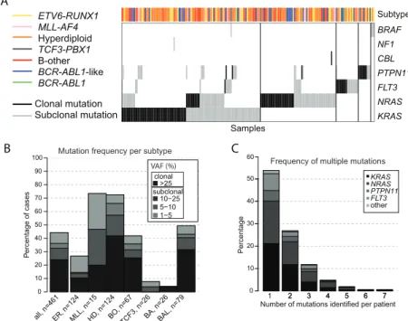
JAK2 mutation status was analyzed in 461 newly diagnosed BCP-ALL cases representing all major subtypes seen in children, with a distribution that is comparable to the general pediatric BCP-ALL population. JAK2 exons 16, 20, 21 and 23 were examined by targeted amplicon sequencing at a median read depth of 673, 577, 711 and 944, respectively. Analyses revealed that 3.5 % (16/461) of these BCP ALL cases harbored JAK2 mutations, which were detected in 7.6 % (6/79) of BCR-ABL1-like cases, 11.9 % (8/67) of non-BCR-ABL1-like B-other cases, and 1.6 % (2/124) of high hyperdiploid cases. No JAK2 mutations were detected in MLL-AF4 (0/15), BCR-ABL1 (0/26), ETV6-RUNX1 (0/124) or TCF3-PBX1 (0/26) cases. The variant allele frequency (VAF) ranged from 1.0 % to 56 % (Figure 1A). Seven patients carried two different JAK2 mutations, and one patient even harbored three different JAK2 mutations. Mutations involved amino acid residue R683 in 13 of 16 mutated cases, which is an important amino acid for the JH2 domain mediated negative auto-regulation of JAK2 activity [23]. CRLF2 overexpression was detected in 87.5 % (14/16) of these cases (Figure 1A, Supplementary Figure 1). One CRLF2 low expressing case harbored a subclonal JAK2 mutation, suggesting that CRLF2 overexpression might be subclonal as well. The other case harbored a JAK2 R923H with a VAF of 50 %, suggesting that this mutation in the kinase domain is not associated with CRLF2 overexpression.
The screen for JAK2 fusion genes was confined to 153 BCP-ALL cases, negative for sentinel BCP-ALL associated lesions (MLL-rearranged, BCR-ABL1, ETV6-RUNX1, TCF3-PBX1, high hyperdiploid), as JAK2 translocations were previously reported in this group of patients.12, 13, 24 No JAK2 translocations were detected in 76 non-BCR-ABL1-like
Ch
ap
ter 2
Patient Protocol Risk group(years)Age (x10WBC 9/L)SyndromeDown (d29/d33)MRD Subtype Translocation Mutation 1 (VAF) Mutation 2 (VAF) Mutation 3 (VAF) CRLF2 status
A20 ALL9 HR 14 75 <10^-3 BCR-ABL1-like p.R873N 40% p.R683S 2% p.R683G 2% High A35 ALL9 NHR 1 44 NA BCR-ABL1-like p.R683T 56% p.R683G 13% High A38 ALL9 HR 6 128,8 NA BCR-ABL1-like p.R683G 32% p.T875N 10% High h g i H % 1 E 4 1 9 K . p % 2 G 3 8 6 R . p r e h t o -B A N s e Y 0 5 4 4 S -R H 3 0 L L A O C 9 2 1 A w o L % 1 G 3 8 6 R . p d i o l p i d r e p y H h g i H A N 9 3 4 S -R H 3 0 L L A O C 9 5 1 A h g i H % 4 2 G 3 8 6 R . p r e h t O -B A N 3 , 4 3 S -R L 3 0 L L A O C 0 6 1 A h g i H % 8 G 3 8 6 R . p % 0 1 N 3 7 8 D . p r e h t o -B A N 0 3 9 S -R H 3 0 L L A O C 7 6 1 A h g i H % 4 G 3 8 6 R . p % 0 3 T 3 8 6 R . p r e h t o -B 3 -^ 0 1 0 1 7 R -R L 3 0 L L A O C 8 7 1 A
A186 COALL97 HR-S 11 9,9 10^-3 BCR-ABL1-like p.T875N37% High A251 COALL97 HR-S 7 115 NA BCR-ABL1-like p.R683G63% High A282 ALL10 SR 3 21 <10^-3 B-Other p.R683S 25% p.R683G 7% High h g i H % 1 4 G 3 8 6 R . p r e h t O -B 3 -^ 0 1 < s e Y 6 , 3 3 R M 0 1 L L A 5 1 3 A h g i H % 5 2 S 3 8 6 R . p % 7 2 G 3 8 6 R . p r e h t o -B 3 -^ 0 1 s e Y 0 9 3 8 R M 0 1 L L A 9 5 3 A
A404 ALL10 MR 8 222,6 <10^-3 BCR-ABL1-like p.R683G26% High h g i H % 5 2 G 3 8 6 R . p d i o l p i d r e p y H h g i H 3 -^ 0 1 s e Y 9 , 1 2 R M 0 1 L L A 3 3 4 A w o L % 0 5 H 3 2 9 R . p r e h t O -B 3 -^ 0 1 < 1 , 2 1 1 1 R M 0 1 L L A 1 2 5 A
A31 ALL9 HR 1 53,8 <10^-3 BCR-ABL1-like PAX5-JAK2 Low A204 COALL03 HR-S 14 4,5 <10^-3 BCR-ABL1-like PAX5-JAK2 Low A286 ALL10 SR 1 3,4 <10^-3 BCR-ABL1-like PAX5-JAK2 Low A214 COALL03 HR-S 11 19,5 10^-3 BCR-ABL1-like TERF2-JAK2 Low A216 COALL03 HR-S 5 204 <10^-3 BCR-ABL1-like BCR-JAK2 Low
JA K2 Tr an sl oc at io n JA K2 M ut at io n 0 5 10 15 Follow-up in years A B Remaining BCP-ALL
JAK2wtB other and BCR-ABL1-like
JAK2 lesions n=279 n=141 n=21 p=0.04 p=0.04 0 2 4 6 8 0 20 40 60 80 100
Time from initial diagnosis (years)
Cumulativ
e incidence of
relapse and non
response (%)
Figure 1: JAK2 aberrations in BCP-ALL patients
(A) Type of lesions, clinical characteristics and follow-up of JAK2 lesion positive patients. Treatment
protocol and risk group assigned to each patient per protocol have been listed (HR-S: High Risk Standard. HR: High Risk. MR: Median Risk. SR: Standard Risk. LR-S: Low Risk Standard. LR-R: Low Risk Reduced. NHR: Non-High Risk). WBC indicates white blood cell count. Minimal residual disease (MRD) levels at day 29/33 of treatment of COALL and DCOG protocol, respectively. Type of translocation or mutation is listed. VAF indicates the variant allele frequency (%). CRLF2 status indicates gene expression below (low) or above (high) the 90th percentile levels. Right panel: Bar plot represents years from diagnosis to event or last contact. In blue: cases in complete clinical
remission. In red: cases with an event (relapse or death). (B) Cumulative incidence of relapse curves
for patients with JAK2 lesions (green line), JAK2 wildtype BCR-ABL1-like and B-other cases (grey
line), and JAK2 wildtype remaining BCP-ALL cases (black line; ETV6-RUNX1, high hyperdiploid,
TCF3-PBX1). Patients were treated according to ALL8, ALL9, ALL10, COALL97 or COALL03 protocol. Cumulative incidence of relapse (CIR) was estimated using a competing risk model. Relapse and non-response were considered as event, and death as competing event. Non-response
was counted as event at day 79. The Gray’s test was applied to test for equality of CIRs (JAK2 lesion
versus remaining BCP-ALL p=0.04; JAK2 wildtype B-other/BCR-ABL1-like versus remaining
activating fusion genes were identified. The cases involved three PAX5-JAK2 cases, one BCR-JAK2 case and one TERF2-JAK2 case (Figure 1A). The PAX5-JAK2 and BCR-JAK2 fusions contained identical exons as reported before [12, 13]. The TERF2-JAK2 case displayed an in frame fusion of TERF2 exon 10 to JAK2 exon 19. All JAK2 fusion genes harbored an intact JH1 kinase domain (Supplementary Figure 2). Gene expression data revealed high expression levels of JAK2 in these cases (Supplementary Figure 3). Absence of the cytokine receptor-binding FERM domain in JAK2 fusion protein suggests that they signal independent of a cytokine receptor.
Clinical characteristics and prognosis of patients harboring
JAK2 lesions
Ten out of sixteen (62.5%) JAK2-mutated patients remained in continuous complete remission at more than 5 years of follow up. The median time to relapse in the six other patients was 2.1 years [range 0.71-7.8 years]. Minimal residual disease (MRD) data were available for nine out of fourteen patients. The four patients with high MRD levels (≥10-3) at day 29/33 of treatment (time point 1 of COALL and DCOG protocol, respectively) relapsed, whereas the remaining five mutated patients with low MRD levels remained in continuous complete remission (p=0.008, Fisher exact test).
Three out of the five cases harboring JAK2 fusion genes remained in continuous complete remission at more than 5 years of follow up, whereas two cases suffered from a relapse within 2.4 years of diagnosis (Figure 1A). Both patients who relapsed were assigned to the High Risk arm of the COALL-03 study protocol because of unfavorable age (>10 years) or high white blood cell count at diagnosis (>50 WBC/nl).
Cumulative incidence of relapse in these JAK2 aberrant patients did not differ from JAK2 wildtype BCR-ABL1-like and B-other cases. Both displayed an unfavorable outcome compared to remaining BCP-ALL cases (p=0.04; Figure 1B). These findings underline the clinical relevance of JAK2 lesions. Mutations and translocations represent biologically distinct entities, but both may be targetable by JAK-inhibitors.
Leukemic cells with JAK2 lesions can be targeted by JAK
inhibitors
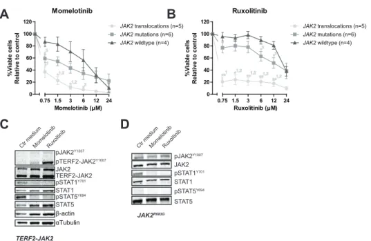
Primary leukemic and patient-derived-xenograft (PDX) cells (Supplementary Figure 4, reference 25) were exposed to momelotinib and ruxolitinib. JAK2 translocated cells were more sensitive to both momelotinib and ruxolitinib compared to JAK2 wildtype cases (P<0.05; Figure 2A-B, Supplementary Figure 5). JAK2 mutated cells were less sensitive to these inhibitors than JAK2 fusion positive cells, and were only marginally more sensitive than wildtype cells (P<0.05). Leukemic cells without genetic JAK2 aberrations were resistant to ruxolitinib, but showed some sensitivity to momelotinib. Importantly, normal bone marrow mononuclear cells were resistant to both inhibitors (Supplementary Figure 5). Both JAK inhibitors effectively reduced levels of phosphorylated STAT5 Y694 and/or STAT1 Y701 (Figure 2C-D, Supplementary Figure 6).
The marginal sensitivity for both inhibitors and the low levels of phosphorylated STAT5 in JAK2 mutated cells may be explained by lack of human TSLP ligand to activate the CRLF2 pathway in our culture conditions. Addition of human TSLP sensitized JAK2 mutated cells to ruxolitinib, but not to momelotinib (P<0.01, Figure 3A-B, Supplementary Figure
Ch
ap
ter 2
Figure 2: The efficacy of JAK inhibitors on JAK2 translocated and mutated cells
(A-B) Leukemic (PDX or primary patient) cells were incubated for four days with to an increasing
concentration range of momelotinib or ruxolitinib, after which cell viability was measured using an MTT assay. Sensitivity of exposed cells was calculated relative to vehicle treated controls. Individual
samples were tested in duplicate. Mean±SEM of five JAK2 translocated cases, six JAK2 mutated cases
and four JAK2 wildtype cases is shown. Cell viability of samples was compared using independent
sample T-test. **p≤0.01, *p≤0.05, 1JAK2 translocations versus JAK2 wildtype, 2 JAK2 translocations
versus JAK2 mutations, 3JAK2 mutations versus JAK2 wildtype. (C-D) TERF2-JAK2 and JAK2
R683G PDX cells were exposed for four hours to vehicle control medium, 1.5 μM momelotinib or 0.75 μM ruxolitinib, after which (phosphorylated) TERF2-JAK2, JAK2, STAT1 and STAT5 levels were analysed using western blot (25 μg lysate).
7A-B). TSLP exposure did not further sensitize JAK2-fusion positive cells, confirming cytokine-independent signaling (Figure 3C D, Supplementary Figure 7C-D). These results confirm that JAK2 signaling triggered by JAK2 fusion proteins is independent of cytokine receptor activation. JAK2 wildtype leukemic cells were not sensitized to JAK inhibitors by TSLP treatment (Figure 3E-F, Supplementary Figure 7E F). In the presence of TSLP both JAK2 translocated and JAK2 mutated cells were sensitive to JAK inhibitors (Figure 3G-H). At the protein level, TSLP exposure upregulated the levels of phosphorylated STAT1 Y701 and STAT5 Y694 in JAK2 R683S mutated cells, whereas no effect was observed in JAK2 fusion positive and JAK2-wildtype leukemic cells (Figure 3I-K). Notably, TSLP triggered the phosphorylation and hence activation of the MEK/ ERK pathway in JAK2 R683S mutated cells, but not in JAK2 R683G mutated cells (Figure 3I, 3L), suggesting that this activation is context-dependent. Phosphorylation of STAT1 Y701 and STAT5 Y694 was inhibited by momelotinib and ruxolitinib (Figure 3L).
JAK2 inhibition results in accumulation of phosphorylated
JAK2
B
A
D
C
Momelotinib 0 20 40 60 80 100 120 JAK2 translocations (n=5) JAK2 mutations(n=6) JAK2 wildtype (n=4) 0.75 1.5 3 6 12 24 Momelotinib (μM) % Vi ab le ce lls R el at iv e to c on tr ol Ruxolitinib 0 20 40 60 80 100 120 JAK2 translocations(n=5) JAK2 mutations (n=6) JAK2 wildtype (n=4) 0.75 1.5 3 6 12 24 Ruxolitinib (μM) % Vi ab le ce lls R el at iv e to c on tr olCtr mediumMomelotinibRuxoltinib
JAK2R683G pTERF2-JAK2Y1007 TERF2-JAK2 STAT5 pSTAT5Y694 STAT1 pSTAT1Y701
Ctr mediumMomelotinibRuxoltinib
pJAK2Y1007 JAK2 TERF2-JAK2 *1 *1,2 *1,2 *2 *3 **1 *1,2 **1,2**1,2 **1,2 *3 *3 STAT5 pSTAT5Y694 STAT1 pSTAT1Y701 pJAK2Y1007 JAK2 β-actin αTubulin
Exposure of primary leukemic cells, harboring TERF2-JAK2 or PAX5-JAK2, to momelotinib and ruxolitinib resulted in accumulation of phosphorylated JAK2 Y1007 fusion proteins (Figure 2C, Supplementary Figure 6). Wash out of both inhibitors induced a slight rebound effect with upregulation of pSTAT1 Y701 and pSTAT5 Y694 in TERF2-JAK2 cells (Figure 4A).
This rebound effect was also observed in the JAK2 V617F -positive leukemic cell line HEL (Supplemental Figure 7). Phosphorylated JAK2 Y1007 accumulated upon exposure to ruxolitinib. Removal of the inhibitor resulted in reactivation of JAK2 signaling, observed by a clear increase in phosphorylated STAT 5Y694 levels within 4 hours (time point 100 hours; Supplementary Figure 7A-B). The inhibitory effect of momelotinib was more transient compared to ruxolitinib, resulting in an earlier reactivation of JAK2, observed by high levels of phosphorylated STAT5 Y694 after 48 hours of momelotinib exposure (Supplemental Figure 7A).
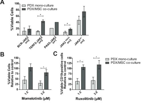
Mesenchymal stromal cells protect against JAK inhibitors
Leukemic cells reside in the bone marrow microenvironment, where they disrupt normal hematopoietic stem cell niches.26 This abnormal niche protects ALL cells against
chemotherapy.27,28 To study whether the bone marrow microenvironment protects
against JAK inhibitors, we mimicked this niche by co-culturing PDX cells with bone marrow mesenchymal stromal cells (MSCs) derived from a leukemia patient. Survival of leukemic cells was improved in co-cultures together with MSCs compared to leukemic cells cultured without MSCs (Figure 5A). In these PDX/MSC co-cultures, JAK inhibitors decreased leukemic cell survival (Supplementary Figure 9A-G). However, leukemic cells were more resistant to ruxolitinib in PDX/MSC co-culture compared to culture without MSCs (P<0.05). A similar trend was observed for momelotinib (Figure 5B-C, Supplementary Figure 9H-I).
Different outgrowth pattern in xenografts
The outgrowth patterns of primary leukemic cells (>90% blast purity) in three NSG mice
Figure 3 (opposite page): The effect of TSLP stimulation on the efficacy of JAK inhibitors
Cells (PDX or primary ALL) were pre-incubated for 1 hour with or without 25 ng/ml TSLP, after which cells were exposed for four days to indicated concentrations of momelotinib or ruxolitinib. Cell viability was measured using an MTT assay. Sensitivity was calculated relative to vehicle treated
controls. Individual samples were tested in duplicate. (A-B) Efficacy of momelotinib and ruxolitinib
on JAK2 mutated cells with or without TSLP pre-incubation. Mean±SEM of six independent
samples is shown. (C-D) Efficacy of momelotinib and ruxolitinib on cells with JAK2 translocations.
Mean±SEM of two independent samples is shown. (E-F) Efficacy of momelotinib and ruxolitinib
on JAK2 wildtype PDX cells. Mean±SEM of three independent samples is shown. (G-H) Combined
graph of the efficacy of momelotinib (G) and ruxolitinib (H) on TSLP stimulated cells with
JAK2 mutations (n=6), JAK2 translocations (n=2), or JAK2 wildtype cells (n=3). Mean±SEM of
independent samples is shown. Cell viability of samples was compared using the independent
sample T-test. **p≤0.01, *p≤0.05. 1JAK2 translocations versus JAK2 wildtype, 2JAK2 wildtype versus
JAK2 mutations. (I-K) Western blot of JAK2 R683S, TERF2-JAK2 and JAK2wt PDX cells with or
without TSLP stimulation (25 ng/ml for 1 hour). (L) JAK2 R683G cells were pre-incubated for 1
hour with or without 25 ng/ml TSLP, after which cells were exposed for four hours to vehicle control medium, 1.5 μM momelotinib or 0.75 μM ruxolitinib. Levels of (phosphorylated) JAK2, STAT1, STAT5, MEK1/2 and ERK1/2 were analysed using western blot.
Ch
ap
ter 2
B
A
D
C
F
E
H
G
I - + J - + K - + 25 ng/ml TSLP LJAK2R683S TERF2-JAK2 JAK2 Wildtype
JAK2 STAT5 STAT1 pMEK1/2S217/221 MEK1/2 ERK1/2 25 ng/ml TSLP pJAK2Y1007 JAK2 pSTAT5Y694 STAT5 pSTAT1Y701 STAT1 pMEK1/2S217/221 MEK1/2 ERK1/2 pERK1/2T202/204 -actin 25 ng/ml TSLP TERF2-JAK2 STAT5 STAT1 pMEK1/2S217/221 MEK1/2 ERK1/2 JAK2 pJAK2Y1007 pJAK2Y1007 pTERF2-JAK2Y1007
pSTAT1Y701 pSTAT1Y701
pSTAT5Y694 pSTAT5Y694
pERK1/2T202/204 pERK1/2T202/204 -actin -actin pSTAT1Y701 pSTAT5Y694 STAT1 JAK2 pJAK2Y1007 pERK1/2T202/204 ERK1/2 Tubulin STAT5 pMEK1/2S217/221 MEK1/2 JAK2R683G 25 ng/ml TSLP - + + + JAK2 Mutation 0 20 40 60 80 100 120 0.75 1.5 3 6 12 24 No TSLP (n=6) 25 ng/ml TSLP (n=6) Momelotinib ( M) %V ia bl e c ell s Re la tive to cont ro l * JAK2 Mutation 0 20 40 60 80 100 120 0.75 1.5 3 6 12 24 No TSLP (n=6) 25 ng/ml TSLP (n=6) Ruxolitinib ( M) %V ia bl e c ell s Re la tive to cont ro l ** ** ** ** JAK2 Translocations 0 20 40 60 80 100 120 0.75 1.5 3 6 12 24 No TSLP (n=2) 25 ng/ml TSLP (n=2) Ruxolitinib ( M) %V ia bl e c ell s Re la tive to cont ro l JAK2 Wildtype 0 20 40 60 80 100 120 0.75 1.5 3 6 12 24 No TSLP (n=3) 25 ng/ml TSLP (n=3) Momelotinib ( M) %V ia bl e c ell s Re la tive to cont ro l JAK2 Wildtype 0 20 40 60 80 100 120 0.75 1.5 3 6 12 24 No TSLP (n=3) 25 ng/ml TSLP (n=3) Ruxolitinib ( M) %V ia bl e c ell s Re la tive to cont ro l Momelotinib In presence of 25 ng/ml TSLP 0 20 40 60 80 100 120 JAK2 mutations JAK2 translocations JAK2 wildtype 0.75 1.5 3 6 12 24 Momelotinib ( M) %V ia bl e c ell s Re la tive to cont ro l *1 *1 Ruxolitinib In presence of 25 ng/ml TSLP 0 20 40 60 80 100 120 JAK2 mutations JAK2 translocations JAK2 wildtype 0.75 1.5 3 6 12 24 Ruxolitinib ( M) %V ia bl e c ell s Re la tive to cont ro l *2 *1,2 * 1,2 *1,2 *1,2 *2
Ctr medium Momelotinib Ruxoltinib
Ctr medium JAK2 Translocations 0 20 40 60 80 100 120 No TSLP (n=2) 25ng/ml TSLP (n=2) 0.75 1.5 3 6 12 24 Momelotinib ( M) %V ia bl e c ell s Re la tive to cont ro l
per patient was determined by paired-end deep-sequencing of JAK2 hot spot regions (exon 16, 20, 21 and 23; median read depth 554, 465, 411 and 593, respectively). PDX cells originating from a JAK2 R863G mutated case had a different VAF profile compared to the original patient sample (Figure 6A, Supplementary Figure 10A). The primary sample contained a major JAK2 R683G clone at a VAF of 63% and a minor KRAS G12D clone at VAF 14%. In two out of three PDX samples generated, the VAF of the JAK2 R683G mutation increased to 98% (PDX1) and 99% (PDX3), whereas in the remaining PDX sample the VAF decreased to 49% (PDX2). In contrast, the KRAS G12D mutation increased to a VAF 23% in this PDX2 sample, whereas this mutation was not detected in PDX1 and PDX3. The reduced VAF of the JAK2 clone in PDX2 did not result in a decreased efficacy of momelotinib or ruxolitinib (Figure 6B). However, levels of pMEK1/2 S217/221 and pERK1/2 T202/Y204 in this sample were increased compared to the other two PDX samples (Figure 6C). Exposure to both JAK inhibitors did not decrease the levels of phosphorylated MEK and ERK.
Sanger sequencing of JAK2 R683S and JAK2 R683T PDX models also indicated a change in the VAF of PDX cells compared to the primary sample. In PDX cells from three NSG mice injected with JAK2 R683S cells, the A/T peak ratio at nucleotide position 2049 differed, suggesting heterogeneity in frequency of JAK2 mutations between samples (Supplementary Figure 10B). Strikingly, two mice injected with JAK2 R683T mutated leukemic cells developed JAK2 wildtype leukemia (Supplementary Figure 10C). Although the JAK2 mutation was lost, CRLF2 expression levels remained high (Supplementary Figure 10D-E). TSLP stimulation activated the JAK2 pathway signaling (Supplementary Figure 10F), but cells were not sensitive for JAK2 inhibition (Supplementary Figure 10G-H).
Discussion
This study aimed to evaluate the clinical need (frequency of lesions and prognostic value) and potential of JAK inhibitors in pediatric BCP-ALL. For this purpose pediatric BCP-ALL patients were screened for JAK2 lesions. JAK2 point mutations were found in 3.6% of our BCP-ALL patients, of which the majority were JAK2 R683 mutations. These mutations were solely detected in BCR-ABL1-like, B-other and high hyperdiploid patients, but not in MLL-AF4, BCR-ABL1, ETV6-RUNX1 or TCF3-PBX1 patients. JAK2 translocations were detected in the poor prognostic BCR-ABL1-like group, but not in non-BCR-ABL1-like B-other cases. The prognosis of patients with JAK2 aberrations was as poor as JAK2 wildtype BCR-ABL1-like and non-BCR-ABL1-like B-other patients. JAK2 mutations were not detected as frequently in our DCOG/COALL sequencing cohort as reported for COG high-risk cohorts.5, 10 Two independent classifiers are used
to describe BCR-ABL1-like BCP-ALL.29-31 Differences in genetic ancestry between the
American COG and European DCOG/COALL cohorts likely affected the signatures to classify patients as BCR-ABL1-like. Hence, genetic differences, or more specifically the lack of Hispanic/Latino cases10, 32 might explain the lower frequencies of JAK2 mutations,
as well as the difference in treatment outcome.
A targeted approach was used to detect JAK2 translocations. All cases with high JAK2 expression levels harbored one of the known fusion genes, making it therefore unlikely
Ch
ap
ter 2
Figure 4: Accumulation of pJAK2 Y1007 results in a rebound effect of JAK2
(A) TERF2-JAK2 PDX cells were incubated for four hours with or without 1.5 μM momelotinib or
0.75 μM ruxolitinib, after which cells were washed to remove the JAK inhibitors. Half of the cells were exposed for another 1.5 hours to 1.5 μM momelotinib or 0.75 μM ruxolitinib, whereas the other cells were incubated in vehicle control (Ctr) medium. Protein expression levels were examined
by western blot (25 μg lysate). (B) HEL cells were incubated with or without 1.5 μM momelotinib or
0.75 μM ruxolitinib for 4 hours, 24 hours, 48 hours, 72 hours and 96 hours, after which