Action of a Transposable Element in Coding Sequence Fusions
James A. Shapiro* and David Leach?
*Department of Biochemistry and Molecular Biology, University of Chicago, Chicago, Illinois 60637, and ?Department of Molecular Biology, University of Edinburgh, Edinburgh EH9 3JR, Scotland
Manuscript received April 16, 1990 Accepted for publication June 12, 1990
ABSTRACT
T h e original Casadaban technique for isolating fused cistrons encoding hybrid /3-galactosidase proteins used a Mucts62 prophage to align the upstream coding sequence and lac2 prior to selection. Kinetic analysis of araB-lac2 fusion colony emergence indicated that the required DNA re- arrangements were regulated and responsive to conditions on selection plates. This has been cited as an example of “directed mutation.” Here we show genetically that the MuA and integration host factor (IHF) transposition functions are involved in the formation of hybrid araB-lac2 cistrons and propose a molecular model for how fusions can form from the initial strand-transfer complex. These results confirm earlier indications of direct M u involvement in the fusion process. T h e proposed model explains how rearranged Mu sequences come to be found as interdomain linkers in certain hvbrid cistrons and indicates that the fusion Process involves a spatially and temporally coordinated sequence of biochemical reactions.
C
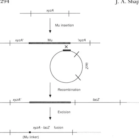
ASADABAN (1976) described a technique for fusing any Escherichia coli coding sequence tol a d . The technique used a transposable Mucts62 pro- phage as portable genetic homology to align the de- sired xyzA coding sequence for protein X and the lac2
cistron (Figure 1). T h e theory of the method was that a subsequent spontaneously arising in-frame deletion would then create an xyyrA-lac2 hybrid coding se- quence directing the formation of an X-P-galactosid- ase fusion protein. T h e kinetics of appearance of colonies carrying fusions between the E. coli araB cistron and lacZ were unexpectedly complex and in- dicated that the underlying genetic process was regu- lated and responsive to conditions on the selective medium (SHAPIRO 1984). T h e cellular events under- lying the kinetics of araB-lac2 fusion colony appear- ance have become an important issue in the “directed
mutation” controversy (CAIRNS, OVERBAUCH and
MILLER 1988; MITTLER and LENSKI 1990). In this paper, we present genetic evidence for a direct role of M u transposition functions in the formation of hybrid araB-lac2 coding sequences and suggest a mo- lecular model of how such functions might act in the fusion process.
Two previous results already suggested that Mu transposition functions played a direct role in the formation of araB-lac2 coding sequence fusions. One was the observation that enhanced repression of the Mucts62 prophage in the presence of a second
Muc+pApl prophage in the pre-fusion strain MCS2
also repressed the appearance of araB-lacZ fusion colonies (SHAPIRO 1984). Another indication of an active Mu role came from sequence analysis of fusions
Genetics 126: 299-299 (October, 1990)
made to a variety of upstream coding sequences; these results showed that Mutts62 excision was often incom- plete, leaving oligonucleotide segments derived from Mu termini as linkers between lac2 and its new 5’
sequence (summarized in SHAPIRO 1987). In some cases (discussed in more detail below), these linker regions were inverted. Concerted excision/inversion events may actually be rather common among trans- posable elements because their occurrence has also been inferred from sequence analysis of reversion events in maize and Antirrhinum (COEN et al. 1989).
MATERIALS A N D METHODS
Bacterial strains: The basic pre-fusion strains MCS2 and
its derivatives MCSl235 and MCS1237 have been described (SHAPIRO 1984). Strain MCS2 was derived from MC4143 (F- araD139 araB::+Mucts62 A[lacIPOZYA, argFlU169Ja relA rpsL) by homology-dependent lysogenization with Xpl (209, U118) as schematized in Figure 1 and described by CASADABAN (1976). These strains differ only in that MCS1235 and MCS1237 have the Mucts62pApl prophage (rather than Mucts62) located between araB and the decap- itated 1acZcistron. The MuA2098::mini-TnIU mutation har- boring a transposon insertion at coordinate 2 kb on the M u
294
I xyzA I
Mu insertion
A. Shapiro and D. Leach
1
g
I
/, MCS2 (2 subclones)0
0
i
M C S 21
RecombinationI
Excision1
.
I
...xyzA - lacZ fusion
(Mu Ilnker)
F I G U R E I .-The CASADARAN ( 1 976) technique for fusing lacZ to
any other coding sequence in the E . coli genome. Homologous
recombination substrates for aligning the two coding sequences are
the end of a MLI prophage inserted i n the correct orientation into
the chosen cistron ( x y z A ) and a terminal fragment of M u located
upstream of a decapitated lacZ cistron in a Xplac bacteriophage.
T h e reciprocal recombin;ttion event depicted integrates the Xplac
and positions lacZ downstream of xyzA to generate the prefusion
structure. There is no promoter for lacZ transcription in the Xplac,
and lacZ has an ochre triplet at codon 18, so that neither transcrip-
tion nor translation can occur without a fusion to upstream se-
quences. An appropriate excision event removes all blocks to tran- scription and translation between the start of xyzA and the region that contains the sequence for the catalytically significant domain
o f @-galactosidase downstream of lacZ codon 18. A small number
of Mu-derived nucleotides are frequently found in the hybrid
coding sequence and constitute the " M u linker" between the xyzA
and l a c 2 donlains.
tant that was also Km"Aps and phage-defective. In both MCS1330 and MCS1366, as mentioned below, the mini- T n 10 marker was located in the Muds62 prophage between
araB and lacZ because it was deleted in the formation of
araB-lac2 fusions. Dilysogenic derivatives of MCSl330 and MCSl366, such as MCSl380, were isolated by transducing the defective lysogens with P1 grown on an arg:: Mucts62pApl strain, selecting for Ap', and screening for
Tc", Arg- and phage production phenotypes. Derivatives of MCS2, MCS1235 and MCS1237 carrying the himA42, himAA~2-Tc,hipll5,hipl57,hu~Al6::KANandhu~Bll::CAM
mutations were isolated by P1 transduction, selection for linked antibiotic resistance markers, and screening (in the case of the himA and hip mutations) for loss of thermosen- sitivity and phage production.
Microbiological methods and scoring of fusion colo- nies: The basic procedures have been described (SHAPIRO 1984). Briefly, subclones of the various pre-fusion strains were grown overnight in TYE broth ( 1 % tryptone, 0.5% yeast extract, 0.5% NaCI, pH 7) at room temperature, 10'- 1 On bacteria of each culture were plated as confluent lawns on L-arabinose
+
lactose selective agar, and the plates were incubated at 32" with daily scoring for the emergence of fusion colonies. The exact titer of each culture was not determined because the kinetics of fusion colony appearance are independent of inoculum size over several orders off
0
0 1 0 1 0 2 0 30
Days132 Days/32
FIGURE 2.-The effect of a M u A defect on fusion colony for- mation. T h e abscissa indicates the number of days of incubation at
32" on selective agar before scoring, and the ordinate indicates the cumulative total number of fusion colonies appearing at each scor-
ing. Although not a l l the separate symbols can be seen in this
computer-generated plot of the data, the results are for six platings
of MCSl330 subclonal cultures and four platings of MCS1366
subclonal cultures, each harboring the MuA2098::mini-TnIU mu- tation.
magnitude (SHAPIRO 1984). In all experiments, replicate cultures were plated and yielded similar results. The data are presented in the form of graphs showing the cumulative colony totals versus days of incubation at 32" (rather than as histograms of new colonies appearing each day) in order to facilitate comparisons between cultures of different ge- netic constitutions.
EXPERIMENTAL RESULTS
Defects in Mu transposition functions inhibit fu- sion colony formation: We took advantage of the recent isolation of a mini-Tn10 insertion into the MuA cistron of the Mud11 168 1 element (SHAPIRO and HIG-
GINS 1989) to examine more directly the role of Mu transposition functions in coding sequence fusion. When this mutation was introduced into the pre- fusion strain MCSP, the appearance of fusion colonies was dramatically inhibited, confirming an active role for the MuA transposition function in the formation of araB-lac2 fusions (Figure 2). In these experiments, no colonies were observed on any plates before the 4th day of incubation, indicating that no fusion events had occurred during growth in TYE broth prior to plating. T h e MCS2 control cultures produced colo- nies starting at day 5 , and all three cultures had over
150 colonies by day 10. T h e MuA-defective cultures
MCSl330 and MCS1366 produced very few colonies
after 22 and 18 days of incubation, respectively. At later times, more fusion colonies did appear on the MCS1330 and MCSl366 plates, but reliable quanti- tative data were difficult to obtain from these older plates because it was difficult to distinguish small fusion colonies from non-fusion papillae. As expected, the late-appearing fusion colonies derived from the MuA2098::mini-TnlO strains had lost the trans- poson tetracycline-resistance marker together with the Mucts42prophage.
0
A
r+
2001 00
0
1 0 20
Days132
FIGURE 3.-The consequences of
mutations affecting I H F and HU
T h e abscissa and ordinate values are as described in the legend to Figure
hupA16::KAN 2. (A) T w o MCS2 cultures are
hupB1l::CAM marked IHF+ and five IHF-defective
B proteins for fusion colony formation.
m A 8 2
and hupB11::CAM mutations (Figure 3). T h e himA and hip loci encode subunits of the IHF protein which is necessary for normal M u transcription (KRAUSE and HICCINS 1986) and which can also play a direct role in the transposition process in vitro (SURETTE and CHACONAS 1989). T h e IHF-defective strains were also inhibited in fusion formation, although not as severely as the MuA derivatives. Reversion of the himA and hip mutations did not accompany Mu excision because fusion derivatives of these strains all tested IHF- (de- termined by sensitivity to Mucts62pApl lysates), and the IHF- phenotype did not affect expression of araB-
lacZ hybrid cistrons because him A and hip fusion strains grew normally on arabinose-lactose selective agar and required arabinose induction for growth. T h e hupA and hupB mutants were moderately re- tarded in fusion colony emergence, consistent with redundancy of these loci for HU protein function. No Hup+Him+ control is shown in Figure 3B because no parental pre-fusion strain was included in the partic- ular series of platings from which these data are taken. However, other control platings gave higher results for Hup+Him+ strains as shown in Figures 2 and 3A.
T h e level of the hupA16::KAN and hupBl1::CAM effects on fusion formation can be estimated by com- paring colony counts after 9 days of incubation from different experiments: for Hup+Him+, there was a mean of 154 (range 130-168, eight cultures); for
hupA16::KAN, there was a mean of 47 (range of 33- 63, four cultures); and for hupBl1:: CAM, there was a mean of 82 (range of 72-9 1, two cultures).
In order to confirm that the inhibition to fusion colony emergence in the MuA2098::mini-Tn10 strains was specific for the MuA defect, we constructed strains with a MuA+ prophage elsewhere in the bac- terial chromosome. This was accomplished by intro- ducing a transposition-competent arg::Mucts62pApl prophage into the genome by PI transduction, select- ing for the ampicillin-resistance marker of the Mu derivative and confirming the arginine requirement. T h e resulting dilysogens showed complementation by producing many more fusion colonies than did their
1 0
Days132
a
E
0
C
ul
-
-
.-
c
-
mI-
-
20
50
-
40-
30
-
20-
cultures are marked himA, hip. These
latter had the hip115 (two cultures),
hip157 (two cultures) and himA42
mutations. (B) Four different cul-
tures had the HimAA82-Tc mutation,
four cultures had the hupA16::KAN
mutation, and two cultures had the hupB1Z::CAM mutation.
MCS1330
0 (3 subcloneq
0 1 0 20 30
Days132
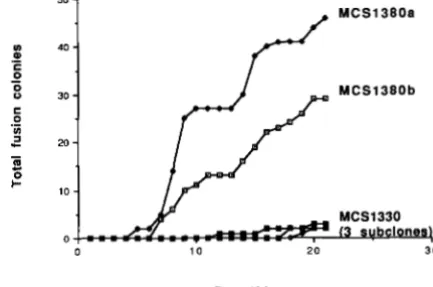
FIGURE 4.-Complementation of the M u A fusion defect. T h e
abscissa and ordinate values are as described in the legend to Figure
2. Strain MCS1380 was derived from the MuA2098::miniTnIO
mutant MCS1330 by introduction of an arg::Mucls62pApl pro-
phage as described in MATERIALS A N D METHODS. T h e results for
two independent dilysogenic cultures are shown.
MuA-defective parents, although complementation resulted in the appearance of fewer fusion colonies than obtained from a M u A + pre-fusion strain (Figure 4). T h e reduced yield of colonies from these dilyso- gens (as compared to the parental MuA+ pre-fusion strains) appeared to result from a lower level of pro- phage derepression when two copies of the Mucts62 repressor locus were present in the genome. We have previously reported enhanced repression of Mucts62 derivatives in other kinds of dilysogens. Addition of a second Mucts62pApl prophage into the parental
MCS2 strain, which had M u A + in cis to araB and lacZ, resulted in delayed and reduced fusion yields (SHAP-
IRO 1984). In addition, the MudII 168 1 cts62dlac fu-
296 J. A. Shapiro and D. Leach
namely that coincident derepression of the active and defective prophages was lethal to potential fusion progenitors. In order to assess whether fusions arose in bacteria that had undergone some general process of M u excision, 26 araB-lacZ fusion derivatives of the arg::Mucts62pApl strains were purified and tested. Because they all retained an active Mucts62pApl pro- phage, no such general excision process had occurred. T h e multiphasic nature of fusion colony appearance in these complemented MuA2098::mini-TnlO/
Mucts62pApl strains was intriguing and merits further investigation because it may shed light on the physio- logical events which lead to M u activation and conse- quent fusion formation. A similar multiphasic pattern was reported for strain MCS2 plated on selective agar enriched with low levels of glucose (SHAPIRO 1984).
We do not know the basis for late fusion colony appearance in lawns of the MuA2098::mini-TnlO bac- teria. Although it is possible that an alternative, Mu- independent fusion pathway was involved, it should be kept in mind that mini-Tn10 insertions are subject to precise excision. Thus, fusion colonies could have arisen from bacteria in the MCS1330 and MCS1366 lawns carrying M u A + revertant prophages. We were, however, unable to detect any tetracycline-sensitive M u A + bacteria in old lawns of MCS1330 and MCSl366 on minimal arabinose-lactose agar by rep- lica-plating of isolated colonies; thus, there was no evidence for a high frequency excision event under selection conditions similar to the excision of bgl::IS103 on salicin-containing plates reported by HALL (1 988).
MOLECULAR MODEL AND DISCUSSION
The results just presented are strong evidence that M u transposition functions play an active role in the emergence of araB-lacZ fusions. Because the transpo- son insertion in the MuA cistron did not produce an absolute block to araB-lacZ fusions, the possibility remains that other kinds of biochemical activities may have come into play and produced fusions after pro- longed incubation. Nonetheless, the initial fusion waves seen on plates seeded with MCS2 must have involved MuA activity because they were eliminated by the MuA2098::mini-Tn10 mutation (Figure 2).
T h e IHF protein involved in expression of M u trans- position/replication functions was also seen to be in- volved in fusion events, although himA and hip muta- tions did not have as strong an effect as the MuA block.
Coding sequence fusion as a complex biochemical event: Sequence analysis had provided one of the initial clues to the role of M u transposition functions in cistron fusions. A number of hybrid 0-galactosidase coding sequences prepared by the Casadaban tech- nique contained nucleotides from the S (or attR) end
Mu S terminus:
AC"CC%CGc G " T T T G CKTTTw;cA AGKXTA'ITT AcGcmTGA PATCGAAAGC
TGFAGcGGcG CACGAAAAAc GCGPAAGCGT TCACGATAPA TGCGAAAACT T T A G c m c G
<""""_ """"""""""
"""____
_____"_"
"__""__
m a l F fusion 6-3:
G A C G C G C T T T C G C G T l T T T C G T Z G c C K T T C A A C C C P A C T T
asp ala l e u ser arg phe ser cys ala ala ser t h r g l n l e u CTGCGCGAAAGCGCAPAAAGCACGCGGCGAAGTTGGGTTGAA
> 222 222 222
FFF F-- __- --- --- -__
___ ___
_-____
__
FIGURE 5.-Sequence of a malF-lacZ fusion isolated by the CAS-
ADARAN technique (FROSHAUER and BECKWITH 1984). The top
sequence is the S terminus of the Mucts62 prophage which abutted
malF nucleotides in the pre-fusion strain. The bottom sequence is the junction between malF and lacZ connected by a Mu-derived linker in the hybrid malF-lacZ cistron. Base-pairs derived from malF are indicated by F, base-pairs from lacZ by Z, and baSe-pdirS from Mu by dashes and (for the terminal base-pair) an arrowhead.
of Mu as a linker between the amino terminal domain of the hybrid cistron and the lac2 sequences (summa- rized in SHAPIRO 1987). These Mu nucleotides were frequently rearranged in a particular way: they were inverted from their position in the prefusion strain, and the terminal sequences previously adjacent to the amino terminal domain of the upstream coding se- quence now abutted the lac2 portion of the hybrid cistron. T h e sequence of malF-lacZ fusion 6-3 (FRO-
SHAUER and BECKWITH 1984) illustrates this structure
(Figure 5 ) . These inverted Mu nucleotides could not have come from a simple Mu-lacZ deletion, and the fact that one of the inversion breakpoints was located precisely at a Mu terminus indicated a role for MuA activity, which is known to cleave the phosphodiester bond between the last Mu nucleotide and the adjacent
chromosomal nucleotide (CRAIGIE and MIZUUCHI
1987).
Molecular model for the role of Mu transposition
functionsincodingsequencefusions:Rearrangements
like the ones reported by FROSHAUER and BECKWITH
A
Mu-Dependent Fusions
B
Isband trenafer
n::
n ::
::
:i
1
Replication initlatlon.,"."* vi
...
I
Strand transferVI VI'
VI' "IX"
I
Strand transfer product1
Cut noncodlng strand, patch and ligate .""""""_"-.._.I.
"_
"P """" A. I .... !.," .czxl". I. .,.Fusion product with Inverted S end linker
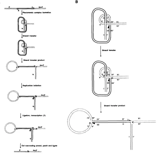
FIGURE 6.-A molecular model for MuA activity in genetic fusions. (A) The top line schematizes the region of a pre-fusion strain with the
Mucts62 prophage located between an upstream transcription unit (P = promoter) and a decapitated lac2 cistron. The two ends of the M u
prophage are indicated c (closest to the repressor cistron, solid arrowhead) and S (closest to the S cistron, open arrowhead). The first step in
the fusion process is the same as the first step in Mu prophage replication and involves the formation of a plectonemic complex bringing
together the two M u termini and the target sequence, which in this case is located in the 5' region of the lacZ cistron, in the appropriate
geometry. The strand transfer reaction results in the ligation of 3' hydroxyl groups from each Mu extremity (here indicated by the open
and solid arrowheads) to 5' phosphate groups spaced 5 base pairs apart at the target sequence; strand transfer also leaves two exposed 3'
hydroxyl groups i n the flanking target DNA which may serve as primers for leading strand chain elongation (indicated by arrowheads) as
well as two 5' phosphate groups which were formerly attached to the Mu extremities. These two steps require Mu A and B and the E. coli
HU proteins (CRAIGIE and MIZUUCH~ 1987). The strand transfer product can be redrawn as a branched molecule with upstream and lac2
sequences in close proximity. If the 3' hydroxyl group on the bottom strand of the lacZ sequence is used for chain elongation, strand
displacenlent will result as indicated (Replication Initiation), and the newly replicated Mu S extremity may then be ligated to the free 5'
phosphate group on the bottom strand of the upstream sequence. This ligation will produce a continuous strand which may serve as a
template for transcription from the promoter (P) in the upstream sequence. Either with or without such transcription, the displaced Mu
D N A sequences may be endonucleolytically cleaved, leading to exonucleolytic resection and removal of the stalked structure. Polymerase
patching using the intact coding strand as a template and religation would then generate a complete fusion product containing the rearranged
Mu S terminal sequences. (B) The easiest way to follow the strand transfers is by labeling segments of the duplexes in the plectonemic
complex and comparing them with the opened strand transfer product. In this cartoon, the arrangement of the plectonemic complex is
depicted with the segments carrying Mucts62 termini passing above (1-1' to 11-11') and below (111-111' to IV-IV') the segment carrying the
J. Shapiro and D. Leach
upstream promoter. In this way, a hybrid P-galactosid- ase could be synthesized from a cell which did not yet contain a stable fusion structure. Removal of the noncoding strand from the transcribed region (per- haps facilitated by R-loop formation), polymerase I patching, and ligation would complete formation of the stable fusion.
The model in Figure 6 provides a straightforward role for MuA activity, consistent with the protein’s known biochemical properties, in coding sequence joining by the CASADABAN technique. It also explains
the structures of complex fusion events with inverted
M u linkers that are difficult to understand on other kinds of break-and-join models. Several additional fusion structures have been described (SHAPIRO
1987). These include fusions with no Mu linkers, fusions with MUS terminus linkers in their original orientations, and at least one fusion with internal rearrangement of the M u linker (FROSHAUER and BECKWITH 1984). We have no detailed explanation for the last type of fusion. T h e model in Figure 6 can readily explain the fusions without a M u linker by postulating that no chain elongation occurs before ligation and patching. T o explain the fusions with MUS terminus linkers in their original orientation requires additional assumptions. (They are not ex- plained by reversing the orientation of M u termini with respect to the target sequence in the plectonemic complex because that leads to fusions containing Muc terminus linkers.) Since MuA appears to be required for most (if not all) fusions, we may assume that an incomplete strand transfer reaction can lead to exo- nucleolytic degradation in both directions from a cleavage in the M u c terminus-lac2 region and that subsequent ligation and patching will produce the fusions without disrupting the linkage between the MUS terminus and the upstream coding domain. T h e model in Figure 6 has one further feature which may prove important in understanding the regulation of fusion events and the emergence of fusion colonies: a hybrid transcription template can be formed before all DNA rearrangements have been completed. It is possible that the hybrid RNA molecules could serve
as templates for guiding the fusion process, possibly by reverse transcription (as suggested by CAIRNS, OVERBAUCH and MILLER 1988) or by other molecular mechanisms that remain to be defined.
T h e observation that Mu transposition functions play an active role in coding sequence joining is con- sistent with the recent results of MITTLER and LENSKI (1 990) and helps to clarify the kinetics of fusion colony appearance. One important step in the DNA re- arrangements needed to generate a fused araB-lacZ coding sequence is the activation of M u transposition functions, and this activation could occur either dur- ing incubation on the selection medium (SHAPIRO
1984) or during prolonged aeration in glucose-mini- mal medium (MITTLER and LENSKI 1990). Such acti- vation would be independent of the presence of ara- binose and lactose and probably would involve proc- esses similar to those which lead to the periodic derepression of a related Mud11 1681 cts62dZac ele- ment in colonies growing on glucose-minimal agar (SHAPIRO and HIGGINS 1989). Once Mu transposition functions are present in the pre-fusion strain, the events leading to the DNA rearrangements cartooned in Figure 6 would require the construction of a mul- ticomponent nucleoprotein complex and the accurate execution of a coordinated series of biochemical re- actions. There could be many possibilities for regula- tion and specificity at this stage of the fusion process,
as suggested by CAIRNS, OVERBAUGH and MILLER
(1 988), and the results of MITTLER and LENSKI (1 990) do not exclude a role for substrate-directed events in steps such as the choice of lac2 target sequences. Only further research will decide whether the strong form of the directed mutation hypothesis (ie. that substrate plays a direct informational role in guiding adaptively useful DNA rearrangements) is correct for the for- mation of araB-lac2 fusions. T h e weak form of the hypothesis ( i e . , that selective conditions can stimulate the occurrence of DNA rearrangements needed for proliferation) has been confirmed in this system by all investigators.
Generality of mutational systems involving trans- posable elements: In discussing the relevance of the
araB-lac2 fusion system to general theories of muta- tion, it has been argued that the presence of a Mucts62 prophage constitutes an artificial or exceptional ele- ment. Such arguments have been raised in evolution- ary discussions ever since the first report of transpos- able elements and the proposal that they are major agents of genetic change (MCCLINTOCK 1950). Now- adays we know that transposable elements are not exceptional but are ubiquitous in the genomes of all organisms that have been studied (BERG and HOWE
1989). In Drosophila, moreover, the large majority of spontaneous mutations involve transposable elements (GREEN 1988), and there are certain naturally occur- ring situations, like hybrid dysgenesis (ENGELS 1989), where transposable elements can bring about major changes in genome structure. Thus, it is not realistic to exclude cases involving transposable elements from general discussions of genetic mutability. One of the salient features of all transposable elements studied is that their DNA rearrangement activities are subject to multiple levels of regulation (BERG and HOWE
We thank PAT HIGGINS for sending us strains carrying the T n 10-
linked himA and hip mutations, DAVID FRIEDMAN for the interrupted
himA, hupA and hupB alleles, NANCY COLE for technical assistance,
and J A C ~ R SHAPIRO for help in preparing Figure 6 on the Macintosh
computer. This research was supported by grant DMB-8715935
from the National Science Foundation.
L I T E R A T U R E C I T E D
BERG, D. E., and M. M. HOWE (editors), 1989 Mobile DNA. Amer-
CAIRNS, J., J. OVERRAUGH and S. MILLER, 1989 T h e origin of
CASADARAN, M. J., 1976 Transposition and fusion of the lac genes
to selected pronloters in Escherichia coli using bacteriophages
lambda and Mu. J . Mol. Biol. 1 0 4 541-555.
<:OEN. E. S., T . P. ROBBINS, J. ALMEIDA, A. HUDSON and R.
C A R P E N ~ E R , 1 9 8 9 Consequences and mechanisms of trans-
position in Antirrhinum majus, pp. 413-436 in Mobile D N A ,
edited by D. E. BERG and M. M. HOWE. American Society for
Microbiology, Washington, D. C.
CRAIGIE, R., and K. MIZUUCHI, 1987 Transposition of Mu DNA:
joining of target DNA can be uncoupled from cleavage at the
ends of M u . Cell 51: 493-50 1.
ENGELS, W . R., 1989 P elements in Drosophila melanogaster, pp.
437-484 in Mobile D N A , edited by D. E. BERG and M. M.
HOWE. American Society for Microbiology, Washington, D.C.
FROSHAUER, S., and J. R. BECKWITH, 1984 T h e nucleotide se-
quence of the gene for malF protein, an inner membrane
component of the maltose transport system of Escherichia coli.
J. Biol. Chem. 259: 10896-10903.
GREEN, M. M . , 1988 Mobile DNA elementsand spontaneousgene
mut;~tion. Banbury Rep. 30: 41-50.
ican Society for Microbiology, Washington, D.C.
m u t ~ n t s . Nature 335: 142-145.
HALL, B. G., 1988 Adaptive evolution that requires multiple
spontaneous mutations. I. Mutations involving an insertion
sequence. Genetics 120: 887-897.
KRAUSE, H. M., AND N. P. HIGGINS, 1986 Positive and negative
regulation of the Mu operator by M u repressor and E. coli
integration host factor. J. Biol. Chem. 261: 3744-3752.
McClintock, B., 1950 T h e origin and behavior of mutable loci in
maize. Proc. Natl. Acad. Sci. USA 3 6 344-355.
MITTLER, J., and LENSKI, R. E., 1990 Further experiments on
excisions of M u from Escherichia coli MCS2 cast doubt on
directed mutation hypothesis. Nature 344: 173-1 75.
SHAPIRO, J. A,, 1979 Molecular model for the transposition and
replication of bacteriophage Mu and other transposable ele-
ments. Proc. Natl. Acad. Sci. USA 76: 1933-1937.
SHAPIRO, J. A,, 1984 Observations on the formation of clones
containing araB-lacZ cistron fusions. Mol. Gen. Genet. 194:
79-90.
SHAPIRO, J. A., 1987 Some lessons of phage Mu, pp. 251-258 in
The Bacteriophage M u , edited by N. SYMONDS, A. TOUSSAINT, P. VAN DE PUTTE, and M. HOWE. Cold Spring Harbor Labo- ratory, Cold Spring Harbor, N.Y.
SHAPIRO, J. A , , and N. P. HIGGINS, 1988 Variation of 0-galacto-
sidase expression from Mudlac elements during the develop-
ment of E. coli colonies. Ann. Inst. Pasteur 1 3 9 79-103.
SHAPIRO, J. A., and N. P. HIGGINS, 1989 Differential activity of a
transposable element in E. coli colonies. J. Bacteriol. 171: 5975-
5986.
SURETTE, M., and G. CHACONAS, 1989 A protein factor which
reduces the negative supercoiling requirement in the Mu DNA
strand transfer reaction is Escherichia coli integration host fac-
tor. J . Biol. Chem. 264: 3028-3034.