Copyright 0 1997 by the Genetics Society of America
The
Caklp
Protein
Kinase
Is
Required at G1/S and GZ/M
in
the Budding
Yeast
Cell
Cycle
Ann
Sutton"and Richard
Freimant
*Department of Biochemistry and Cell Biology and +Graduate Program in Genetics, State University of New York, Stony Brook, New York 11 794
Manuscript received February 17, 1997 Accepted for publication May 21, 1997
ABSTRACT
The CAKl gene encodes the major CDK-activating kinase ( C A K ) in budding yeast and is required for activation of Cdc28p for cell cycle progression from G2 to M phase. Here we describe the isolation of a mutant allele of CAKl in a synthetic lethal screen with the Sit4 protein phosphatase. Analysis of several
different cakl mutants shows that although the G2 to M transition appears most sensitive to loss of Caklp function, Caklp is also required for activation of Cdc28p for progression from GI into S phase. Further characterization of these mutants suggests that, unlike the CAK identified from higher eukaryotes, Caklp of budding yeast may not play a role in general transcription. Finally, although Cakl protein levels and in vitro protein kinase activity do not fluctuate during the cell cycle, at least a fraction of Caklp associates with higher molecular weight proteins, which may be important for its in vivo function.
P
ROGRESSION through two key transition points in the cell cycle, from G1 to S and from G2 to M, is regulated by the activation and inactivation of cyclin- dependent kinases (CDKs). Only one CDK, Cdc28p, is necessary for normal cell cycle progression in the bud- ding yeast Saccharomyces cerevisiae, and in the fission yeast Schizosaccharomyces pombe the cdc2 protein kinase is re- quired. Higher eukaryotes have multiple CDKs; differ- ent CDKs function at different points in the cell cycle (reviewed in NASMITH 1993; NORBURY and NURSE 1993; SHERR 1993). The Cdc28p/cdc2/CDK monomer is in- active; its activity is controlled in the cell cycle through a program of inhibitory and activating phosphorylation events and through the association with different classes of regulatory molecules including cyclins and inhibitors (reviewed in SOLOMON 1993; COLEMAN and DUNPHY 1994; ELLEDGE and HARPER 1994; PETER and HERSKO-In S. cermisiue, exit from GI into S is controlled at a point late in GI called Start and requires the association of Cdc28p with G1-specific cyclins, Clnlp, Cln2p and Cln3p (NASH et al. 1988; HADWICER et al. 1989; RICHARD- SON et al. 1989; CROSS and BLAKE 1993). Association of Cdc28p with Rtype cyclins (Clbs) is necessary for cell cycle progression after Start. Six Rtype cyclins (Clbl- 6p) have been identified, which fall into three classes based on sequence similarity and patterns of RNA accu- mulation during the cell cycle. Of these, Clb2p is the most important for progression from G2 into M; strains deleted for CLB2 have a growth defect and a G2/M delay (SURANA et al. 1991; FITCH et al. 1992; RICHARDSON WITZ 1994b).
Corresponding authw; Ann Sutton, Department of Biochemistry and Cell Biology, State University of New York Stony Brook, NY 11794 5215. E-mail: [email protected]
Genetics 147: 57-71 (September, 1997)
et al. 1992). Clbl,2p/Cdc28p kinase activity has also been proposed to promote the switch in G2 from apical, tipdirected bud growth to isotropic bud growth. As a consequence, cells that are defective in the activation of Clb2p/Cdc28p show hyperpolarized bud growth and an elongated cell morphology (LEW and REED 1993).
58 A. Sutton and R. Freiman
phorylation on threonine 170 or an assembly factor of 36 kD (DEVAULT et al. 1995; FISHER et al. 1995; TASSAN et al. 1995). Recently, these proteins have also been identified as components of the transcription factor TFIIH and shown to phosphorylate the C-terminal re- peat domain (CTD) of the largest subunit of RNA poly- merase I1 in vitro (ROY et al. 1994; SERIZAWA et al. 1995; SHIEKHATTAR et al. 1995). The Cdk7/cyclin H protein complex may therefore have a role in both transcription and in cell cycle progression. Kin28p is a close homo- logue of Cdk7 in budding yeast, and it interacts with a homologue to cyclin H (VALAY et al. 1993). However, although Kin28p appears to be involved in transcription
(CISMOWSKI et al. 1995; VALAY et al. 1995), is associated with TFIIH (FEAVER et al. 1994) and has CTD kinase activity, it does not have detectable CAKactivity (FEAVER et al. 1994; CISMOWSKI et al. 1995). This result suggests that in S. cerevisiae, in contrast to higher eukaryotes, CTD kinase and CAK are encoded by different genes.
Sit4p is a protein phosphatase in S. cerevisiae, which, like Cdc28p, is required for progression from GI into S phase (SUTTON et al. 1991). The Sit4p protein phos- phatase is involved in several aspects of cell cycle control in S. cermisiae (SUTTON et al. 1991; FERNANDEZ-SARABIA et al. 1992). Sit4p is required late in GI for the normal accumulation of CLNl and CLNZ RNA, and conse- quently, temperature-sensitive sit4 strains arrest in late GI at the nonpermissive temperature (SUTTON et al. 1991). However, Sit4p has additional function(s) essen- tial for bud emergence and cell cycle progression (SUT-
and ARNDT 1995). In an effort to identify the additional function(s) for Sit4p in cell cycle progression, we under- took a genetic approach. This approach was based on the fact that the phenotypes of sit4 strains are depen- dent on an additional locus SSDl. SSDl is polymorphic in laboratory strains; Asit4, ssdl-d or Asit4, A s s d l cells are inviable, while Asit4, SSDl-v cells are viable, but with a growth defect and a GI delay (SUTTON et al. 1991). The function of Ssdlp in cells is unknown; how- ever, it has been implicated in a wide variety of cellular processes, including growth control (CVRCKOVA and NASMITH 1993), morphogenesis (COSTIGAN et al. 1992; LEE et al. 1993) and RNA metabolism (STETTLER et al. 1993). The screen that we devised was designed to iden-
tify genes that, when mutated, cause a requirement for SIT4 for viability in an SSDl-v background. This syn- thetic lethal screen was expected to identify genes whose products have functions in the cell cycle that overlap those of Sit4p.
We have recently shown that one of the genes that we identified in the SIT4synthetic lethal screen encodes the major CAK from budding yeast (KALDIs et al. 1996). Using a combination of genetics and biochemistry, we demonstrated that Caklp from S. cerevisiae is a protein kinase with unusual structural features, only distantly related to Cdk7/M015 and other CDKs. In contrast to TON et UZ. 1991; FERNANDEZ-SARABIA et UZ. 1992; DOSEFF
Cdk7/M015, Caklp can function as a monomer. Caklp phosphorylates and activates Clb2p/Cdc28p in vitro and in vivo (KALDIs et al. 1996). Analysis of a tem- perature-sensitive allele of U K l , cakl-22, showed that the majority of cakl-22 cells arrest at the nonpermissive temperature at G2/M. Further, cakl mutations showed genetic interactions with mutations in CLB2, which en- codes the major mitotic cyclin in budding yeast. The genetic interactions and phenotypic defects of the cakl- 22 mutants are consistent with an essential role for Caklp in the activation of Cdc28p for the G2 to M transition (KALDIS et al. 1996). The identification of Caklp as the major CAK in budding yeast has also re- cently been described by others (ESPINOZA et al. 1996; THURET et al. 1996). In this paper, we describe our further characterization of the properties and functions of Caklp of budding yeast. Our analyses of the pheno- types of two different cakl mutants and strain back- grounds show that although the G2 to M transition is most severely affected by loss of Caklp function, Caklp also has an essential role in activation of Cdc28p for the GI to S transition. Analysis of these mutants provides no evidence that Caklp is required for activity of other CDKs in budding yeast.
MATERIALS AND METHODS
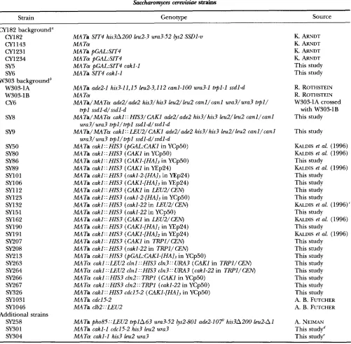
Strains and media: Table 1 shows the genotypes of the
yeast strains used in this study. Unless indicated otherwise, all strains were grown in WD medium (1% yeast extract, 2% Bacto-peptone and 2% glucose), which was supplemented with adenine at 0.3 mM and tryptophan at 0.1 pg/ml. W- gal contained 2% galactose instead of glucose. Wethanol contained 3% ethanol instead of glucose. For experiments in which cells were incubated with 35Smethionine, the synthetic minimal medium was yeast nitrogen base (Difco) supple- mented with adenine at 0.3 mM, tryptophan at 0.1 pg/ml and leucine at 0.2 pg/ml, with 2% galactose as carbon source. For
the acid phosphatase assays, cells were grown in SC (ARNDT
et al. 1989).
Isolation of pdlmutants: Strains CY1231 and CY1234 were grown to stationary phase in W-gal. Cells were incubated for
70 min at room temperature with 30 pl/ml EMS (Sigma). After stopping the mutagenesis with sodium thiosulfate, cells were diluted and plated onto W-gal and subsequently screened for growth on W-gal, but no growth on WD.
Cloning of M l : The CAKl gene was cloned from aYCp50 library containing yeast genomic inserts (ROSE et al. 1987) by transformation of the pGAL:SZT4 cakl-1 mutant and selection
on SGuracil medium. Plasmids that allowed growth on glu- cose were recovered and grouped by restriction pattern analy- sis. Two different groups with overlapping restriction frag-
ments were obtained. The chromosomal locus of one cloned insert was marked with the URA3gene in strain (31231. After crossing to the cakl-1 mutant (SY5), linkage analysis was used to show that the Ura+ phenotype segregated 100% (1 7 tetrads tested) with ability to grow on glucose.
A 3.9-kilobase (kb) region of DNA containing the comple-
menting region of CAKI was inserted into pUC118 in both orientations and sequenced. Subsequent to our sequencing of the CAKI gene, the DNA sequence was released as part of the S. cereuisim genome project. CAKl corresponds to open
Role of Caklp in Budding Yeast 59
TABLE 1
Saccharomyces cereuisiae strains
Strain Genotype Source
CY182 background" CY1 82
CY1 143 CY1231 CY1 234 SY5 SY6
W303 backgroundb W303-1A
W303-1B CY6
SY8
m
SY50 SY80 SY86 SY89 SY1 01
SYl06
5x1 12 SY123 SY132 SY151 SY162 SY190 SY191
sy207
SY208
SY213
SY263 SY264 SY266 SY267 SY326 SY1031 SY1046
SY258 SY301 SY304
Additional strains
MATa SIT4 his3A200 leu2-3 ura3-52 lys2 SSDl-v MA Ta
MA Ta pGAL:SIT4 MATa pGAL:SIT4
MATa pGAL:SIT4 cakl-1 MATa SIT4 cakl-1
MATa add-1 his3-11,15 leu2-3,112 canl-100 ura3-1 t q l - 1 ssdl-d MA Ta
MATa/MATa ade2/ade2 hi53/hi53 leu2/leu2 canl/canl ura3/ura3 t q l /
trpl ssdld/ssdl-d
MATa/MATa cakl:: HIS3/CAKl ade2/ade2 his3/his3 bu2/leu2 canl/canl
ura3/ura3 trpl/trpl ssdl-d/ssdl-d
MATa/MATa cakl ::LEUZ/CAKl ade2/ade2 his3/his3 leu2/leu2 canl/canl
ura3/ura3 trpl/trpl ssdl-d/ssdl-d
MATa cakl :: HIS3 (pGAL:CAKl in YCp50)
MATa cakl :: HIS3 (CAKl in YCp50)
MATa cakl : : HIS3 ( CAK1-[HAI3 in YCp50)
MATa cakl: : HIS3 (CAKl in YEp24)
MATa cakl: : HIS3 (cakl-Z-[HA]j in YEp24)
MATa cakl: : HIS3 ( CAK1-[HAl3 in YEp24)
MATa cakl: : HIS3 (CAKl in LEU2/ CEN)
MATa cakl: : HIS3 (cakl-2-[HA]j in YCp50)
MATa cakl : : HIS3 (cakl-22 in LEU2/ CEN) MATa cakl: : HIS3 (cakl-22 in YCp50)
MATa cakl : : HIS3 ( CAKl in LEU2/ CEN) MATa cakl:: HIS3 ( CAK1-[HAlI in YEp24)
MATa cakl:: HIS3 (CAKl-[HA]2 in YEp24)
MATa cakl:: HIS3 (CAKl in TRPl/CEN)
MATa cakl :: HIS3 (cakl-22 in TRPl/ CEN)
MATa cakl : : HIS3 (pGAL:CAK1-[HAI3 in YCp50)
MATa cakl ::LEU2 clnl ::HIS3 cln3:: URA3 ( M I in TRPl/CEN)
MATa cakl ::LEU2 clnl ::HIS3 cln3:: URA3 (cakl-22 in TRPI/CEN) MATO cakl ::HIS3 cln2:: TRPl (CAKl in YCp50)
MATa cakl ::HIS3 cln2::TRPl (cakl-22 in YCp50)
MATa cakl:: HIS3 cdc15-2 (CAKl-[HA], in YCp50)
MATa cdc15-2 MATa clb2::LEUZ
MATa pho85::LEU2 t q l A 6 3 ura3-52 lys2-801 ade2-107 his3A200 leu2-A1 MATa cakl-1 cdc15-2 his3 leu2 ura3
MATa cakl-1 his3 leu2 ura3
K. ARNDT
K. ARNDT
K. ARNDT K. ARNDT
This study This study
R. ROTHSTEIN
R. ROTHSTEIN W303-1A crossed
with W303-1B This study
This study
KALDIS et al. (1996)
KALDIS et al. (1996) This study
KALDIS et al. (1996) This study
This study This study This study
KALDIS et al. (1996)' This study
KALDIS et al. (1996) This study
KALDIS et al. (1996)
This study This study This study This study This study This study This study This study A. B. FUTCHER A. B. FUTCHER
A. NEIMAN This studyd This study"
a Strains below are isogenic to CY182 except as indicated.
*
Strains below are isogenic to W303-1A except as indicated. "Essentially identical to SY143 (KALDIs et al. 1996). e Obtained from sixth back-cross of SY6 to W3031B.Obtained from cross of SY304 with SYl031.
Mutagenesis of CAKk Deletion of CAKl: A 685-base pair (bp) XbaI-Nszl fragment from the CAKl OW was replaced with a 1.8-kb BamHI fragment containing the HIS3 gene to create plasmid SB14. SB14 was digested with SphI (site is in vector, adjacent to cloned insert) and EcoRI and the
Acakl ::HIS3 fragment transformed into diploid strain CY6. The diploid transformants were sporulated and tetrads dis- sected. Each tetrad (>40 tested) gave rise to two viable His- colonies and two microcolonies (50-100 cells) presumed to be His+. To create cakl ::LEU2, the XbaI-Nsd fragment of
CAKl was replaced with the 2.2-kb SalI-XhoI fragment of LEU2.
Epitope tagging of Caklp: Oligonucleotide-directed muta- genesis (KUNKEL 1985) was used to create a NotI restriction site immediately upstream of the termination codon for CAKl
cloned in plasmid pUC118. To create CAKl-[HA],, a NotI
60 A. Sutton and R. Freiman
Oligonucleotidedirected mutagenesis was used to create the mutant cakl-2 protein in which lysine (K) at position 31 was changed to arginine (R)
.
The HA epitope-tagged version of cakl-2p (cakl-2p-HA) was obtained by replacing a 830-bp EcoRV fragment of SB27 (see above) with an EcoRV fragment containing the K31R mutation. The constructs were con- firmed by DNA sequencing.The CAKl wild-type and mutant genes were cloned into the BamHI site of plasmids YCp50, YEp24, LEU2/CEN or
TRPl/CEN as described (KALDIS et al. 1996). Plasmids were transformed into diploid strain SY8 (CAKl/cakl ::HIS3) or SY9 (CAKl/cakl ::LEU2) followed by sporulation and dissec- tion to obtain cakl ::HZS3 or cakl ::LEU2 haploid cells con- taining the appropriate plasmids.
Preparation of cellular extracts and Western immunoblot-
ting: Cell extracts were prepared as previously described (SUTTON et al. 1991). Preparation of samples for Western analysis was as described (SUTTON et al. 1991). Following elec- trophoresis, samples were transferred to Immobilon-P mem- brane (Millipore) using a Milliblot-SDE system (see Figure 5 )
or to nitrocellulose using a BioRad Trans-Blot SD Electropho- retic Cell (see Figure 6). Proteins were detected by ECL ( A m - ersham) according to instructions from the manufacturer.
Immunoprecipitation and in vitro kinase assay: For 35S la- beling of proteins, cells were grown as previously described (SUTTON et al. 1991). For the experiments described in Figure 5, cell extracts were prepared from "S-labeled cells grown in synthetic minimal medium with galactose or from cells grown to early-logarithmic phase (boo = 0.5) in WD and immuno- precipitation carried out essentially as described (SUTTON et
al. 1991). For each sample, 300 pl of extract (8 mg/ml pro- tein) were incubated with 2 pl of 12CA5 monoclonal antibody (MAb) ascites for 1 hr and subsequently mixed with 30 p1 of a suspension of protein A-Sepharose beads (Pharmacia). After rocking for 1 hr at 4", the beads were washed four times as described (SUTTON et al. 1991). The samples from the 35S- labeled cells were then prepared for electrophoresis as de- scribed (SUTTON et al. 1991). The beads for the samples to be analyzed for kinase activity were washed an additional two times with kinase reaction buffer (50 mM Tris, pH 7.5, 10 mM MgCI2, 1 mM DTT) and then all remaining liquid was removed by aspiration. For the in vitro kinase reaction, 4 pl of 2X kinase reaction buffer, 1 pl y3'P-ATP (3000 Ci/mM), 2 pl ATP ( 3 p M ) and 1 pl H 2 0 was added to the washed beads, which were then incubated for 15 min at 37". The reaction was stopped by adding 20 pl of 2 X gel sample buffer (SILHAVEY
et al. 1984). The samples were heated to 95" for 5 min, centri- fuged at 16,000 X g for 3 min and loaded onto an SDS- polyacrylamide gel. Following electrophoresis, the gel was fixed in 25% methanol, 10% acetic acid, dried and visualized by autoradiography. Phosphatase inhibitors were found not to affect the results of the protein kinase assay and were there- fore omitted.
For the protein kinase assays described in Figure 6, cell extracts were prepared and immuoprecipitation of Caklp-HA was as described above except that for the cdcl5 synchroniza- tion experiments, 200 pl of extract (0.9 mg/ml protein) was used for each immunoprecipitation with 0.5 pl of 12CA5 MAb, and for the elutriation experiment, 100 pl of extract (0.4 mg/ml protein) was used with 0.5 p1 of 12CA5 MAb. After incubation of the extracts with the antibody and subse- quently with protein A beads, the beads were washed four times as described (SUTTON et al. 1991), two times in CAK assay buffer (80 mM Tris, pH 7.3,20 mM EGTA, 15 mM MgC12, 10 mM D m , 1 mg/ml ovalbumin, 1.25 p~ each of leupeptin, antipain, chymostatin and pepstatin) and then remaining liq- uid was removed by aspiration. For the in vitro kinase assay, 10.5 pl of CAK assay buffer, 1 p1 yJ'P-ATP (3000 Ci/mM), 1.6
~1 ATP (100 p M ) , 0.4 p1 1 M MgCI2 and 1 pl Cdk2 (KALDIS et al. 1996) was added to the beads. After incubation for 30 min at 24", the reactions were stopped with 20 p1 2X gel sample buffer and processed as described above.
Liquid acid phosphatase assays: Quantitative liquid acid phosphatase assays were done essentially as described in O'NEILL et al. (1996). Cells were grown in high phosphate (SC) medium to 11600 of between 0.4 and 1.2.
Northern analysis and probes: Northern analysis was per- formed as described (FERNANDEZ-SARABIA et al. 1992). The probes used were the 750-bp XhoI-Hind111 fragment of CLN2,
the 2.7-kb EcoRI fragment of RNRI, the 2-kb HindIII-XhoI fragment of CLB2, the 600-bp EcoRI-Hind111 fragment of
ACTl, a 1.2-kb fragment from YEp24 for URA3 and a 1.1-kb XhoI-Hind111 fragment of LIDC28. The probe for CLB3 RNA was a 1.2-kb BamHI fragment that was generated from restric- tion digestion of a CLB3 gene that has a naturally occurring BamHI site at the 5' end of the ORF and a genetically engi- neered BamHI site near the 3' end of the ORF (provided by
B. FUTCHER) .
Immunofluorescence: 4'-6'-diamidino 2-phenylindole (DAPI) staining was carried out as previously described (SUT- TON et al. 1991). Cells were viewed with a Zeiss Axioskop microscope with a X100 objective and Kodak Tmax p3200 film was used for photography.
Cell cycle synchronization: Cells were synchronized with a-
factor as described (FERNANDEZ-SARABIA et d . 1992) using a-
factor at a final concentration of 0.012 mM. Cells with a tem- perature-sensitive cdc15-2 allele were synchronized by shifting an exponentially growing culture from 24 to 37" for 3.3 hr. Cells were then released from the block by shifting cultures to a 24" water bath. Centrifugal elutriation of cells was as described (SUTTON et al. 1991) except that the cells were grown in W-ethanol. Approximately 10'" cells from an expo- nential culture were loaded into the elutriator. Cells were prepared for flow cytometry as described (FERNANDEZ-SARABIA
et al. 1992) and were analyzed with a Coulter EPICS Elite cytometer. Cell volume distributions were obtained using a Model ZM Coulter channelizer as described (NASH et al.
1988).
RESULTS
Identification of the CAKl gene in a synthetic lethal screen with SIT* In an ssdl-d strain, SIT4 is essential. In an SSDl-v background, loss of Sit4p function does not cause lethality (although cells grow slowly) (SUTTON et al. 1991). SSDl-v strains of opposite mating type in which SIT4 was under control of the GAL1 promoter (CY1231 = MATa pGAL:SIT4 SSDl-v, CY1234 = MATa pGAL:SIT4 SSDl-v) were created. In galactose medium, these strains grew well because SIT4 was expressed (Fig- ure 1A). In glucose medium, expression of SIT4 was repressed. However, the strains were viable because of SSDl-v, but grew at a slow growth rate (Figure 1A). Strains cy1231 and CY1234 were grown to stationary phase in YP-gal. The cells were mutagenized with EMS. Following mutagenesis, cells were diluted and plated onto YP-gal medium. Approximately 40,000 colonies of each mating type were screened by replica plating f o r
Role of Caklp in Budding Yeast 61
A
Galactose
Glucose
p a L:SIT4
p d / 3 - 1
pGA L:SIT4
WT
B
WTF I G ~ ~ R I ~ . l.-rcnkI-I mutants require SIT4 for viability. (A) A strain i n which SIT4 is rlndcr control of the
G \ I J promoter (pC;,\I,:SlT4 = CY1 234) grows as well as a wiltl-type strain (\\T = CY1 143) on YP-gal me- dium (galactose), Ixlt also grows (slowly) on JTD (glrtcosc). If the pG\I.:
SIT4strain also has a muta- tion in PI)I.3/G4h'l (i)GAl,:
SIT4 pd13-I = .W.5), i t grows well on galactose, but docs not grow o n glu-
mutation (pd13-I = SY6) does not have a mqjor cf- fect on the growth rate of
SIT4 cells on either galac- tose or glucose. Cells were grown a t 30" for 2 days (glucose) or 3 days (galac-
a n elongated morpI1oloU. tose). (B) m k l - 1 cells haw
'
Micrographs of wild-ppeI
( M T = CY182) and mltf-1 cells (.SY6) rlsing Nomarski optlcs and D A N staining of DNA. Bar, 8 pm. pG4L:SIT4,pdl3
-
1nornarskl
c a k l -
I
7L-i..
-
COSC. Thc idI3- l / r ~ l < I - l
nomarskl
I
.
DA PI
this screen. The genes containing these mutations were named PDI, for phosphatase deficient lethals. For most of the complementation groups, only one member was isolated, indicating that the screen was not saturating. O n e of the mutations chosen for fk-ther study (pd13-1) caused a slight growth defect when SIT4 was expressed, b u t n o growth when SIT4 was not expressed (Figure 1A). Because we have recently determined that
1'D1,3
encodes an unusual protein kinase that is the major CAK in S . c m m i ~ i n p ( KMDIS PI n/. I!?%), we have re- named this gene C A K I .
cakl-2 cells are delayed in cell cycle progression late in GP: T o study the phenotypes of rtrltl-1 strains, we crossed the /IGAI,:SIT~ m l t l - I strain (SY.5) to the paren- tal strain in which SIT4 was under control of it? own promoter (CY1 82). Following sporulation and dissec- tion of tetrads, we obtained a SI774 m l z l - I strain (SY6).
This m l t l - 1 strain had a striking, elongated cell shape cornpared to wild-type strains (Figure 1B). Further- more, an asynchronous population of c d l - 1 cells showed an enrichment in budded cells compared to wild type (69% compared to 44%). DAPI staining of the DNA for rtrltl-I cells showed that many of the bud-
ded cells had an undivided nucleus at the neck between the mother and bud (Figure 1 B) and staining of micro- tubules with an antitubulin antibody revealed that these cells had a short spindle spanning this nucleus (data not shown). The phenotypes of the cnltl-1 mutants are v e n similar to those of strains deleted for the gene
encoding the major mitotic cvclin CIB2 (SCRAM PI n/. 1991). This result suggests that the cnltl-1 mutation, like the cnkl-22mutation (KAI.DIS PI
NI.
1996), may primarily affect the ability of the cells to progress through GL' into M.T h e m l r l - I mutation is not temperature sensitive, but caused a growth defect at 30" in a wild-type SIT4 back- ground compared to an isogenic CAKI strain (120 min doubling time in YPD compared to 80 min for wild tvpe). Flow cvtometn analysis of asynchronous m l t l - 1 cells showed an enrichment in cells that have a 2N DNA content compared to the isogenic CAKI strain (Figure 2A). Such an enrichment in cells with a 2N DNA con- tent could result from a shortened GI phase or an elon- gated G., o r M phase in the cnlrl-1 mutant. Flow cytome-
62 A. Sutton and R. Freiman
A
Asynchronous
B
Synchronous
7 5
I
W T c a k l - 1 W T c a k l - 1
C
a-factor arrest/release
*-wi l
d type
CL NZ
j c a k l - 1
l w i l d type RNR 1
' rl)llta) c . I r*.CWr c a k 7 - 1
1
wild typeCL B3
y c a k l - 1
P
CLB2
k
I
wild type
wild type
0 1 8 3 6 5 4 7 2 90 108 1 2 6 1 5 3 min after
9 2 7 4 5 6 3 8 1 99 1 1 7 1 4 4 1 6 2 r e ' e a S e
D
cdc
I
5
arrest/release
CL N 2 wild type
cakl-7
CL B2
wild type
c a k l - 1
ACT 1
1
1
wild typecakl-7
0 3 0 60 90 120 1 5 0 1 8 0 F l e : z r
1 5 7 5 4 5 1 0 5 1 3 5 1 6 5
FKXXI;. 2.-Thc cnkl-1 nlrltation causes a delay a t Gp/M. (A) Flow cytometry analysis of the DNA content of asynchronous wild-type (CY182) antl cnltl-I (.S'6) cells. ( R ) Exponentially growing cultures of CY182 and SY6 were synchronized in GI by treatmrnt wirh a-factor a t 30" for 3.3 h r . T h e a - k c t o r was removed by filtration and samples collected at the indicated times
(minutes) for flow cytomctn. (C) Northcrn analysis of RNA from CY182 a n d SY6 synchronized with a-factor as in R. At the indicated tirnrs after rclcase from a-fxtor, samples were taken and RNA isolated. Five micrograms of RNA was loaded onto cach law, antl wild-type and cnlrl-1 samples were analyzed on the same gel. (D) Northern analysis of RNA from cdc15 (SY1031) a n d rdcl5 d l - I (SY3Ol) strains synchronized in 14 with a cdcl5induced arrest at 37". At the indicated times after release to
24', samples were taken and RSA isolated and analyzed as in ( C ) . The G,/S transitions (50% of cells have new buds) for cdcl5
cells were a t .52 and 1.50 min and for cdc15 c n l r l - I cells was a t 70 min.
that the mutants have an elongated G2 o r M, as a short- ened GI phase would be expected to result in a smaller cell size. To confirm this, w e synchronized wild-type and mutant cells bv treatment with a-factor. Both strains arrested aftcr 3 h r in a-factor with a 1N DNA content
(Figure 2B). Upon release from a-factor, both strains completed S phase (had a 2N DNA content) within a
few minutes of each other (Figure 2 8 , = 45). However, the rtrkl-1 strain exhibited an extensive delay after S phase; the wild-type strain progressed through almost
two cell cycles during the time the mutant cells com- pleted only o n e cycle (Figure 2B). These data show that the cokl-I mutation causes a cell cycle specific growth delay in G2 and/or M phase.
Role of Caklp in Budding Yeast 63
show periodic transcription during the cell cycle. The results of this analysis are presented in Figure 2C. RNA for the G1 cyclin genes CLNI, CLN2 and HCS26, as
well as RNA for a number of genes required for DNA synthesis, including RNRI, accumulate to maximal lev- els late in G1 (ELLEDGE and DAVIS 1990; WITTENBERG et al. 1990; CROSS and TINKELENBERG 1991). The rate of accumulation of RNA for CLN2 and for RNRl after release from a-factor was similar in the CAKl and c a k l -
I
strains (Figure 2C). The pattern of RNA accumulation for these transcripts also confirms that the wild-type strain progressed through two cell cycles while the mu- tant only completed one cycle during the course of this analysis. RNA for the B-type cyclins, Clb3p and Clb4p, accumulates in S phase and remains high until ana- phase (FITCH et al. 1992; RICHARDSON et al. 1992). The cakl-1 cells showed a slight delay in the accumulation of CLB3 RNA compared to wild-type cells (Figure 2C). CLB3 RNA began to accumulate at27
min after release from the a-factor block in wild-type cells, but not until 36 min after release in the mutant cells. RNA for Clblp and Clb2p accumulates late in G2, peaks in early M and decreases in anaphase (SURANA et al. 1991; FITCH et al. 1992; RICHARDSON et al. 1992). The cakl-1 strain showed a prolonged delay in the accumulation of CLB2 RNA. The wild-type strain achieved maximal levels of CLB2 RNA within 54 min after a-factor release, while culzl-1 cells obtained maximal levels only after 108 min (Figure 2C). These results demonstrate that while the cakl-1 mutant is unaffected in the timing of the GI to S transi- tion, and is only slightly delayed in the rate of accumula- tion of CLB3 RNA in S phase, it is very defective in the rate of accumulation of CLBB transcripts. The cakl-1 mutation also causes a defect in the maximal level of CLBB RNA, wild-type cells showed higher levels of CLBB RNA than did mutant cells in this and other analyses (Figure 2C and data not shown). The pattern of accu- mulation ofCLBl
RNA paralleled that of CLB2 RNA in these analyses (data not shown). The kinetics of accu- mulation and disappearance of RNA for RNRl was al- most the same in the wild-type and cakl-l mutant cells, indicating that the apparent delay in G2 in the cakl-I cells did not result from a loss of cell cycle synchronyof this strain upon release from a-factor. While RNRl RNA declined at about the same rate in the mutant and wild-type cells, CLN2 RNA declined more slowly in the cakl-1 cells. This observation is consistent with the find- ing that Clb2p is required for the repression of CLN2 but not of RNRl transcription (AMON et al. 1993). The results from this analysis therefore demonstrate that the delay in progression through the cell cycle caused by the cukl-1 mutation occurs primarily in late GB, at or near the time at which CLBl and CLBB RNA begn to accumulate. This result is consistent with a role for Caklp in the activating phosphorylation of Clbl,2p/ Cdc28p for the G2 to M transition. The transcription of CLBl and CLBB is dependent on a positive feedback
loop, which requires activated Clb1,2p/Cdc28p for its function (AMON et al. 1993). The inability of cakl-1 mu- tants to activate Cdc28p late in G2 can therefore explain the reduced levels of CLBB RNA in this analysis as well as the delay in entry into M phase.
The experiment described above showed no effect of the cakl-I mutation on the timing of CLN2 RNA accumulation. It is possible that lack of an effect on the timing of CLN2 accumulation was an artifact of the method of synchrony used. Therefore, we repeated the Northern analysis using cakl-1 and CAKI cells that were synchronized by release from a block in anaphase rather than by a block in late G1. To do this, we con- structed cakl-1 and CAKl strains that carried a tempera- ture-sensitive allele of the W C I 5 gene (cdc15-2). Both strains arrested synchronously as large budded cells after 3 hr at the nonpermissive temperature. Budding index and Northern analysis revealed that wild-type cells underwent two cell cycles while the c a k l mutant progressed through less than one cycle following re- lease from the cdcl5 block (Figure 2D). In this experi- ment, the cakl-1 cells did exhibit a slight delay (-15 min) in the accumulation of CLN2 RNA compared to
CAKl cells, as well as a 15-20-min delay in the emer- gence of new buds at the GI to S transition (Figure 2D). This result suggests that the cakl-1 mutation affects either the progression of cells out of M phase or pro- gression through G1. However, as in the a-factor experi- ment, the most severe defect was in the accumulation of CLBB RNA, which only begins to accumulate in the last time points of the experiment, 90 min after the time of maximal accumulation of CLB2 RNA in the CAKI strain. This analysis confirms the conclusion that cakl-I mutations have the most profound effects on cell cycle progression at the G2 to M transition.
64 A. Sutton and R. Freiman
function by deletion has the most profound effect on progression through G2 o r M phase. However, although the majority (6.5%) of cnizl-22 cells arrest at GJM at the nonpermissive temperature, SO% of the cells arrest as large unbudded GI cells (KAI.DIS Pt a/. 1996), sug- gesting that in these cells the activation of Cdc28p a t G I / S is also defective. If Caklp is required to activate Cdc28p for the G I to
S
transition as well as for G2 toM,
then there might he genetic interactions between mutations in CAKI and mutations in genes encoding the G I cvclins. We have already shown that cnkl-22 mu- tations caused lethality at the permissive temperature when combined with AcB2 (KAIDIS P( nl. 1996), provid-ing further evidence for a function of C a k l p in activa- tion of the mitotic form of the Cdc28p kinase. T h e results of similar analyses combining the cnkl-22 muta- tion with deletions of the genes encoding the G, cyclins are shown in Figure SA. Possibly because of the func- tional redundancy of the GI cvclins, no additional phe- notypic defects were detected when we combined the cnkl-22 mutation with deletion of either CLNI o r CLN3 alone (data not shown). However, we did see genetic interactions when we crossed the enkl-22 strain with a
strain containing deletion of both CI,NI and C I A 3 (Fig- ure SA). T h e triple mutant was barely able to grow at SS", while the Aelnl,3strain and the cnkl-22strain grew nearly as well as wild type at this temperature (Figure SA). Further, when we crossed the cnkl-22 strain with
a strain containing deletion of C I N 2 , we found that the double mutant was nearly inviable at SO" (Figure 3B). These results strongly argue for a role of C a k l p in the activation of the G I cyclin/Cdc28p complexes. T h e growth defects caused by combining cnkl-22 with cln mutations are not as severe as that seen for the cnkl-22 Aclh2 strain ( K A I ~ S r/ nl. 1996), consistent with the fact that the cnlzl mutants we have analyzed are more defective in the G? to M transition than in the GI to S transition. That the cnlrl-22 mutant is most defective in activation of the mitotic cvclin/Cdc28p kinase is further supported by the Fact that while the temperature sensi- tivity of cnlzl-22 strains is partially suppressed by expres- sion of CLB2 from the cell cycle-independent AIlH pro- moter ( K A I . I ~ d al. l996), expression of CLN2 from this same promoter is unable to suppress the temperature sensitivity of enlrl-22 strains. Expression of C I N 2 from the ADH promoter was also unable to augment the growth rate of colzl-22 ADH:C12B2 cells (data not shown).
Additional support for a role of C a k l p in the activa- tion of G I cyclin/Cdc28p protein kinase complexes comes from the ohsenmion that cnkl mutant cells are more sensitive to growth arrest by a-factor than are wild- type cells (Figure 3C). At the semipermissive tempera- ture of SO", the cnkl-22 strain showed strong growth inhibition by concentrations of cy-factor that did not affect the growth of the isogenic wild-type strain (Figure SC). This effect was not a result of the fact that the cnkl-
A
c a k l - 2 2
B
c a k l - 2 2
WT
A c l n l , ~ c l n 3 , c a k l - 2 2
WT
~ c l n 2 , c a k l - 2 2
Aclnl, A cln3
A cln2
FIGURE 3.-ccnlzl mutants are defective in Cln/CdcSSp func- tion. (A) Wild-type (sY207), A c l n I A c l n 3 (SY263), A c l n l A c l n 3 c n k l - 2 2 (SY264). and cnkl-22 (.SY208) strains were grown a t 33" for 2 days o n YPD. (B) Wild-type (SYSO), A c l n 2
(sY266), A c l n 2 r a k l - 2 2 (SY267) and cnkl-22 (SYllil) strains were grown at 30" for 2 days o n YPD. (C) Wild-type (sY1 12). A c l b 2 (SY1046) and cnkl-22 (sY132) strains were tested for
sensitivity to growth arrest by a-factor by spotting 10 pl of an
exponentially growing culture ( 3 X 10:' cells) o n YPD plates containing the indicated concentrations of a-factor. Plates were incubated at 30" for 40 hr.
22 strains grow more slowly than the wild-type strains; a
Role of Caklp in Budding Yeast 6.5
1993; TYERS and FVTCHER 1993; PETER and HERSKOM~TZ 1994a), this result suggests that these complexes are
already functionally impaired in the cakl mutant back- grounds.
The cakl-22 mutation does not affect the functions of other CDKs in budding yeast: Our previous bio- chemical analyses showed that Caklp is required for activation of the Cdc28p protein kinase through phos- phorylation of threonine 169 in its T-loop (KALDIS et
nl. 1996). The phenotypes of the cakl mutants we have analyzed can all be explained by defects in Cdc28p func- tions. However, S . cmvisine has other CDKs, which are closely related to Cdc28p and which may also require an activating phosphorylation by Caklp. One of these CDKs is Kin28p, which is 38% identical to Cdc28p and has a putative phosphorylatable threonine (threonine 162) in a position analogous to threonine 169 of Cdc28p (SIMON e/ nl. 1986). Kin28p most closely resem- bles vertebrate Cdk7/M015, and, like Cdk7/M015, is a component of the transcription factor TFIIH (FEAVER e/ nl. 1994), phosphorylates the CTD of the large sub- unit of RNA Polymerase
I1
in vitro (FEAVER et nl. 1994; CISMOWSKI P / nl. 1995) and functions in general tran-scription (CISMOMWU et nl. 1995; VAIAY et 01. 1995). Unlike Cdk7/M015, however, Kin28p does not have in ui/ro CAK activity (FEAVER e/ nl. 1994; CISMOWSKI rt al. 1995). In
S.
rmmisiae, CAKI was recently identified by others as a multicopy suppressor of a kin28 mutant, and cnkl mutations cause lethality when combined with kin28 mutations (VAIAY rt nl. 1995; THURET et nl. 1996). Therefore, Caklp may be required for the activation of Kin28p for its role in transcription as well as for the activation of Cdc28p for its role in cell cycle progres- sion. The phenotypes of the cnkl mutants analyzed to date, as well as the terminal phenotype of strains de- leted for CAKI, are heterogeneous and therefore do not rule out an additional role of Caklp in transcrip tion. If Caklp is required to activate Kin28p for i t s func- tion in general transcription, then cakl mutants should also be defective in general transcription. To test this, we shifted the rnkl-22 strain to the nonpermissive tem- perature and isolated RNA from fractions taken at dif- ferent times after the shift. We have previously shown that activation of Cdc28p by Caklp is severely dimin- ished within 1 hr after shift of cnkl-22 strains to the nonpermissive temperature and that <lo% of Cdc28p activity remains after .5 hr at nonpermissive temperature (KAL.DIS et nl. 1996). The RNAs were examined by Northern analysis for steady-state levels of transcripts (Figure 4). In the case of URA3, CDC28 and A C T l RNAs, little o r n o difference in abundance between CAKI and mk1-22 strains was detected (Figure 4). In contrast, in a kin28 mutant, these transcripts dropped to <25% of their normal steadystate levels within 1.5 hr after shift to nonpermissive temperature (CISMOWSKI et 01. 1995). Only in the case of CLB2 was there a de- crease in RNA levels in the cakl-22 strain after shiftCDC28
um3
,
CLB2
ACTl
h r a t 3 7 O 0 2 3 4 5 6 7 1 0 2 3 4 5 6 7
FIGURE 4.-Effect of cnkl mutations on general transcrip- tion. Exponential cultures of wild-type (sYl(i2) and r n k l - 2 2
(SY132) strains were shifted to the nonpermissive tempera- ture of 37" a t = 0. At the indicated times after shift, samples were collected, RNA was isolated and examined by Northern analysis with probes specific for the indicated genes. Each lane contained 5 1-18 of RVA.
to 37" (Figure 4). This effect on CLB2 transcription is probably the indirect result of the fact that active Cdc28p is required for the maximal expression of CIA32 (AMON et al. 1993). Our analysis of the pattern of RNA accumulation during the cell cycle in the c a k l - 1 mutant (Figure 2C and D) also did not reveal any differences in the steady-state levels of RNA.. examined, with the exception of CLB2 RNA. Therefore, there is no e\<- dence from analyses with two different cnkl mutants to suggest that Caklp is required for the activation of Kin28p for its role in transcription.
Another CDK in S. cernJisine is Pho85p, which is >50% identical to Cdc28p (TOH-E e/ nl. 1988) and has a serine in the site analogous to threonine 169 and a threonine just upstream. In the presence of high levels of inorganic phosphate, the Pho80p/Pho85p cyclin/ CDK complex phosphorylates and inactivates the tran- scription factor Pho4p (HIRST et nl. 1994; KAFFMAN et al. 1994). Inactivation of Pho4p prevents the expression of PHO5, which encodes the secreted acid phosphatase. If Caklp is required to activate Pho80p/Pho83p, then inactivation of Caklp should cause an increase in acid phosphatase levels. To test whether Caklp is required for the activation of Pho85p, we examined the effect of
a shift of cakl-22 cells to nonpermissive temperature on the levels of acid phosphatase. M'ild-type (SY162) and cakl-22 (.SYl32) cells were grown in high phosphate medium (SC) at 24 o r 37". mkl-22 cells grown either at 24" or
7
h r after shift to 37" had an activity of 1.1 relative to wild-type cells grown at 24", wild-type cells grown for7
hr at 37" had an activity of 0.95 relative to wild-type cells at 24". A strain with a deletion of pH085( 5 6 A. Sutton and R. Freiman
A
B
nificant effect on the activity of Pho85p in the acidI h n y - 7 "n I h -
Z
several high molecular weight proteins: Caklp can Caklp autophosphorylates in vifro and associates with0 u v u v function as a monomer in vi/ln (KALDIS PI nl. 1996), and
A" H L H L H copy # the bulk of the CAK activity in
S.
r~rmisinr elutes from'<> gel filtration columns as a monomer ( E s ~ w o z x Pt 01.
a u phosphatase pathway.
a
2 2
a ac
-s x
m m
v u
x x x z x
m m m m m
c a w 1 -
-
6k'Caklp-
L
4
-
C a k l p4 4 -
-
C
..
.>;;
2 8-
5s-met 1 2 3 4 s
IP-kinase
1 2 3 4
IP-kinase
1 2 3 4
Western
F K X W .',.-Itlentification of Caklpassociated proteins. (A) Extracts wrrc prepared from a strain containing an HA-epi- tope tagged version of Chkl p expressed from the GAL1 pro-
moter ( ( ; a k l p H X = SY21.7) and an isogenic strain in which
<>tklp was not HA-tagged (Caklp = SY.50) that had been grown in synthrtic minimal medium with galactose as carbon source i n thr presence o f :%nethionine. Immunoprecipita- tions wrc- donc using the 12CA5 MAh directed against the HA cpitopc. Ilnmunoprecipitates were subjected to electro- phorrsis through an 8% polyxrylamide gel, followed by fluo-
rography. Arrows point t o high molecular weight bands spe- cific t o the inlmunoprecipitate from the CaklpHA strain. Nrlmhers rcfer t o the approximate molecular mass (kD) of stmclarrls r u n on the same gel. ( R ) Extracts were prepared from the following strains: SYlOl (cn/tl-2-[HAJ7 in YEp24), S Y I 2.7 (cnk 1-2-[ H A J in Y<:p.50), sY89 ( CAKl in YEp24), SY86
( C A K l - [ If,.\] ; in Y(;p:iO) antl SYlO(5 ( C A K l - [ H A ] ; in YEp24).
Following i m ~ n u n o p r e c i p i ~ t i ~ n with the 12CA.5 MAh, in vih-0 kinase assays werr clone, antl the samples subjected to electro- phoresis a s in h antl autoradiography. Arrows point to high molrcular wcight phosphoproteins specific to immunoprecip itatcs from strains expressing Chklp-HA or cakl-2pHA from
1996; KALDIS et 01. 1996; TI-IURET Pt 01. 1996). Neyerthe-
less, it is possible that a fraction of Caklp associates with regulatory subunits required for its in vivo func- tion(s). M7e therefore used immunoprecipitation to test whether Caklp associates with other proteins. In the first analysis, we examined Caklpassociated proteins by immunoprecipitation of Caklp from cells that had been incubated in the presence of :'.%"ethionine. In this analysis, Caklp, tagged at i t s carboxyl terminus with three tandem copies of the hemagglutinin antigen ( H A ) epitope (CaklpHA), was produced at high levels from the CALI promoter. The results of this analysis are shown in Figure 5A. The heavily labeled band at 49 kD corresponds to HA-tagged Caklp. Some, but not necessarily all, bands of faster mobility than Caklp-HA may correspond to degradation products of Caklp, as similar bands are seen in Western analysis of CaklpHA (see Figure 5D). Two higher molecular weight proteins of -85 and 92 kD are specific to the sample in which the immunoprecipitation was from a strain containing HA-tagged Caklp. The intensities of these bands are weak, suggesting that only a small amount of C a k l p HA associates with these proteins. We were unable to detect these co-immunoprecipitating proteins when CaklpHA was expressed from its own promoter.
In a separate analysis, we examined the pattern of CaklpHA-associated proteins by immunoprecipitation of CaklpHA followed by an in vifro kinase assay (Figure 5B). Immunoprecipitates from a strain containing U K I - H A on low copy number plasmid YCp50 showed a phosphorylated protein of 49 kD (Figure 5B, lane 4) that was not detected using a strain in which Caklp was not tagged (Figure 5B, lane 3) and that increased in intensity when CAKI-HA was on high copy number plas- mid YEp24 (Figure 5B, lane 5). The 49-kD band corre- sponds to phosphorylated CaklpHA as demonstrated by mobility shifts that result when an identical in vitro phosphorylation reaction is done using immunoprecip itates from cells in which Caklp contains only a single
the high copy number YEp24 plasmid. The heavily labeled band of - I f 0 kD detectable in lanes 2.5 and 4 is not specific to the HA-tagged Caklp samples. (C) Extracts were prepared from strains with either 0 ( S Y W ) , 3 (SY106), 1 (,SY190) or 2
(SY191) tandem copies of the HA epitope at the carboxyl terminus of Cakl p. Samples were analyzed by immunoprecipi- tation and in vitro kinase assay as in R. Only the part of the autoradiograph with the different versions of Caklp is shown.
( D ) Western analysis of extract5 from strains used in R. The 12CA3 MAh was used to detect CaklpHA. A nonspecific pro-
Role of Caklp in Budding Yeast 67
(Figure 5C, lane 3) or two (Figure 5C, lane 4) copies of the HA epitope. This result suggests that Caklp-HA autophosphorylates in vitro or that Caklp-HA coprecipi- tates with a protein kinase that phosphorylates it. We repeated the in vitro kinase assay using immunopreci- tates from a strain expressing a mutant version of CaklpHA (cakl9pHA). This protein contains a con- servative change in which an invariant lysine in domain 11 (K31) is changed to arginine. This lysine is critical for the phosphotransfer reaction and mutations in this residue result in a loss of protein kinase activity in a variety of protein kinases (HANKS et al. 1988). The K31R mutation in Caklp-HA severely reduced the amount of phosphorylation of cakl-2p-HA (Figure 5B, compare lane 1 to lane 5). Western analysis showed that the decrease in level of in vitro phosphorylation of cakl-2p- HA in this experiment did not result from a decrease in the amount of cakl9pHA in cells (Figure 5D, compare lanes 3 and 4). These results suggest that most of the phosphorylation of Caklp-HA in vitro results from auto- phosphorylation rather than phosphorylation from a coprecipitating protein kinase. Interestingly, strains ex- pressing the cakl-2p protein from either a low or high copy number plasmid as the only version of Caklp in the cell have growth rates and phenotypes indistinguish- able from wild-type strains. The analysis in Figure 5B also reveals several phosphoproteins in addition to CaklpHA that are specific to immunoprecipitates from strains containing HA-tagged Caklp. The most abun- dant of these migrate faster than Caklp-HA, and at least some of them may correspond to Caklp-HA degrada- tion products. A series of phosphoproteins in the mo- lecular mass range of 150-200 kD can be detected in the immunoprecipitate of cakl-2p-HA (Figure 5B, lane 1) as well as in the immunoprecipitate of Caklp-HA expressed from the high copy number plasmid (Figure 5B, lane 5). These proteins migrate just above and just below a major nonspecific phosphoprotein of variable intensity seen in all the samples and are not detected in the immunoprecipitates of cells in which CaklpHA is not tagged (Figure 5B, lane 3) and are clearly differ- ent from those detected by in vivo labeling of proteins with "%methionine (Figure 5A). Whether these pro- teins are regulatory subunits for Caklp or in vivo sub- strates of Caklp or a co-immunoprecipitating protein kinase awaits further characterization.
In vitro Caklp activity does not fluctuate during the cell cycle: It is not known whether phosphorylation of Cdc28p at threonine 169 is regulated during the cell cycle. It has recently been shown that a mutant Cdc28 protein in which threonine 169 has been changed to glutamic acid (which may mimic constitutive phosphor- ylation) can bind to Clb2p, but not to ClnSp, and is unable to allow progression through GI (LIM et al. 1996). It has therefore been suggested that dephos- phorylation of Cdc28p at threonine 169 may be re- quired for association with GI cyclins prior to progres-
sion through Start (LIM et al. 1996). If so, then Cakl protein levels and/or activity might decrease in early GI, when Cdc28p must be dephosphorylated and in- crease in late G1 when Cln1,2p/Cdc28p complexes are formed and must be activated. Cell cycle changes in Cakl protein and activity have previously been exam- ined in cells that were synchronized by a-factor arrest/ release (ESPINOZA et al. 1996). The levels of Cakl pro- tein and activity did not change significantly during the course of this experiment. However, in this analysis, cells were only followed from late GI into M phase, and any differences in Caklp levels or activity in late M or in early GI would have been missed. We therefore took
two additional approaches to test for cell cycle-specific changes in Caklp activity. In the first, we synchronized cdcl5 cells containing Caklp-HA by an arrest/release regime similar to that described above. Samples were taken as the cells progressed synchronously out of M phase and through 1.5 cell cycles. We measured the levels of Caklp in the cells by Western analysis and the levels of Caklp activity by immunoprecipitation fol- lowed by an in vitro kinase assay with Cdk2 as substrate. We have previously shown that vertebrate Cdk2, as well as Cdc28p, is phosphorylated by Caklp in vitro, and that the level of phosphorylation of Cdk2 in vitro reflects the amount of active Caklp protein kinase (UDIS et
al. 1996). In this analysis, we were unable to detect any significant differences in the levels of Cakl protein or in Caklp protein kinase activity at different points dur- ing the cell cycle (Figure 6A). Because this analysis might have missed a decrease in Caklp activity in early G1, we repeated the analysis using cells that had been fractionated by centrifugal elutriation. This procedure, which separates cells on the basis of their volume, pro- vides an enriched source of small, early GI cells. None of the fractions of unbudded cells, which ranged in mean cell volume from
17
fl to 28 fl (Figure 6B, frac- tions 1-8) showed significantly different levels of Caklp protein kinase activity than budded cells (Figure 6B, fractions 10- 11) or an asynchronous population of cells(Figure 6B, cycling),
DISCUSSION
68 A. Sutton and R. Freiman
A
c d c l 5
a r r e s t / r e l e a s eG1 I S
I
Cdk2
I P - k i n a s e
Ca k l
time after m O 3 0 6 0 9 0 1 2 0 1 5 0 1 8 0 r e l e a s e 1 5 4 5 7 5 1 0 5 1 3 5 1 6 5
*
W e s t e r n0 0
B
elut riat ioncell size ( f L ) 1 7 2 8
I
4 2 G1 I S.I .
,
~ "."J."-1"
1
-
" - - ----
CdkPfraction #t 1 2 3 4 5 6 7 8 9 1 0 1 1 m
L C
u
*
0
-
-
0
I P - k i n a s e
F[(xw< 6.-CeIl cycle analysis of Caklp activity. (A) A ctiCl5 strain with an HA-tagged version of Caklp (.SY326) was ar- rested in M by growth for 3.3 h r a t 3;". Following release
from the rdr15 hlock by shift to 24", samples were taken at the indicated times (min). Extract5 were prepared and ana- lyzed for i n v i / m kinase activity of immunoprecipitated Caklp w i t h Cdk2 a s exogenous substrate and for CaklpHA protein Icvcls by Western using the 12CX5 MAh. The upper hand in
the Western is not specific (see Figure .XI). The G , to S
transitions (50% cells have new hurls) occurred at 45 and 155 min. ( R ) A strain expressing C a k l p m (SY86) was grown
in YP-ethanol and suhjectcd to centrifugal elutriation. Cell extracts werc prepared from fractions and analyzed hy immu-
noprecipitation of CAlpHA followed hy in 7ri/ro kinase assay with C d k 2 a s suhstrate as in A. The average cell volume of
each fi-action was determined using a Coulter channelyzer. Cells in fractions 1-8 werc largely r~nbutlcled, - 5 0 % of the cells i n fraction 9 were hudded. The n o tag sample was pre- pared from a n exponential culture of strain SY80 ( C A K I in Y<:p.'0).
(ci7~1-4) in which 70% of cells arrest in G I at the non- permissive temperature (TIIVRI.:T et 01. 1996). T h e dif- ferences behveen effects of o u r cnkl mutations on cell cycle progression with those obtained with the rivl-4 mutation may result from the nature of the specific alleles isolated o r from differences in the strain back- grounds used.
FACS analyses of svnchronous mkl-I mutants repro- ducibly show a broad
S
phase peak (Figure 2B) and a slight delay in the accumulation duringS
phase of C I J 3 RNA (Figure 2C). These results suggest that cnkl mu- tants mav be defective inS
phase progression as well. T h e terminal phenotypes of cells containing deletion of CAKI are heterogeneous, as reported in this study and by others (ESPISOZA e/ ( I / . 1996; THURET p t a/. l996),but the majority of cells are either singly o r multiply budded, often with a filamentous phenotype similar to
that reported for strains in which the Rtype cyclins (Clbl-Gp), but not the G I cyclins, are inactivated (SCHWOR et (11. 1994). T h e in vitro activity of both Cln2p/Cdc28p (DESHAIES and KIRSCHNER 1995) and Clb2p/Cdc28p (KALDIS et nl. 1996) requires phosphory- lation of threonine 169. We suspect that the in 7li71o activity of Cdc28p throughout the cell cycle also re- quires this phosphorylation and that Caklp provides the only CAK activity required for the in vivo function
Does Caklp have other functions in the cell in addi- tion to activation of Cdc28p? S. cmmisine has several protein kinases, including Kin28p and Pho85p, which are homologous to Cdc28p, depend on cyclins for activ- ity and have serine or threonine residues in the site analogous to threonine 169 of Cdc28p. These protein kinases, like Cdc28p, may require phosphorylation at these sites for activation. We saw n o effect of cnkl muta- tions on the levels of RNAs whose steady-state levels depend on Kin28p function. It is possible that the cnkl- 22 mutation specifically affects the ability of Caklp to activate Cdc28p, but not its ability to activate Kin28p; however, another mutant allele of CAKI, cnkl-I, also showed n o defects in general transcription. However, if the turnover rate of phosphorylated Kin28p is slower than that of phosphorylated Cdc28p, effects of rnkl mutations on Kin28p function would not be ohsewed in o u r analysis. We also cannot rule out the possibility that while phosphorylation of Kin28p by Caklp might be required for the transcription of some genes, un- phosphorylated Kin28p is active for transcription of the genes measured in this analysis. CAKI o n a high copy number plasmid was found to suppress the temperature sensitivity of a kin28 mutant, and cnkl mutations are lethal when combined with a kin28 mutation (VAIAY et
nl. 1995). While this could result if C a k l p is required for Kin28p function, other explanations are possible. For example, while kin28 mutants are defective in tran- scription o f many genes, they may be most sensitive to reduction in transcription of genes whose RNAs are very unstable. These would include the CLN and C I B