Eye and Brain
Dove
press
R E v i E w
open access to scientific and medical research
Open Access Full Text Article
infectious optic neuropathies: a clinical update
Rim Kahloun Nesrine Abroug imen Ksiaa Anis Mahmoud Hatem Zeghidi Sonia Zaouali Moncef Khairallah
Department of Ophthalmology, Fattouma Bourguiba University Hospital, Faculty of Medicine, University of Monastir, Monastir, Tunisia
Correspondence: Moncef Khairallah Department of Ophthalmology, Fattouma Bourguiba University Hospital, 5019 Monastir, Tunisia Tel +216 7346 1144
Fax +216 7346 0678 Email [email protected]
Abstract: Different forms of optic neuropathy causing visual impairment of varying severity have been reported in association with a wide variety of infectious agents. Proper clinical diag-nosis of any of these infectious conditions is based on epidemiological data, history, systemic symptoms and signs, and the pattern of ocular findings. Diagnosis is confirmed by serologic testing and polymerase chain reaction in selected cases. Treatment of infectious optic neu-ropathies involves the use of specific anti-infectious drugs and corticosteroids to suppress the associated inflammatory reaction. The visual prognosis is generally good, but persistent severe vision loss with optic atrophy can occur. This review presents optic neuropathies caused by specific viral, bacterial, parasitic, and fungal diseases.
Keywords: optic neuropathy, viruses, bacteria, parasites, fungi, vaccination
Introduction
Optic nerve involvement with variable visual impairment has been associated with a wide variety of infectious disorders.1–3 It may present as anterior optic neuritis, also
called papillitis (swollen optic disc), retrobulbar optic neuritis (normal optic disc), neuroretinitis (optic disc edema with macular star), anterior ischemic optic neuropathy, or as another form of optic neuropathy.
The pathogenesis of infectious optic neuropathies remains speculative. Direct involvement of the optic nerve by a pathogen and indirect involvement with inflam-matory, degenerative, or vascular mechanisms might contribute to the development of optic nerve involvement.1–3
The purpose of this article is to review optic neuropathies caused by specific viral, bacterial, parasitic, and fungal diseases.
Viral optic neuropathies
Herpes viruses
Herpes simplex virus (types 1 and 2)
Optic neuropathy can occur in association with herpes simplex virus encephalitis, as well as with acute retinal necrosis (ARN) syndrome. ARN syndrome is defined by a combination of clinical features, including areas of retinal necrosis, occlusive vasculopathy, vitritis, and anterior chamber inflammation. This entity is character-ized by a high rate of complications, including retinal detachment, optic nerve or macular involvement, and fellow eye disease.4 Optic neuropathy has been reported in
11%–57% of ARN cases.4–6 Optic nerve involvement in ARN may occur before, after,
Eye and Brain downloaded from https://www.dovepress.com/ by 118.70.13.36 on 22-Aug-2020
For personal use only.
Number of times this article has been viewed
This article was published in the following Dove Press journal: Eye and Brain
Dovepress
Kahloun et al
or simultaneously with retinal necrosis and usually causes a rapid and severe vision loss.7–9 It may present as papillitis
(Figure 1), neuroretinitis, retrobulbar optic neuropathy, or optic disc atrophy that may develop several weeks after acute ARN.4 Several mechanisms have been postulated for
the pathogenesis of optic nerve involvement in ARN, includ-ing intraneural vasculitis, compressive ischemia of the optic nerve, and inflammation and necrosis due to direct herpes virus infection.4
Acyclovir appears to be efficacious in the treatment of ARN-associated optic neuropathy. The role of systemic corticosteroids in improving visual outcome is not well established. However, it is important to ensure that the infec-tious disease has been properly covered with antiviral therapy prior to initiation of corticosteroid therapy.4
varicella zoster virus
Varicella zoster virus (VZV) is responsible for two dis-tinct clinical entities, ie, varicella zoster and herpes zoster. Varicella, often occurring in childhood, is the primary infec-tion, and herpes zoster, most commonly seen among elderly or immunocompromised patients, is due to recurrent disease. Papillitis associated with varicella has been reported to occur in children and adults, during or after the onset of varicella rash.10–16 Visual loss is usually bilateral and can be severe.
A few cases of papillitis preceding the onset of varicella rash have been reported.13 Visual outcome of varicella-associated
papillitis is generally good, with complete restoration of visual acuity, although there may be residual optic disc pallor.10–13 However, severe persistent visual loss has been
reported.14,15
The role of corticosteroid therapy in the management of varicella-associated optic neuritis is controversial. Systemic corticosteroids have been advocated to accelerate visual
recovery, but there are reports of patients who recovered spontaneously without corticosteroid therapy or had severe residual visual loss despite corticosteroid therapy.10,14,15 The
role of antiviral therapy is not established either.
Optic neuropathy in the form of anterior or retrobul-bar optic neuritis is a rare complication of herpes zoster ophthalmicus. It may occur simultaneously to the acute vesicular rash or, more frequently, as a postherpetic com-plication, weeks to months after disease onset.17–26 Visual
acuity may vary from severe bilateral impairment to moder-ate unilmoder-ateral impairment, with a normal or edematous optic disc. Good recovery from herpes zoster ophthalmicus optic neuritis with systemic acyclovir and corticosteroid therapy has been reported; however, cases of visual loss due to optic disc atrophy may occur.18 Giant cell arteritis is the main
differential diagnosis of VZV-associated optic neuropathy, mainly in elderly patients without skin rash. A normal eryth-rocyte sedimentation rate and C-reactive protein level along with a negative temporal artery biopsy can rule out giant cell arteritis. Optic nerve involvement in herpes zoster might be caused by direct infection of the nerve or an ischemic pro-cess due to inflammatory thrombosis.18 Other VZV-related
ocular conditions that may be accompanied by optic nerve involvement include ARN syndrome and progressive outer retinal necrosis (PORN). PORN is a necrotizing herpetic retinopathy usually seen in immunocompromised patients and is caused by VZV. Optic nerve involvement has been reported in 17% of eyes with PORN including optic disc edema and optic disc atrophy.24 Retrobulbar optic neuropathy
has been reported to precede the development of PORN.25,26
Eyes with PORN-associated optic neuropathy have a poor visual outcome despite aggressive antiviral therapy.
Cytomegalovirus (herpesvirus 5)
Cytomegalovirus (CMV) papillitis has been reported in 4%–14% of patients with acquired immune deficiency syn-drome and CMV retinitis.27–30 Several cases of CMV optic
neuropathy in immune-compromised patients unrelated to CMV retinitis have also been described, including isolated optic neuritis, retrobulbar optic neuritis associated with meningoencephalitis and bilateral PORN, and bilateral ret-robulbar optic neuritis following haploidentical hematopoietic stem cell transplantation.31–35 A few cases of bilateral CMV
papillitis without associated retinitis have been reported in young immunocompetent patients, with good recovery after antiviral therapy with or without associated corticosteroid therapy.36–38 The prognosis of CMV-associated papillitis
remains guarded despite aggressive antiviral therapy with or
Figure 1 Fundus photograph of the left eye of a patient with HSv1-associated acute retinal necrosis shows marked optic disc edema associated with peripheral areas of retinal necrosis and retinal hemorrhages.
Abbreviation: HSv1, herpes simplex virus type 1.
Eye and Brain downloaded from https://www.dovepress.com/ by 118.70.13.36 on 22-Aug-2020
Dovepress infectious optic neuropathies without associated corticosteroid therapy, with final visual
acuity less than 20/68 in almost all patients.27–30
Epstein-Barr virus (herpesvirus 6)
The Epstein-Barr virus causes infectious mononucleosis in childhood and adolescence. It is also associated with Burkitt’s lymphoma, primary cerebral lymphoma in patients with acquired immune deficiency syndrome, and nasopharyngeal carcinoma. Optic nerve involvement in Epstein-Barr virus infection is generally bilateral and may include papillitis, retrobulbar optic neuritis, and neuroretinitis.39–41 A few cases
of chiasmal involvement have been also reported.42,43 Most
cases had a good visual outcome after oral or intravenous corticosteroid therapy.39–41
Human immunodeficiency virus
Human immunodeficiency virus (HIV) is a retrovirus and a member of the genus Lentivirus within the family
Retroviridae.2 Primary HIV optic nerve involvement is rare
and may be the presenting manifestation of the disease.44
However, it should remain a diagnosis of exclusion to be considered only after ruling out opportunistic infections and neoplastic conditions.
HIV optic neuropathy may be unilateral or bilateral and may present as retrobulbar optic neuropathy, papil-litis, ischemic optic neuropathy, or optic disc pallor.2,44–50
Inflammatory, vascular, and degenerative mechanisms have been postulated to play a role in the pathogenesis of HIV-associated optic neuropathy. Histopathologic studies of affected optic nerves have demonstrated axonal degeneration and demyelination, and glial changes involving hypertrophic astrocytes, vacuolated oligodendrocytes, and mononuclear phagocyte series cells.51,52 These findings suggest that optic
nerve degeneration may be mediated by HIV-infected macrophages. HIV-associated optic neuropathy may be treated with antiretroviral drugs, corticosteroids, and tumor necrosis factor antagonists.44,45,50
Arboviruses
west Nile virus
West Nile virus (WNV) is an enveloped single-stranded RNA virus of the family Flaviviridae, genus Flavivirus that is transmitted to humans by an infected mosquito vector of the genus Culex, with wild birds serving as its reservoir.53
Most human infections are subclinical or manifest as febrile illness.53 However, severe neurologic disease, frequently
associated with advanced age and diabetes, was reported to occur in less than 1% of patients.53A bilateral or rarely
unilateral multifocal chorioretinitis with linear clustering of chorioretinal lesions is the most common finding, occurring in almost 80% of patients with acute WNV infection associ-ated with neurologic illness.54,55
Optic nerve involvement, with or without associated chorioretinitis, has been described in association with WNV infection.2,53–61 It may present in the form of optic neuritis,
neuroretinitis, optic disc swelling, optic disc staining on fluorescein angiography, or papilledema due to intracranial hypertension caused by meningoencephalitis.2,53–62
To date, there is no effective treatment for WNV infec-tion. In cases of severe systemic disease, intensive supportive therapy is indicated. The overall prognosis of optic nerve involvement in WNV infection is good, although persistent visual loss may occur due to optic atrophy.
Chikungunya
Chikungunya fever is an emergent infectious disease caused by Chikungunya virus, and transmitted by the bite of the infected Aedes mosquitoes. Systemic involvement includes acute fever with headache, fatigue, myalgia, diffuse macu-lopapular rash, bleeding from the nose or gums, peripheral edema, joint pain neurological signs, acute hepatic failure, and multiorgan failure.56 Ocular involvement is common,
and may include episcleritis, anterior uveitis, retinitis, retinochoroiditis, mild vitritis, occlusive vasculitis, central retinal artery occlusion, exudative retinal detachment, and optic nerve involvement.56 Optic neuropathy is one of the
most important causes of acute vision loss in patients with Chikungunya. It may occur simultaneously with systemic infection, suggesting a direct viral mechanism, or later in the course of the disease, suggesting an immune-mediated reaction.63 Various clinical forms of optic neuropathy have
been described including unilateral or bilateral papillitis, retrobulbar neuritis, and neuroretinitis.56,63–69 The overall
visual outcome of Chikungunya-associated optic neuritis is good, and corticosteroid therapy seems to accelerate recovery when initiated at an early stage of the disease.66,67
Dengue fever
Dengue fever is an arthropod vector-borne disease caused by the Dengue virus, a Flavivirus transmitted by the Aedes mosquito.56 Systemic disease may range from mild febrile
ill-ness to life-threatening disease, such as Dengue hemorrhagic fever and Dengue shock syndrome.56 Ocular involvement may
include subconjunctival hemorrhage, anterior uveitis, vitritis, retinal hemorrhages, retinal vascular sheathing, yellow sub-retinal dots, sub-retinal pigment epithelium mottling, foveolitis,
Eye and Brain downloaded from https://www.dovepress.com/ by 118.70.13.36 on 22-Aug-2020
Dovepress
Kahloun et al
retinochoroiditis, choroidal effusion, panophthalmitis, oculo-motor nerve palsy, and optic nerve involvement.56,70 Optic
nerve involvement may include neuroretinitis, optic disc swelling, and optic neuritis.56,70–78 The reported incidence of
optic neuritis ranges from 0% to 1.5%.71 Optic neuritis may
be bilateral or unilateral, isolated, or associated with Dengue maculopathy. Spontaneous visual recovery is possible in Dengue fever-associated optic neuritis, but severe permanent visual loss has been reported.72,76 Self-limited single cases of
bilateral neuroretinitis and neuromyelitis optica have been also reported.77,78
Rift valley fever
Rift valley fever is an arthropod-borne viral disease caused by Bunyaviridae and transmitted to humans through a bite by infected mosquitoes or through direct contact with infected animals.56 Systemic involvement includes fever with a
bipha-sic temperature curve, headache, arthralgia, myalgia, and gastrointestinal disturbances.56 Severe clinical presentations
may include a hemorrhagic fever with liver involvement, thrombocytopenia, icterus and bleeding tendencies, and a neurological involvement with encephalitis after a febrile episode with confusion and coma.56
Ocular involvement includes anterior uveitis, macular or paramacular necrotizing retinitis, retinal hemorrhages, vitritis, retinal vasculitis, and optic nerve involvement.56,79,80
Optic nerve involvement includes optic disc edema, reported in 15% of patients in the acute phase, and optic atrophy, reported in 20% of cases during follow-up.79
Other viruses
Influenza
A few cases of optic neuritis in the setting of influenza infec-tion have been reported. The visual outcome was good after corticosteroid therapy.81–85 Neuroretinitis and neuromyelitis
optica with a self-limited course have also been reported.83,85
Optic nerve involvement may be related to direct viral infec-tion or due to an autoimmune event triggered by infecinfec-tion. The association with other neurological complications, including extrapyramidal syndrome, Guillain-Barré syndrome, myelitis and myositis,82 and improvement after systemic corticosteroid
therapy may argue in favor of the latter hypothesis.
Mumps
Mumps is an acute contagious viral disease of the parotid salivary glands, characterized by swelling of the affected parts, fever, and pain beneath the ear, which commonly affects children. Optic nerve involvement is rare in mumps,
and may include papillitis, retrobulbar optic neuritis, and neuroretinitis.86–90 Optic nerve involvement is usually
bilateral and occurs 2–5 weeks after parotiditis.86,87 Visual
impairment is usually severe, with recovery during the fol-lowing month, but there may be permanent vision loss with optic atrophy.86
Rubella
Rubella is a common infectious disease caused by the rubella virus. The disease is generally mild in children but has seri-ous consequences in pregnant women, causing fetal death or congenital rubella syndrome. Symptoms include rash, low fever, nausea, and mild conjunctivitis. The rash, occurring in 50%–80% of cases, starts on the face and neck and then progress down the body. Swollen lymph glands behind the ears and in the neck are the most characteristic clinical fea-ture of rubella infection. Infected adults, more commonly women, may develop arthritis and painful joints that usually last from 3 to 10 days.
A few cases of optic neuritis related to rubella infection have been reported.91–93 A delayed onset of optic neuritis
after the initial infection and a prompt response to corticoster-oid therapy may suggest involvement of an immune process in the pathogenesis of post-rubella optic neuritis.
Measles
Measles is a highly contagious infection caused by the measles virus. Signs and symptoms of measles include cough, runny nose, sore throat, fever, and a red, blotchy skin rash. Optic nerve involvement, including optic neuritis and ret-robulbar optic neuropathy, is a rare complication of measles that may affect children or adults with or without associated encephalomyelitis.94–98 Optic neuritis usually occurs about
1 week after the onset of initial symptoms. The prognosis is generally favorable with recovery of good visual acuity after corticosteroid therapy.94–98
Bacterial optic neuropathies
Cat scratch disease
Cat scratch disease (CSD), or ocular Bartonellosis, is a worldwide zoonotic infectious disease caused by the Gram-negative bacillus Bartonella henselae, and is transmitted to humans by the scratches, licks, or bites of an infected cat, particularly a kitten.99 The systemic illness, which occurs
mainly in children and young adults, is typically self-limited and usually presents as a flu-like syndrome and a tender lymphadenitis involving the lymph nodes draining dermal or conjunctival sites of inoculation.100 Ocular involvement
Eye and Brain downloaded from https://www.dovepress.com/ by 118.70.13.36 on 22-Aug-2020
Dovepress infectious optic neuropathies can occur in 5%–10% of patients with CSD.101 The eye can
be involved either with the primary inoculation complex, resulting in the Parinaud oculoglandular syndrome or by hematogenous spread leading to an array of ocular manifesta-tions, including neuroretinitis, inner retinitis, and occlusive vasculitis.99,101
Neuroretinitis was found to be the most characteristic and common posterior segment manifestation of CSD, occurring in 49%–71% of cases.102–105 Conversely, CSD is
the most common identifiable cause of neuroretinitis (two thirds of cases).99,100 The ocular condition is usually unilateral,
although bilateral cases have also been reported. The onset of visual symptoms usually follows the inoculation by approxi-mately 4 weeks and the systemic symptoms by 2–3 weeks. Typically, the patient presents with decreased vision, with visual acuity ranging from 20/20 to light perception. A rela-tive afferent papillary defect, dyschromatopsia, and a visual field defect are usually seen. Mild anterior chamber and vitreous inflammation is also common. Fundus examination typically shows optic disc edema associated with a partial or complete macular star (Figure 2). The optic disc edema occurs approximately 1 week prior to the development of stellate maculopathy, which therefore may be absent at the time of initial presentation. The optic nerve involvement leads to peripapillary retinal thickening and, frequently, an exudative retinal detachment.106 Intraretinal hemorrhages
or telangiectatic vessels may be seen.102 A multifocal inner
retinitis or chorioretinitis, typically juxtavascular in loca-tion, may accompany the disc swelling.103,104 Rarely, a large
inflammatory mass or exudate of the optic nerve head may be seen.107 Fluorescein angiography shows leakage from
the optic disc with no evidence of capillary abnormality in the macular area.108 Optical coherence tomography may be
helpful in detecting subclinical serous retinal detachment. Neuroretinitis usually has a self-limited course. Most patients recover excellent visual acuity over a period of several weeks to months (20/40 or better in 65%–80% of eyes).104,105,109,110
However, significant visual morbidity may occur.105 The
macular star usually resolves in approximately 8–12 weeks, but it may be present for up to 1 year. A few patients may be left with mild pallor of the optic disc.111 Retinal pigment
epithelial changes may also develop after resolution of macu-lar hard exudates. The diagnosis of CSD is based on clinical findings and laboratory tests, including indirect fluorescent antibody test, enzyme-linked immunoassay, Western blot, and polymerase chain reaction (PCR)-based assays.99,100,112–114
Until now, there are no guidelines for the treatment of CSD or its ocular complications. Treatment is recommended
Figure 2 (A) Fundus photograph of the right eye of a patient with cat scratch disease showing optic disc edema and a complete macular star consistent with a diagnosis of neuroretinitis. (B) Late-phase fluorescein angiogram shows optic disc leakage with no abnormalities in the macular area. (C) Optical coherence tomography shows peripapillary serous retinal detachment.
for severe ocular or systemic complications of B. henselae infection. A typical regimen consists of doxycycline 100 mg twice daily for 4–6 weeks in immunocompetent patients and 4 months in immunocompromised patients.115–117 It seems to
shorten the course of infection and hasten visual recovery.104
However, a few other studies suggest that there is no asso-ciation between final visual acuity and the use of systemic antibiotics.105,110 The role of corticosteroids in the treatment
of ocular CSD remains unclear.111 Some authors recommend
Eye and Brain downloaded from https://www.dovepress.com/ by 118.70.13.36 on 22-Aug-2020
Dovepress
Kahloun et al
the early use of corticosteroids as they may hasten recovery and other authors failed to support the use of corticosteroid therapy in CSD optic neuropathy.105,118
Tuberculosis
Tuberculosis (TB) is an infectious disease caused by
Mycobacterium tuberculosis, mainly affecting the lungs,
and is histologically characterized by caseating granulomas. Intraocular TB usually occurs in the absence of evident active systemic disease.119 TB may affect all ocular tissues and
may manifest as anterior granulomatous uveitis, choroiditis, retinal vasculitis, subretinal abscess, endophthalmitis, pan-ophthalmitis, or optic neuropathy.119,120 Optic nerve
involve-ment is a common complication of ocular TB. It may result from direct mycobacterial infection, by contiguous spread from the choroid or hematogenous dissemination, or from a hypersensitivity to the infectious agent.120 The clinical
spec-trum of tuberculous optic neuropathy is wide, with papillitis (51.6%), neuroretinitis (14.5%), and optic nerve tubercle (11.3%; Figure 3) being the most common clinical form.121
Associated posterior uveitis or panuveitis may be seen in almost 90% of cases. Extraocular tuberculosis, particularly pulmonary and meningeal, could be associated in more than one third of patients.121 There are reports of compressive optic
neuropathy, anterior ischemic optic neuropathy, optic atrophy, and optic chiasmatic arachnoiditis.122–132 TB should also be
considered in the differential diagnosis of apparently iso-lated papillitis or neuroretinitis, particularly in patients from endemic areas. In such cases, indocyanine green angiography may show subclinical choroidal involvement.121 Tubercular
choroidal lesions may develop later as well in the course of optic neuropathy.127 Unilateral optic disc swelling may be
secondary to tubercular posterior scleritis.122 Patients with
central nervous system TB, particularly tubercular menin-gitis (hydrocephalus), optic chiasmatic arachnoiditis, and optochiasmatic tuberculoma, may develop papilledema and, in advanced cases, bilateral optic atrophy.123,124,132
The diagnosis of tuberculous optic neuropathy is often presumptive, based on suggestive ocular features, positive results of ocular or systemic investigations, exclusion of other specific causes of uveitis, and a positive response to anti-tubercular treatment.119
Management of ocular TB involves the use of anti-tubercular treatment for 9–12 months.119 The use of
adjunc-tive systemic corticosteroid therapy may help reduce the inflammatory reaction, but its beneficial effect and safety remains controversial.120,121,133 Tuberculous arachnoiditis may
be treated with neurosurgical decompression of the anterior
visual pathways.131 Visual outcomes of tuberculous optic
neuropathy are generally good, with 76.7% of eyes achieving final visual acuities of 20/40 or better, and complete or partial recovery of visual field defects in 63.2% of eyes.121
Syphilis
Syphilis is a sexually transmitted disease caused by the spiro-chete Treponema pallidum, and is known as “the great mim-icker” due to its wide variety of clinical presentations.134,135
A broad spectrum of ophthalmic manifestations may occur in both acquired and congenital syphilis, including uveitis, scleritis, episcleritis, dacyroadenitis, interstitial keratitis, vitritis, chorioretinitis, retinal vasculitis, serous retinal
Figure 3 (A) Fundus photograph of the left eye of a patient with ocular tuberculosis shows a juxtapapillary choroidal granuloma with associated papillitis. Note the exudative retinal detachment surrounding the choroidal granuloma and the macular exudates. (B) Late-phase fluorescein angiogram shows leakage of the optic disc and granuloma.
Eye and Brain downloaded from https://www.dovepress.com/ by 118.70.13.36 on 22-Aug-2020
Dovepress infectious optic neuropathies detachment, optic neuropathy, and cranial nerve palsies.135
Ocular involvement is strongly suggestive of central nervous system disease and should be considered synonymous with neurosyphilis.134,135 Unilateral or bilateral optic neuropathy
may occur in secondary and tertiary syphilis, often with minimal or no anterior segment inflammation. It may mani-fest as papillitis, perineuritis, chiasmal syndrome, gumma of the optic disc, neuroretinitis, and optic disc cupping.136–142
Serodiagnosis is usually based on the results of both non-treponemal antigen tests, such as the Venereal Disease Research Laboratory and rapid plasma reagin, and specific treponemal antigen tests, such as the fluorescent treponemal antibody absorption assay and T. pallidum particle agglutina-tion test.143 PCR analysis of intraocular and/or cerebrospinal
fluid may be useful to confirm syphilitic infection.144 The
rec-ommended treatment for ocular syphilis, as for neurosyphilis, involves intravenous penicillin G or intramuscular procaine penicillin for 10–14 days along with oral probenecid.145
Systemic or periocular corticosteroids may be a useful adjunct to antimicrobial agents.146
Lyme disease
Lyme disease (LD) or Lyme borreliosis is an emerging tick-borne infection caused by Borrelia burgdorferi. The spirochete is transmitted to humans by tick bites of the genus
Ixodes during the peak season of May to September.147 The
disease has a bimodal distribution, with peaks in children aged 5–14 years and in adults aged 50–59 years.147,148 Three
clinical stages of LD are described, including early (local), disseminated, and late (persistent) stages.147 A protean of
ocu-lar manifestations may occur and vary with each stage. It may include conjunctivitis, keratitis, posterior scleritis, dacryoad-enitis, orbital myositis, uveitis, retinal vasculitis, multifocal choroiditis, and neuro–ophthalmic manifestations.148
Optic neuropathy has been reported to occur in the early and disseminated stages of LD, most often with bilateral involvement. Besides papilledema associated with menin-goencephalitis in children, optic nerve involvement, includ-ing papillitis, neuroretinitis, ischemic optic neuropathy, optic atrophy, and chiasmal syndrome, has been described in patients with positive Lyme serology, but causality links remains controversial.149–156 In endemic areas, where residents
are often seropositive for Borrelia but are asymptomatic, a causal relationship between the disease and the optic neuropathy is difficult to establish.156 The diagnosis of LD
is based on history, clinical presentation, and supportive serology. Furthermore, other causes of the disease should be excluded. Lack of standardization of cut-off value and
cross-reactivity with other spirochetes may lead to false posi-tive and false negaposi-tive test results. The Centers for Disease Control and Prevention recommend a two-step protocol for the diagnosis of active disease or previous infection: enzyme-linked immunosorbent assay for immunoglobulin (Ig)M and IgG, followed by Western immunoblot testing.157 PCR
analysis of a variety of tissues, including ocular fluids, can be useful. Cerebrospinal fluid pleocytosis with demonstration of intrathecal synthesis of specific antibodies is a mainstay of the diagnosis of Lyme neuroborreliosis in Europe.147 The
route and duration of antibiotic treatment for presumed optic nerve involvement in LD has not been established. However, optic neuropathy associated with LD is best regarded as a manifestation of central nervous system involvement and requires intravenous antibiotic therapy with ceftriaxone (2 g intravenously once daily) for at least 3 weeks.
Rickettsioses
Rickettsioses are zoonoses due to a group of obligate intra-cellular small Gram-negative bacteria and are distributed worldwide. Most of them are transmitted to humans by the bite of contaminated arthropods, such as ticks.158 A recent
syndromic classification distinguishes three major catego-ries of rickettsial diseases: the exanthematic rickettsioses syndrome with a low probability of inoculation eschar; the rickettsioses syndrome with a probability of inoculation eschar; and their variants.159 Systemic disease is characterized
by the triad of high fever, headache, and skin rash, with or without associated inoculation eschar, termed “tache noire” or dark spot.
Ocular involvement in rickettsioses is common, with retinitis, retinal vasculitis, and optic nerve involvement being the most common ocular manifestations.160–164 Other
findings have been described, including Parinaud oculoglan-dular syndrome, conjunctivitis, keratitis, non-granulomatous anterior uveitis, panuveitis, cranial nerve palsies, and endophthalmitis.160–164 Rickettsial optic nerve involvement
may present in the form of optic disc swelling, optic disc staining on fluorescein angiography, optic neuritis, neurore-tinitis (Figure 4), ischemic optic neuropathy, papilledema, and optic atrophy (Figure 5).165–171
The exact mechanism of optic neuropathy is unknown, but it may be due to an immune-mediated inflammation or ischemia from endothelial injury and tissue necrosis, reflecting the tropism of rickettsial organisms for optic disc vasculature.170,171 Diagnosis of rickettsial infection is made
on the basis of epidemiological data, history, systemic symp-toms and signs, and the pattern of ocular involvement. It is
Eye and Brain downloaded from https://www.dovepress.com/ by 118.70.13.36 on 22-Aug-2020
Dovepress
Kahloun et al
usually confirmed by positive indirect immunofluorescent antibody test results. Although ophthalmic manifestations of rickettsial disease have a self-limited evolution in most patients, severe persistent visual loss may occur, mainly due to optic neuropathy.160,170,171 The role of antibiotic therapy,
as well as that of oral corticosteroids, in the course of optic neuropathy remains unknown.159,165
Q fever
Q fever is a worldwide zoonosis caused by Coxiella
burnetii, an obligate Gram-negative intracellular bacteria.172
Transmission to humans occurs primarily through inhala-tion of aerosols from contaminated soil or animal waste.
Other rare routes of transmission include tick bites, ingestion of unpasteurized milk or dairy products, and human transmission.172 The disease has several
manifesta-tions, and may be acute or chronic.
Ocular involvement, including optic neuropathy, has rarely been described in the course of Q fever.173–180 The
mechanism of optic nerve involvement may be an autoim-mune or post-infectious phenomenon.179 In most reported
cases, optic neuritis was bilateral and occurred either in the acute or chronic stage of the disease. Associated neurological manifestations, including confusion, meningoencephalitis, polyradiculoneuropathy, and cranial nerve palsies, may be seen.179,180 The diagnosis of Q fever can be made on the basis
of serological testing. Persistent visual loss has been reported in about half of cases.179,180 The role of antibiotic and steroid
therapy in the management of optic neuritis associated with Q fever remains unclear.
whipple’s disease
Whipple’s disease is a rare chronic multisystem disease caused by a Gram-positive bacillus, the Tropheryma
whippleii.181 Ocular involvement, including keratitis, uveitis,
retinal vasculitis, cranial nerve palsies, nystagmus, ptosis, and ophthalmoplegia, occurs in about 5% of patients, usually late in the course of the disease.181–183 Other manifestations
may include supranuclear gaze palsy and oculomasticatory myoarrhythmia. A few cases of optic neuritis, optic disc edema with subsequent optic atrophy, and orbital involve-ment with visual loss have been reported.183–185 All ocular
signs may occur in the absence of gastrointestinal, neuro-logic, or other systemic manifestations.182 The diagnosis of
Whipple’s disease is challenging, mainly based on cytologic and molecular analysis.183 Untreated, the disease can be fatal.
Systemic trimethoprim–sulfamethoxazole associated with rifampin, for at least 1 year, is the treatment of choice in cen-tral nervous system or ocular Whipple’s disease. A prolonged low-dose antibiotic regimen to prevent relapse, neurologic involvement, and death is recommended.183 Corticosteroids
are usually not required to control intraocular inflammation during antibiotic treatment.183
Brucellosis
Brucellosis is a worldwide zoonosis due to facultative intracellular Gram-negative bacteria, Brucella species.186
The disease might be acute or chronic, and a multisystemic involvement occurs in 10%–15% of cases.187 Although
ocular involvement is uncommon in brucellosis, any ocular structure may be involved, with a broad spectrum of clinical
Figure 4 Fundus photograph of the left eye of a patient with rickettsial disease shows optic disc edema and a macular star. Note the presence of foci of inner retinitis in the superior periphery (arrows).
Figure 5 Fundus photograph of the left eye of a patient with a history of rickettsial infection shows optic disc atrophy secondary to ischemic optic neuropathy.
Eye and Brain downloaded from https://www.dovepress.com/ by 118.70.13.36 on 22-Aug-2020
Dovepress infectious optic neuropathies findings, including keratitis, uveitis, choroiditis, episcleritis,
endophthalmitis, dacryoadenitis, and optic neuropathy.188–200
Optic nerve involvement, including optic neuritis and papilledema, has been described in about 10% of patients with ocular brucellosis.189,192,193 It seems that optic
neuropa-thy in brucellosis is secondary to meningeal inflammation (neurobrucellosis) and subsequent axonal degeneration.182,183
Ischemic, vasculitic, and immune-mediated mechanisms have also been suggested.188,199 The diagnosis of brucellosis
relies on clinical features supported by microbiological and serological tests.186,187 The visual prognosis of brucella-related
optic neuropathy is usually good after an appropriate course of antibiotic and steroid therapy. However, severe cases with permanent visual impairment have been described.199
Leptospirosis
Leptospirosis is a waterborne zoonotic infection caused by a Gram-negative spirochete of the genus Leptospira.201 Humans
contract the disease by contact with infected urine, tissues, or water. The systemic disease has a biphasic course, with an initial leptospiremic acute phase followed by the immune phase of illness.201,202 Ocular involvement may occur in both
the acute and second phase of the illness. While in the former phase conjunctival chemosis and scleral icterus are the main ocular findings, the latter immune phase has a broad variety of ocular manifestations, including keratitis, nongranulomatous uveitis, retinal vasculitis, cranial nerves palsies, and optic neuropathy.201–207 Optic nerve involvement may present in
the form of optic disc hyperemia (seen in 3%–64% of cases), optic neuritis, papillitis, or neuroretinitis.202–204,206,207
Diagnosis can be established on the basis of laboratory tests including microagglutination test, isolation of the organism from body fluids, and serological and PCR-based assays.201–203 Despite the lack of evidence, use of systemic
antibiotic therapy is common, whereas corticosteroids are the mainstay of treatment for ocular involvement.202,203,207
Leprosy
Leprosy is a chronic granulomatous infectious disease caused by Mycobacterium leprae. Ocular involvement may include lagophthalmos, corneal involvement, cataract, uveitis, dacryoadenitis, eyelid involvement, and optic nerve involvement.208 Optic nerve involvement in the form of
papil-litis or optic atrophy is a rare complication of leprosy.209–211
The pathogenesis of leprosy-associated optic neuropathy is unclear. It might be the result of direct bacterial infection, autoimmune reaction, ischemia, or a combination of these mechanisms.210,211 Treatment of leprosy relies on systemic
dapsone and rifampin. Corticosteroids have been used for the management of leprosy-associated optic neuropathy.210
Other bacterial agents
Occasional cases of optic neuropathies have been described in other bacterial infections including Ehrlichiosis, anthrax, typhoid fever, pertussis, beta-hemolytic streptococcal infection, and meningococcal, Mycoplasma pneumoniae, Chlamydia, and Klebsiella pneumoniae infections.212–222
Optic neuropathies associated
with orbital infections
The term “orbital infections” refers to an invasive bacterial infection of the periorbital and orbital structures.223 Orbital
infections can develop by extension of infection from adja-cent paranasal sinuses or upper respiratory infection, ocular and adnexal structures, direct inoculation as a result of trauma or surgery, or hematogenous spread in the setting of bacteremia.224 Optic nerve involvement may occur in the
setting of complicated retroseptal infection in the form of orbital abscess or cavernous sinus thrombosis. It may result from direct compression of the optic nerve as well as the nutrient vessels (ischemic optic neuropathy) or dissemination of infection (septic optic neuropathy).225
Clinical findings may include decreased visual acuity, afferent pupillary defect, optic disc swelling, retinal venous dilatation, ptosis, severe directional proptosis, periorbital edema, chemosis, ophthalmoplegia, headache, generalized sepsis, nausea, vomiting, and high fever.223 Cranio-orbital
high-resolution contrast-enhanced computed tomography is the gold standard for diagnosis and management of orbital infections. Treatment involves use of systemic broad-spectrum antibiotics, and is associated with surgi-cal therapy in severe forms of postseptal infection.226 The
role of corticosteroids in the management of complicated orbital infections remains controversial. The prognosis depends on the rapidity of treatment, but persistent visual loss resulting from rapidly progressing optic neuropathy often occurs.
Parasitic optic neuropathies
Toxoplasmosis
Toxoplasmosis is an infection caused by the intracellular parasite Toxoplasma gondii and is distributed worldwide. Ocular toxoplasmosis is the most common infectious posterior uveitic entity. It typically presents in the form of active unifocal retinochoroiditis usually associated with adjacent old retinochoroidal scar and significant vitritis.
Eye and Brain downloaded from https://www.dovepress.com/ by 118.70.13.36 on 22-Aug-2020
Dovepress
Kahloun et al
Atypical presentations of ocular toxoplasmosis mainly include multifocal retinochoroiditis, which is common in immunocompromised individuals, punctate outer or inner retinitis, intraocular inflammation without retinochoroidi-tis, unilateral pigmentary retinopathy simulating retinitis pigmentosa, Fuchs’-like anterior uveitis, and scleritis.227
Reactive optic disc hyperemia usually accompanies active toxoplasmic retinochoroiditis. Lesions adjacent to the optic disc may produce significant morbidity leading to central vision loss or sectorial visual field defects.224 In fact, scars
within one disc diameter of the disc are more likely to be asso-ciated with absolute defects breaking out to the periphery. Other clinical forms of toxoplasmic optic neuropathy include neuroretinitis, papillitis causing vision loss associated with a distant active retinochoroidal lesion, and isolated anterior optic neuritis.228–239
The diagnosis of toxoplasmic optic neuritis may be chal-lenging in the absence of an active or inactive retinochoroidal lesion. Diagnosis of toxoplasmic optic neuropathy requires a high index of clinical suspicion and the use of appropri-ate laboratory investigations. A positive assay for IgG does not confirm the diagnosis of ocular toxoplasmosis, given the high rate of seropositivity in the normal population in most countries. The presence of high IgM and/or IgA titer or a rising IgG titer indicates recently acquired infection. A negative serology can exclude the diagnosis of ocular toxoplasmosis. The Goldmann-Witmer coefficient and the Western blot technique are used to demonstrate local pro-duction of antibodies in aqueous humor or rarely in vitreous fluid. Detection of toxoplasma DNA in ocular fluids by PCR is helpful in atypical cases.
The mechanism of the optic nerve involvement in ocular toxoplasmosis may be the result of direct infection of the optic disc by T. gondii or an indirect inflammatory process.228
Management of toxoplasmosis-associated optic neu-ropathy involves the use of a combination of antiparasitic therapy and corticosteroids. The standard treatment includes pyrimethamine, given in a loading dose of 100 mg on day 1 followed by 50 mg daily (25 mg in children) and sulfadiazine 4 g/day.227 Folinic acid (25 mg per os two or three times a
week) is added to prevent bone marrow suppression, which may result from pyrimethamine therapy. Other therapeutic alternatives include oral or intravitreal clindamycin, spiramy-cin, and azithromyspiramy-cin, and trimethoprim-sulfamethoxazole. The overall visual outcome of toxoplasmosis-associated optic nerve involvement is good after systemic antitoxo-plasmosis treatment and corticosteroids.228 However, scars
within one disc diameter of the optic disc are more likely to be associated with absolute defects leading to considerable field loss (Figure 6).240
Toxocariasis
Toxocariasis is a zoonotic disease caused by the infestation of humans with second-stage larvae of the dog nematode
Toxocara canis or the cat nematode Toxocara cati.241
Ocular involvement typically presents in the form of retinal granuloma in the periphery or posterior pole, but chronic endophthalmitis can also occur.241,242 A few cases of optic
neuropathy in the form of papillitis, retrobulbar optic neuritis, or neuroretinitis have been reported in serologically proven toxocariasis.243–247 Optic disc granuloma has been reported
to occur in 6%–19% of cases.248,249 It appears as a
yellow-ish lesion overlying the optic nerve with associated vitritis. Diagnosis of toxocariasis relies on enzyme-linked immu-nosorbent assay testing, and Goldmann-Witmer coefficient analysis applied to an aqueous humor or vitreous sample may help to establish the diagnosis. Treatment of toxocariasis-associated optic neuropathy involves use of periocular and systemic corticosteroids.245 The role of antihelminthic therapy
is still controversial.
Diffuse unilateral subacute neuroretinitis
Diffuse unilateral subacute neuroretinitis is an infectious ocular disease caused by an unidentified motile nematode capable of infiltrating the subretinal space, causing inflam-mation and retinal degeneration leading to profound vision loss.250 Optic disc involvement may be seen in the earlystage of the disease, with optic disc edema associated with evanescent, multifocal, yellow-white chorioretinal lesions.250,251 The late stage is characterized by profound
visual loss, with optic disc atrophy, retinal vessel nar-rowing, and focal or diffuse retinal pigment epithelium degeneration.250,251 Treatment options include laser therapy
when the nematode is visible and chemotherapy with anthelmintic drugs, such as mebendazole, thiabendazole, or albendazole when a worm cannot be visualized.252
Treatment with corticosteroids has shown transient sup-pression of the inflammation without altering the final outcome of the disease.
Other parasitic infections
Onchocerciasis
Onchocerciasis, also named river blindness or Robles disease, is a parasitic disease caused by the microfilariae Onchocerca
volvulus.253 Ocular involvement includes punctate keratitis,
Eye and Brain downloaded from https://www.dovepress.com/ by 118.70.13.36 on 22-Aug-2020
Dovepress infectious optic neuropathies
Figure 6 (A) Fundus photograph of the left eye of a patient with ocular toxoplasmosis shows a juxtapapillary active area of retinochoroiditis adjacent to a pigmented scar with associated serous retinal detachment. (B) Early-phase fluorescein angiogram shows hypofluorescence of both active and old foci. (C) Late-phase fluorescein angiogram shows peripheral hyperfluorescence and persistent central hypofluorescence of the active focus of retinochoroiditis with late pooling of dye in the subretinal space and optic disc hyperfluorescence. (D) Fundus photograph 6 months later shows a small atrophic retinochoroidal scar that replaced the active toxoplasmic lesion with a localized defect of the retinal nerve fiber layer as wedge-shaped area running toward the optic disc. (E) Goldmann perimetry shows a persistent scotoma.
corneal opacity, anterior uveitis, and chorioretinal changes, with early disruption of the retinal pigment epithelium and focal areas of atrophy. Later, severe chorioretinal atrophy occurs predominantly in the posterior pole with sheathing of the retinal vessels and optic disc atrophy.253 Diagnosis of
onchocerciasis is made by extraction of microfilariae or adult worms from skin or subcutaneous nodules by biopsy or by identification of live microfilariae in the aqueous humor. The disease is treated with ivermectin, given in a single oral dose of 150 µg/kg.253
Eye and Brain downloaded from https://www.dovepress.com/ by 118.70.13.36 on 22-Aug-2020
Dovepress
Kahloun et al
Malaria
Malaria is a mosquito-borne infectious disease caused by protists of the genus Plasmodium. Malaria is widespread in tropical regions around the equator, including much of sub-Saharan Africa, Asia, and the Americas, and is uncommonly seen in developed countries. Ocular involvement in malaria may include retinal hemorrhages and edema, papilledema, disc pallor, vitreous hemorrhage, and cortical blindness.254
Optic neuritis is a rare presentation of the disease, and its diagnosis is difficult.255–257 The pathogenesis of retrobulbar
neuritis is still unknown. It is thought to be possibly the result of tissue hypoxia leading to damage of the optic nerve fibers causing loss of vision. The treatment of optic neuritis due to malaria is not clearly established. Improvement of visual acuity has been reported after malarial treatment associated with corticosteroids.256
Angiostrongyliasis
Angiostrongylus cantonensis is a rare parasitic infection
that results in eosinophilic meningitis. The human may be infected by eating raw freshwater snails or other paratenic hosts.258 Ocular angiostrongyliasis is a very rare condition,
and may include uveitis (with worms in the anterior chamber or in the vitreous), macular edema, retinal edema, necrotic retinitis, panophthalmitis, papilledema, and optic nerve compression.258 Optic neuritis is very rare, and sporadic
cases have been reported.258–262 Optic neuritis caused by
A. cantonensis may be treated by surgical removal of the
para-sites or laser-mediated killing of living worms.258 In addition,
oral administration of steroids may improve visual acuity by reducing intraocular inflammation.258 Anthelmintics, such as
albendazole, are not recommended because dead parasites may cause serious intraocular inflammation.258 The prognosis
for optic neuritis in this condition is not favorable, and only slight improvement of visual acuity occurred after treatment in most cases.258
Echinococcosis
Echinococcosis or hydatid disease is a zoonosis caused by the larval stage of the cestode, genus Echinococcus. Orbital involvement is rare, and the most common symptoms in orbital hydatid cyst are slowly progressive unilateral propto-sis, with or without pain, visual deterioration with or without optic disc edema, periorbital pain, headache, and disturbance in ocular mobility.263–265 Ultrasonography, computed
tomogra-phy, and magnetic resonance imaging are diagnostic imaging techniques. The condition may be treated with albendazole or surgical removal of cysts.263–265
Fungal optic neuropathies
Cryptococcosis
Cryptococcus neoformans is the most common cause of
fungal optic neuropathy, and is related to the acquired immune deficiency syndrome epidemic. Optic neuritis occurs commonly after cryptococcal meningitis and may be either unilateral or bilateral.266–270 The optic nerve
dam-age might result from direct invasion of the nerve by the organism, inflammation, ischemia from vasculitis, increased intracranial pressure, or a combination of these factors. A rapid onset of a few hours to a few days is attributed to direct invasion of the optic nerve and its inflammation. A retrobulbar optic neuropathy can also occur.270
Com-monly, patients are already being treated with amphotericin B and/or fluconazole for cryptococcal meningitis and an increase of the dose can be effective in helping to control the optic nerve involvement. Amphotericin B may be given intravitreally and/or intravenously. A slow onset of visual impairment over a few weeks to a few months was attributed to increased intracranial pressure. Antimicrobial treatment may not be effective in such a situation and optic nerve sheath fenestration may be recommended.271
Candidiasis
Candida species are the most common fungal organisms causing endogenous endophthalmitis in immunocompro-mised patients. Ocular involvement may include anterior uveitis with multiple, bilateral, white, well circumscribed foci of chorioretinitis.272,273 The chorioretinal lesions may
be associated with optic disc edema, vasculitis, retinal hem-orrhages, and vitreous exudates with a “string-of-pearls” appearance.272,273 Diagnosis is based on context and clinical
findings and confirmed by positive results on blood or vitre-ous cultures and/or PCR. Treatment relies on systemic and/or intravitreal antifungal agents (amphotericin B, fluconazole, and voriconazole).
Histoplasmosis
Presumed ocular histoplasmosis syndrome (POHS) is a multifocal chorioretinitis presumed to be caused by infec-tion with Histoplasma capsulatum, a dimorphic fungus with both yeast and filamentous forms early in life. Diagnosis of POHS is based on the clinical triad of multiple white, atrophic choroidal scars (histo spots), peripapillary pigment changes, and a maculopathy caused by choroidal neovascularization in the absence of anterior chamber or vitreous inflammation. Optic nerve involvement in POHS is characterized by a ring of peripapillary atrophy with a narrow inner pigment zone
Eye and Brain downloaded from https://www.dovepress.com/ by 118.70.13.36 on 22-Aug-2020
Dovepress infectious optic neuropathies
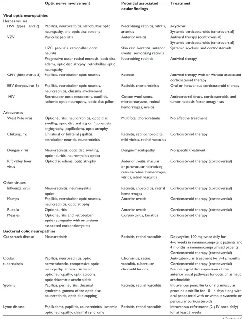
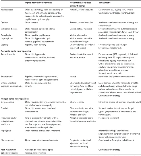
Table 1 Summary of findings in main infectious optic neuropathies
Optic nerve involvement Potential associated
ocular findings
Treatment
Viral optic neuropathies
Herpes viruses
HSv (types 1 and 2) Papillitis, neuroretinitis, retrobulbar optic neuropathy, and optic disc atrophy
Necrotizing retinitis, vitritis, arteritis
Acyclovir
Systemic corticosteroids (controversial)
vZv varicella: papillitis Anterior uveitis Antiviral therapy (controversial)
Systemic corticosteroids (controversial) HZO: papillitis, retrobulbar optic
neuritis
Skin rash, keratitis, anterior uveitis, necrotizing retinitis
Systemic acyclovir and corticosteroids
Progressive outer retinal necrosis: optic disc edema, optic disc atrophy, retrobulbar optic neuropathy
Necrotizing retinitis Antiviral therapy
CMv (herpesvirus 5) Papillitis, retrobulbar optic neuritis Retinitis Antiviral therapy with or without associated corticosteroid therapy
EBv (herpesvirus 6) Papillitis, retrobulbar optic neuritis, neuroretinitis, chiasmal involvement
Retinitis, chorioretinitis Oral or intravenous corticosteroid therapy
Hiv Retrobulbar optic neuropathy, papillitis, ischemic optic neuropathy, optic disc pallor
Cotton-wool spots, microaneurysms, retinal hemorrhages, uveitis
Antiretroviral drugs, corticosteroids, and tumor necrosis factor antagonists
Arboviruses
west Nile virus Optic neuritis, neuroretinitis, optic disc swelling, optic disc staining on fluorescein angiography, papilledema, optic atrophy
Multifocal chorioretinitis No effective treatment
Chikungunya Unilateral or bilateral papillitis, retrobulbar neuritis, neuroretinitis
Retinitis, retinochoroiditis, mild vitritis, retinal vasculitis
Corticosteroid therapy
Dengue virus Neuroretinitis, optic disc swelling, optic neuritis, neuromyelitis optica
Dengue maculopathy No specific treatment
Rift valley fever virus
Optic disc edema, optic atrophy Anterior uveitis, macular or paramacular necrotizing retinitis, retinal hemorrhages, vitritis, retinal vasculitis
Corticosteroid therapy (controversial)
Other viruses
Influenza virus Neuroretinitis, neuromyelitis optica
Retinitis, choroiditis, retinal hemorrhages
Corticosteroid therapy (controversial)
Mumps Papillitis, retrobulbar optic neuritis, neuroretinitis, optic atrophy
Anterior uveitis Corticosteroid therapy (controversial)
Rubella Optic neuritis Anterior uveitis Corticosteroid therapy (controversial)
Measles Optic neuritis and retrobulbar optic neuropathy with or without associated encephalomyelitis
Conjunctivitis, keratitis Corticosteroid therapy
Bacterial optic neuropathies
Cat scratch disease Neuroretinitis Retinitis, retinal vasculitis Doxycycline 100 mg twice daily for 4–6 weeks in immunocompetent patients and 4 months in immunocompromised patients Corticosteroid therapy (controversial) Ocular
tuberculosis
Papillitis, neuroretinitis, optic nerve tubercle, compressive optic neuropathy, anterior ischemic optic neuropathy, optic atrophy, optic chiasmatic arachnoiditis
Choroiditis, retinal vasculitis, tubercular choroidal lesions
Anti-tubercular treatment for 9–12 months Corticosteroid therapy (controversial) Neurosurgical decompression of the anterior visual pathways for optic chiasmatic arachnoiditis
Syphilis Papillitis, perineuritis, chiasmal syndrome, gumma of the optic disc, neuroretinitis, optic disc cupping
Retinitis, retinal vasculitis intravenous penicillin G or intramuscular procaine penicillin for 10–14 days along with oral probenecid with or without systemic or periocular corticosteroids
Lyme disease Papilledema, papillitis, neuroretinitis, ischemic optic neuropathy, chiasmal syndrome
Retinitis, retinal vasculitis intravenous ceftriaxone (2 g iv once daily) for at least 3 weeks
(Continued)
Eye and Brain downloaded from https://www.dovepress.com/ by 118.70.13.36 on 22-Aug-2020
Dovepress
Kahloun et al
adjacent to the disc edge and a white depigmented zone away from the optic disc.274
Aspergillosis
Aspergillus fumigatus is a ubiquitous and saprophytic agent
that becomes pathogenic in case of hypoxic area, which can
explain its higher incidence in the paranasal sinuses in immu-nocompromised patients. A few cases of optic neuritis in the setting of aspergillosis have been reported.275–279 The clinical
presentation can mimic nonspecific optic neuritis, with a possible good response to corticosteroid therapy.278 Several
cases of orbital apex syndrome secondary to aspergillus
Table 1 (Continued)
Optic nerve involvement Potential associated
ocular findings
Treatment
Rickettsioses Optic disc swelling, optic disc staining on fluorescein angiography, optic neuritis, neuroretinitis, ischemic optic neuropathy, papilledema, optic atrophy
Retinitis, retinal vasculitis Doxycycline 200 mg/day for 2 weeks Corticosteroid therapy (controversial)
Q fever Optic neuritis Retinitis, retinal vasculitis Antibiotics and corticosteroid therapy are
controversial whipple’s disease Optic neuritis, optic disc edema,
optic atrophy
vitritis, retinal vasculitis Systemic trimethoprim–sulfamethoxazole associated with rifampin, for at least 1 year Brucellosis Optic neuritis, papilledema vitritis, choroiditis Antibiotics and corticosteroid therapy Leptospirosis Optic disc hyperemia, optic
neuritis, papillitis, neuroretinitis
vitritis, retinal vasculitis, retinal hemorrhages
Antibiotics and corticosteroid therapy
Leprosy Papillitis, optic atrophy Dacryoadenitis, disorder of
the eyelid, uveitis
Systemic dapsone and rifampin Systemic corticosteroids
Parasitic optic neuropathies
Toxoplasmosis Optic disc hyperemia, neuroretinitis, papillitis, isolated anterior optic neuritis
Retinochoroiditis, retinal vasculitis, vitritis
Pyrimethamine (100 mg on day 1 followed by 50 mg daily; 25 mg in children) and sulfadiazine 4 g/day with folinic acid Other alternatives: oral or intravitreal, clindamycin, spiramycin, azithromycin, trimethoprim–sulfamethoxazole Systemic corticosteroids Toxocariasis Papillitis, retrobulbar optic neuritis,
neuroretinitis, optic disc granuloma
vitritis Periocular and systemic corticosteroids
Diffuse unilateral subacute neuroretinitis
Optic disc edema, optic disc atrophy
Chorioretinitis, retinal vessel narrowing, focal or diffuse retinal pigment epithelium degeneration
Laser therapy, when the nematode is visible, and chemotherapy with anthelmintic drugs, such as mebendazole, thiabendazole, or albendazole when a worm cannot be visualized Corticosteroid therapy
Fungal optic neuropathies
Cryptococcus Optic neuritis after cryptococcal meningitis, retrobulbar optic neuropathy
Chorioretinitis intravitreal and/or intravenous amphotericin B
Candida Optic disc edema associated with chorioretinitis
Chorioretinitis, vasculitis, retinal hemorrhages, vitreous exudates
Systemic and/or intravitreal antifungal agents (amphotericin B, fluconazole, and voriconazole)
Presumed ocular histoplasmosis syndrome
Ring of peripapillary atrophy with a narrow inner pigment zone adjacent to the disc edge and a white depigmented zone away from the optic disc
Atrophic choroidal scars, choroidal neovascularization
No specific treatment
Aspergillus Optic neuritis, orbital apex syndrome intensive antifungal therapy with
amphotericin B, surgical excision of involved tissue with sinus exenteration
Mucormycosis Optic nerve infarction and necrosis Proptosis, conjunctival injection, restricted extraocular motility
Amphotericin B, surgical debridement
Post-vaccination optic neuropathies
Anterior or retrobulbar optic neuritis, neuroretinitis
Corticosteroid therapy
Abbreviations: CMV, cytomegalovirus; EBV, Epstein-Barr virus; HSV, herpes simplex virus; HIV, human immunodeficiency virus; HZO, herpes zoster ophthalmicus; vZv, varicella zoster virus.
Eye and Brain downloaded from https://www.dovepress.com/ by 118.70.13.36 on 22-Aug-2020
Dovepress infectious optic neuropathies are reported.279 The pattern of visual field defect depends
on anatomical extension of the infection. The diagnosis of ocular aspergillosis might be difficult especially in the early stage. Repeated radiological examination and orbital biopsy may be required in the event of a high level of clinical suspicion. Management of aspergillosis involves prompt sur-gical excision of the involved tissue with sinus exenteration. Intensive antifungal therapy with amphotericin B is also recommended.
Mucormycosis
Mucormycosis is an opportunistic fungal infection caused by Mucorales (Mucor, Rhizopus, Absidia, and
Cunninghamella).280 It is a potentially lethal infection that
generally affects immunocompromised patients; however, cases in immunocompetent and diabetic patients have been reported. Rhino–orbito–cerebral mucormycosis presents with nonspecific complaints such as headache, low-grade fever, facial swelling, sinusitis, proptosis, conjunctival injection, and restricted extraocular motility. Optic nerve involvement
in mucormycosis includes optic nerve infarction and necro-sis that may result from invasion of the blood vessel walls by the organisms, leading to occlusion or thrombosis of the optic nerve sheath, blood vessels, or ophthalmic artery.281–283
Direct optic nerve infection by mucormycosis may also occur. Treatment involves aggressive surgical debridement of all involved tissues including exenteration of involved orbits, with prolonged administration of amphotericin B.
Post-vaccination optic neuropathies
Optic nerve involvement has been described in associa-tion with vaccinaassocia-tion against various bacterial and virus infections. These include tuberculosis (Bacillus Calmette-Guérin vaccination), influenza virus, hepatitis B virus, hepatitis A virus, yellow fever, measles/rubella vaccines, mumps, diphtheria toxoid, tetanus toxoid, rabies virus, and variola virus.284–301
Post-vaccination optic neuritis is a rare event that may occur hours to weeks after vaccination. The presumed patho-genesis of this event is an immune-mediated mechanism.
Optic neuropathy
Typical optic neuritis Atypical optic neuritis
Stay in or travel to specific endemic areas
Rickettsiosis Q fever Lyme disease
West Nile virus infection Dengue fever
Rift valley fever Chikungunya DUSN Malaria Onchocerciasis
Immunocompromised status
Herpes zoster ophthalmicus Progressive outer retinal necrosis CMV infection
HIV infection
Epstein-Barr virus infection Syphilis
Toxoplasmosis Cryptococcosis Candidiasis Aspergillosis Mucormycosis MRI
Infectious
Noninfectious systemic disease Post-vaccination
Neurological involvement
HSV CMV infection West Nile virus infection Rift valley fever Tuberculosis Q fever
Whipple’s disease Lyme disease Cryptococcosis Skin rash
Rickettsiosis Chikungunya Rubella Measles
Associated systemic involvement
Fever
HSV Varicella CMV infection Chikungunya Dengue fever Rift valley fever Influenza Mumps Rubella Measles
Cat-scratch disease Tuberculosis Rickettsiosis Q fever Brucellosis Leptospirosis Toxoplasmosis Malaria Cryptococcosis
Figure 7 Practical approach to infectious optic neuropathies according to epidemiologic data and associated systemic involvement.
Abbreviations: CMV, cytomegalovirus; DUSN, diffuse unilateral subacute neuroretinitis; HSV, herpes simplex virus; HIV, human immunodeficiency virus; MRI, magnetic resonance imaging.
Eye and Brain downloaded from https://www.dovepress.com/ by 118.70.13.36 on 22-Aug-2020
Dovepress
Kahloun et al
Most cases are bilateral, and include anterior or retro-bulbar optic neuritis and neuroretinitis. The overall prognosis is good, and corticosteroids may hasten visual recovery.284–301
Data from a case-control study show no increased risk of multiple sclerosis or optic neuritis following vaccination against hepatitis B, influenza, tetanus, measles, or rubella.302
Nevertheless, a possible causal relationship between vaccina-tion against virus infecvaccina-tion and development of optic neuritis cannot be completely excluded.303
Diagnostic approach to
infectious optic neuropathy
Optic neuritis is the most common optic neuropathy, which usually affects young adults. It typically presents as an acute, unilateral inflammatory demyelinating disorder of the optic nerve that can be associated with multiple sclerosis. A gradual recovery of visual acuity with time is characteristic of optic neuritis, and the work-up should be limited to cerebromed-ullary magnetic resonance imaging. Atypical optic neuritis may be characterized by bilateral involvement, significant ocular inflammatory reaction, atypical clinical course, and associated systemic involvement. Atypical optic neuropathy may be associated with a wide variety of infectious (Table 1) and noninfectious disorders. Appropriate clinical diagnosis
and laboratory work-up of a patient with infectious optic neuropathy are based on epidemiological data, history, the patient’s immunological status, systemic symptoms and signs, and associated inflammatory involvement that may involve the adnexa, anterior segment, vitreous, retina, and choroid, as well as neuro–ophthalmological involvement (Figures 7 and 8).
Evaluation of patients with suspected infectious optic neuropathy may include a complete blood count, erythrocyte sedimentation rate, C-reactive protein, serological testing, blood cultures, PCR, or antibody assessment in aqueous humor, vitreous, serum, or cerebrospinal fluid, a tuberculin skin test and/or quantiferon, tomodensitometry, and magnetic resonance imaging.
Conclusion
A wide variety of viral, bacterial, parasitic, and fungal agents can cause optic neuropathy, with variable clinical features. Proper clinical diagnosis of any specific infectious condition is based on epidemiological data, history, systemic symptoms and signs, and the pattern of optic nerve involvement and asso-ciated ocular findings, which can be confirmed by laboratory testing. Most infectious agents can be effectively treated with specific anti-infectious drugs with or without associated corti-costeroid therapy, but visual recovery is highly variable. Optic neuropathy
Typical optic neuritis Atypical optic neuritis
Associated retinitis or choroiditis
Acute retinal necrosis CMV infection Tuberculosis Syphilis Rickettsiosis Lyme disease Brucellosis Q fever
Cat scratch disease Toxoplasmosis Toxocariasis
West Nile virus infection Dengue fever Rift valley fever Chikungunya MRI
Infectious Noninfectious systemic disease
Periocular inflammation Post-vaccination
Bacterial orbital infections Orbital echinococcosis Mucormycosis Pattern of optic nerve involvement
Papillitis Neuroretinitis Retrobulbar optic neuropathy
Almost all infections Cat-scratch disease Rickettsiosis Tuberculosis Syphilis Lyme disease Leptospirosis Toxoplasmosis Toxocariasis HSV EBV infection Chikungunya Dengue fever Influenza Mumps
HSV Herpes zoster ophthalmicus PORN CMV infection EBV infection HIV infection Chikungunya Mumps Measles Toxocariasis Malaria Cryptococcosis
Optic atrophy
HSV PORN Rift valley fever Rickettsiosis Whipple’s disease Leprosy Lyme disease DUSN Onchocerciasis
Figure 8 Practical approach to infectious optic neuropathies according to associated ocular findings.
Abbreviations: CMV, cytomegalovirus; DUSN, diffuse unilateral subacute neuroretinitis; EBV, Epstein-Barr virus; HSV, herpes simplex virus; HIV, human immunodeficiency virus; MRi, magnetic resonance imaging; PORN, progressive outer retinal necrosis.
Eye and Brain downloaded from https://www.dovepress.com/ by 118.70.13.36 on 22-Aug-2020