The next step: the influence of acute exercise on cognitive function
during locomotive goal-directed behaviour
Submitted by
Shelley J Duncan
MSc. (1st Class Hons) January 2016
A thesis submitted in fulfilment of the requirements of the degree Doctor of Philosophy
Supervisor: Professor Remco Polman Co-supervisor: Dr Derek Panchuk
Institute of Sport, Exercise and Active Living College of Sport and Exercise Science
i
ABSTRACT
In the performance of goal-directed behaviour, multiple attributes of cognitive function have been investigated. A core confounding factor, however, is that aspects of cognitive function have predominantly been evaluated employing seated or supine paradigms that are not representative of activities of daily living. Therefore the purpose of this PhD thesis was to evaluate differences in and the influence of acute bouts of exercise on cognitive function and gaze behaviour incorporating whole body movement and behaviour. In study 1, a locomotive task was designed and validated to evaluate differences in gaze behaviour during single- and dual-task performance. In study 2, the validated locomotive paradigm was utilised to evaluate differences in neural activity, specifically the N2 and P3 event-related potentials (ERPs) and in study 3, the effects of acute bouts of exercise (aerobic versus resistance) on single- and dual-task performance and neural activity (N2 and P3 ERPs) were evaluated using the locomotive paradigm. A unique aspect of these studies was capturing electroencephalographic (EEG) data during whole body movement and behaviour leading towards a more real world application. Results included: 1) validation of the locomotive paradigm and successful collection of gaze behaviour during single- and dual-task
ii behavioural and neurophysiological measures relating to task difficulty and exercise
iii
DECLARATION
I, Shelley Jane Duncan, declare that the PhD thesis entitled “The next step: the influence of acute exercise on cognitive function during locomotive goal-directed behaviour” is no more than 100,000 words in length, exclusive of tables, figures, appendices, references and
footnotes. This thesis contains no material that has been submitted previously, in whole part, for the award of any other academic degree or diploma. Except where otherwise indicated this PhD is my own work.
iv
ACKNOWLEDGEMENTS
This thesis would not have been possible to put together if it weren’t for the help of many others. This part is an attempt to acknowledge and thank those people.
To my principal supervisor Prof Remco Polman, thank you for taking me on as a PhD student. Thank you for your unwavering support and belief in my ability and capacity to fulfil the requirements of my PhD, and enabling me to develop and pursue my passion within the Neuroscience field of research. Without you, I would not have had the opportunities I experienced throughout my PhD. To my co- supervisor Dr Derek Panchuk, words cannot fully articulate the gratitude I have for your ongoing support, guidance and faith you have had in me. You have provided at times a much needed calming influence and helped me keep my feet on the ground. Your guidance and ability to teach and help me develop my research skills, especially my writing skills was gentle, honest and I had complete faith and trust that you were pushing me in the right direction and helping me develop my academic self-confidence.
To Dr Jacqueline Williams, you were my colleague, friend and someone with whom I hold in the utmost regard. You have been my sounding board and mentor and I will continue to strive to be as ethical, professional and kind in the manner with which I will conduct myself as you have been. To Dr Angela Gosling, my neuroscience saving grace. I am
v Ramon words can’t express, how grateful I am for the support and help you have given me over the past four years and some fun times. To my two amazing research assistants Luca and Rachel, you made my data collection so much easier, thank you for being so supportive and reliable.
To all the participants throughout the three interventions in this thesis, thank you so much for your contribution, without you this PhD would have been impossible. To my family, particularly mum, Sara, Lucy, Jillian, Dave, the Corkin’s/Clark’s and Chandlers, thank you for all the support, kind words and encouragement throughout this time. Thanks for your patience, tolerance and comedy about the fact that you no longer understand my work. Everything you have done has made writing this thesis much easier. Last but not least I need to thank Agnes, who started off as the EEG equipment representative and became my
vi
ABBREVIATIONS
BDNF Brain derived neurotrophic factor
CNS Central nervous system
SPL Superior parietal lobule
HAROLD Hemispheric asymmetry reduction in older adults
EEG Electroencephalography
fMRI Functional magnetic resonance imaging
MEG Magnetoencephalography
EOG Electrooculographic
EMG Electromyographic
ECG Electrocardiographic
ICA Independent component analysis
PCA Principal component analysis
LORETA Low-resolution electromagnetic tomography
sLORETA Standardised low-resolution electromagnetic tomography
ERP Event-related potential
ISI Inter-stimulus interval
QE Quiet eye
GPS Global positioning system
RPE Rating of perceived exertion
HRmax Heart rate maximum
VO2 max Maximum oxygen uptake
1 RM One repetition maximum
vii
AT Anaerobic threshold
W Watts
W.min-1 Watts per minute
n Number
M Mean
µV Microvolts
kΩ. Kiloohm
Hz Hertz
m Meter
m/sec Meters per second
cm Centre meters
mm Millimetre
o
Degree
s Second
min Minute
ms Milliseconds
LMM Linear mixed modelling
viii
PUBLICATIONS AND PRESENTATIONS
This thesis is supported by the following publications and conference presentations:
Papers submitted for publication
1. Duncan, S., Panchuk, D., & Polman, R.: “Validation of a new locomotive single- dual-task paradigm to evaluate task related differences in gaze behaviour and neural activity. Under review in the Journal of Psychophysiology
2. Duncan, S., Panchuk, D., & Polman, R.: “Comparison of acute aerobic and resistance
exercise on brain mechanisms associated with single- and dual-tasks during locomotion”. Under review in the Frontiers of Neuroscience Journal
Conference presentations
1. Poster Presentation (awarded second prize) - Duncan, S., Panchuk, D., & Polman, R.C.J. (2014). Brain mechanisms associated with single and dual-tasks during locomotion. Institute of Sport, Exercise and Active Living (ISEAL) Higher Degree by Research Students Conference, December 2014, Melbourne, Australia
2. Poster Presentation - Duncan, S., Panchuk, D., & Polman, R.C.J. (2014). Brain mechanisms associated with single and dual-tasks during locomotion. 12th International Conference on Cognitive Neuroscience, 27-31 July, Brisbane, Australia
3. Oral Presentation - Duncan, S., Schneider, S., Panchuk, D., & Polman, R.C.J. (2015). The moderating effect of acute bouts of exercise and physical fitness on brain activity during locomotion” –20th
ix
Table of Contents
ABSTRACT ... I DECLARATION ... III ACKNOWLEDGEMENTS ... IV ABBREVIATIONS ... VI PUBLICATIONS AND PRESENTATIONS ... VIII TABLE OF CONTENTS ... IX TABLE OF FIGURES ... XV LIST OF TABLES ... XX
CHAPTER ONE ... 1
INTRODUCTION ... 1
DUAL-TASKING ... 2
COGNITIVE FUNCTION AND EXERCISE ... 3
PURPOSE ... 5
CHAPTER AIMS: ... 6
Chapter 2: Literature review ... 6
Chapter 3: Study One – Validation of a new locomotive single- and dual-task paradigm to evaluate differences in gaze behaviour and neural activity ... 6
Chapter 4: Study Two - Validating the use of EEG to examine neural activity associated with single- and dual-tasks during locomotion ... 7
x Chapter 6: Study Three – Part B - Effect of acute aerobic exercise on neural activity
associated with single and dual-task performance during locomotion ... 8
Chapter 7: Study Three – Part C - Effect of acute resistance exercise on neural activity associated with single and dual-task performance during locomotion ... 8
Chapter 8: Overall discussion ... 8
CHAPTER TWO ... 10
LITERATURE REVIEW ... 10
1.DUAL-TASK PERFORMANCE ... 12
2.VISUOMOTOR COORDINATION ... 15
2.1 Evaluating visuomotor coordination ... 16
3.NEURAL ACTIVITY ... 19
3.1 Evaluating neural activity ... 20
3.2 Event-related potentials ... 25
4.COGNITIVE FUNCTION AND EXERCISE... 30
4.1 Acute aerobic exercise ... 34
4.2 Acute resistance exercise ... 40
4.3 Additional factors in assessing cognitive function and exercise ... 41
5.SUMMARY ... 42
CHAPTER THREE ... 44
STUDY 1–VALIDATION OF A NEW LOCOMOTIVE SINGLE- AND DUAL-TASK PARADIGM TO EVALUATE DIFFERENCES IN GAZE BEHAVIOUR AND NEURAL ACTIVITY ... 44
1.INTRODUCTION... 44
2.METHODS ... 48
xi
2.2 Apparatus ... 49
3.PROCEDURE ... 52
4.DATA PROCESSING AND ANALYSIS ... 53
4.1 Behavioural data ... 53
4.2 Gaze behaviour data ... 53
5.RESULTS ... 55
5.1 Behavioural data ... 55
5.2 Gaze behaviour data ... 55
6.DISCUSSION ... 57
CHAPTER FOUR ... 60
STUDY 2-VALIDATING THE USE OF EEG TO EXAMINE NEURAL ACTIVITY ASSOCIATED WITH SINGLE- AND DUAL-TASKS DURING LOCOMOTION ... 60
1.INTRODUCTION... 60
2.METHOD ... 65
2.1 Participants ... 65
2.2 Apparatus ... 66
3.PROCEDURE ... 69
4.DATA PROCESSING AND ANALYSIS ... 69
4.1 Behavioural data ... 69
4.2 Neural activity data ... 69
5.RESULTS ... 74
5.1 Movement related topographic map identification ... 74
5.2 Behavioural data ... 75
5.3 Neural activity data ... 75
xii
CHAPTER FIVE ... 79
STUDY 3–PART A ... 79
EFFECTS OF ACUTE EXERCISE ON NEURAL ACTIVITY ASSOCIATED WITH SINGLE- AND DUAL -TASK PERFORMANCE DURING LOCOMOTION – AEROBIC VERSUS RESISTANCE EXERCISE... 79
1.INTRODUCTION... 79
2.METHOD ... 84
2.1 Participant ... 84
2.2 Participant screening ... 84
3.MEASUREMENTS ... 85
3.1 Session one: Maximal oxygen uptake testing and strength testing familiarisation ... 85
3.2 Session two: Strength testing ... 86
3.3 Sessions three and four: Study protocol... 86
4.PROCEDURE ... 88
5.DATA PROCESSING AND ANALYSIS ... 90
5.1 Additional EEG data reduction ... 90
6.DATA ANALYSIS: ... 94
6.1 Behavioural data ... 94
6.2 Neural activity data ... 95
7.RESULTS ... 96
7.1 Behavioural data ... 96
7.2 Neural activity data ... 97
8.DISCUSSION ... 104
CHAPTER SIX ... 112
xiii EFFECT OF ACUTE AEROBIC EXERCISE ON NEURAL ACTIVITY ASSOCIATED WITH SINGLE AND
DUAL-TASK PERFORMANCE DURING LOCOMOTION ... 112
1.INTRODUCTION... 112
2.METHODS ... 114
2.1. Data analyses: ... 114
3.RESULTS ... 114
3.1 Behavioural data ... 115
3.2 Neural activity data ... 115
4.DISCUSSION ... 123
CHAPTER SEVEN ... 128
STUDY 3–PART C ... 128
EFFECT OF ACUTE RESISTANCE EXERCISE ON NEURAL ACTIVITY ASSOCIATED WITH SINGLE -AND DUAL-TASK PERFORMANCE DURING LOCOMOTION ... 128
1.INTRODUCTION... 128
2.METHODS AND DATA ANALYSIS ... 129
3.RESULTS ... 130
3.1 Behavioural data ... 130
3.2 Neural activity data ... 131
4.DISCUSSION ... 139
CHAPTER EIGHT ... 145
OVERALL DISCUSSION ... 145
1.SUMMARY OF FINDINGS ... 145
2.LIMITATIONS -ISSUES WITH EEG DATA COLLECTION ... 146
xiv
4.PRACTICAL IMPLICATIONS ... 150
5.CONCLUSION ... 151
REFERENCES ... 152
xv
Table of Figures
Figure 2. 1: ASL Mobile Eye Unit... 17 Figure 2. 2: EEG recording – the electrode is placed on the scalp and measures neural activity within the cortical level of a depth of 0.2 – 0.3 cm (adapted from Nunez & Srinivasan, 2006). ... 22 Figure 2. 3: 32 Channel montage representing a 10/20 electrode configuration (Brain
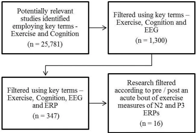
Products, 2009). ... 22 Figure 2. 4: 250 Channel montage representing a 10/5 electrode configuration (Oostenveld & Praamstra, 2001) ... 23 Figure 2. 5: EEG Electrode System Montage (Brain Products, 2009) ... 24 Figure 2. 6: Event-related potential diagram showing the N2 and P3 ERPs of interest. ... 26 Figure 2. 7: “Sensory input is processed, with frontal lobe activation from attention-driven working memory changes (P3a) and temporal/parietal lobe activation from memory updating operations (P3b)” (Polich, 2003). ... 29 Figure 2. 8: Inverted U - physiological response corresponding to cognitive function (adapted from Kashihara et al., 2009). ... 32 Figure 2. 9: Inverted U – exercise intensity corresponding to an increase in the P3 ERP amplitude (Kamijo et al., 2009). ... 32 Figure 2. 10: Article review filter process. ... 36
xvi Figure 3. 4: Comparison of trial completion time between single- and dual-task conditions (* represents p < .05). Data is presented as mean ± SE. ... 55
Figure 4. 1: Eye blink (A) and saccadic eye movement (B) topographies (Plank, 2013). ... 63 Figure 4. 2: A) Movement related artefact contamination using TP10 (mastoid placement) as the online reference. B) Signal comparison using Cz (central midline placement) as the online reference. ... 66 Figure 4. 3: Schematic representation of equipment configuration and direction of signal transmition. ... 68 Figure 4. 4: EEG Move System Transmitter, secured to a GPS harness. ... 68 Figure 4. 5: 32 Channel Montage with key channels examined circled. ... 72 Figure 4. 6: Topographical scalp map comprising the key ERP mean amplitude time windows analysed comparing single- and dual-task neural activity. ... 72 Figure 4. 7: Example of the grand averaged N2 and P3 mean amplitude response time
windows employed to examine the difference in the neural activity over time in response to task difficulty. ... 73 Figure 4. 8: Movement related topographies observed in electrodes TP9 and TP10. ... 74 Figure 4. 9: Comparison of trial completion time between conditions. (* denotes p < .05). Data is presented as mean ± SE. ... 75
xvii Figure 5. 4: Aerobic and Resistance Exercise – Time windows employed to examine the difference in the P3 ERP component peak latency and mean amplitude. ... 93 Figure 5. 5: Example of the grand averaged N2 and P3 mean amplitude response time
windows employed to examine the difference in the neural activity over time in response to task difficulty and exercise type. ... 94 Figure 5. 6: Comparison of trial completion time between pre and post exercise and condition (* represents p < .05). Data is presented as mean ± SE. ... 97 Figure 5. 7: Comparison of the N2 peak latency between pre and post exercise (* represents p
< .05). Data is presented as mean ± SE. ... 98 Figure 5. 8: Topographic scalp map comparison of time window one (300 – 400 ms) lateral
P3a mean amplitude aerobic and resistance exercise. ... 99 Figure 5. 9: Comparison of the lateral P3 mean amplitude across all time windows before and after aerobic and resistance exercise (* represents p < .05). Data is presented as mean ± SE. 99 Figure 5. 10: Topographic scalp map comparison of time window one (400 - 500 ms) lateral
early P3b mean amplitude aerobic and resistance exercise. ... 100 Figure 5. 11: Topographic scalp map comparison of time window one (500 - 600 ms) lateral
late P3b mean amplitude aerobic and resistance exercise. ... 101
Figure 6. 1: Aerobic Exercise Bout - ERP responses with time windows employed to examine the differences in neural activity (N2 and P3 ERP component peak latency and mean
amplitude). ... 114 Figure 6. 2: Comparison of trial completion time between pre- and post-exercise (* represents
p < .05). Data is presented as mean ± SE. ... 115 Figure 6. 3: Comparison of the N2 peak latency between pre and post exercise (* represents p
xviii Figure 6. 4: Comparison of the N2 mean amplitude between pre and post exercise (*
represents p < .05). Data is presented as mean ± SE. ... 117 Figure 6. 5: Topographic scalp map comparison of time window one (300 – 400 ms) lateral
P3a mean amplitude following the aerobic bout of exercise. ... 118 Figure 6. 6: Topographic scalp map comparison of time window one (400 - 500 ms) lateral
early P3b mean amplitude following the aerobic bout of exercise. ... 119 Figure 6. 7: Topographic scalp map comparison of time window one (500 - 600 ms) lateral
late P3b mean amplitude following the aerobic bout of exercise. ... 120
Figure 7. 1: Resistance Exercise Bout - ERP responses with time windows employed to examine the differences in neural activity (N2 and P3 ERP component peak latency and mean amplitudes). ... 129 Figure 7. 2: Comparison of trial completion time between pre and post exercise and condition (* represents p < .05). Data is presented as mean ± SE. ... 130 Figure 7. 3: Topographic scalp map comparison of the N2 mean amplitude 180 – 220 ms post stimulus time window: A) Significant lateral interaction between time and electrode site; B) Significant lateral interaction between task and electrode site. ... 131 Figure 7. 4: Topographic scalp map of the P3a meanamplitude. Scalp maps reflect the
average of time window one 300 – 400 ms post stimulus and the activation during the single- compared to the dual-task condition. ... 132 Figure 7. 5: P3a mean amplitude responses with time window one (300 – 400 ms) employed to examine the difference in the lateral neural activity related to task difficulty. ... 133 Figure 7. 6: Topographic scalp map comparison of time window one (300 - 400 ms) lateral
xix Figure 7. 8: Topographic scalp map comparison of time window one (500 - 600 ms) lateral
late P3b mean amplitude single- compared to dual-task. ... 135 Figure 7. 9: Topographic scalp map comparison of time window three (500 – 600 ms) lateral
late P3b mean amplitude single- compared to dual-task. ... 136 Figure 7. 10: Topographic scalp map of the N2 mean amplitude. Scalp maps reflect the average of 180 – 220 ms post stimulus epoch of an interaction between task and electrode site in the midline electrode Pz (parietal region). ... 137 Figure 7. 11: P3 mean amplitude responses with the three time windows employed to
examine the difference in the lateral neural activity over time in response to resistance
xx
List of Tables
Table 2.1: Articles evaluating acute exercise-related influences on the N2 andP3 ERP
1
Chapter One
Introduction
Over the course of the lifespan the human brain and body experience an array of
changes. This includes the aging process, the influence of illness and disease and the impact
of physical fitness and acute (single) bouts of exercise (Angel, Fay, Bouazzaoui, & Isingrini,
2011; Hogan et al., 2013; Wylie et al., 2009). A decline in the human brain is reported to
occur from the third decade in life (Colcombe et al., 2006), which results over time, in the
progressive loss of the ability to perform activities of daily living (Chou, Hwang & Wu,
2012). Consequently, a key focus of exercise physiology, psychology and neuroscience
research has been to develop a better understanding of these changes and of normal
development and aging to measure decrements in performance, and design interventions that
will attenuate these decrements and improve overall physical and cognitive function
(Colcombe et al., 2006).
2 navigation whilst walking), the capacity to dual-task is of primary importance (Li et al., 2001). Further, with the apparent dual-task costs associated with aging becoming more prominent (Mendelson, Redfern, Nebes, & Jennings, 2010), there is a drive to develop interventions to attenuate these deficits and improve overall cognitive and physical function (Córdova, Silva, Moraes, Simões, & Nóbrega, 2009; Forte et al., 2013). Therefore, for the purpose of this thesis, the two main concepts that will be discussed and manipulated are that of dual-tasking and the relationship between cognitive function and exercise, specifically of an acute nature.
Dual-tasking
A component fundamental to activities of daily living is cognitive processing, including visuomotor coordination (Goodale, 2010; Land & Hayhoe, 2001). For example, the reception, integration and processing of sensory information, that informs goal-directed behaviour. This includes processing of multiple stimuli at one time including a variety of exogenous and endogenous sources of information (Findlay, 2009). The integration of vision and motor performance is specifically pertinent in the efficient and effective hand-eye coordination and performance of time-stressed activities that require tight coupling between vision and action (Land & McLeod, 2000; Panchuk, Davids, Sakadjian, Macmahon, & Parrington, 2013) like catching a ball in flight or a falling glass before it hits the ground.
3 has also been evaluated using brain functioning technology, which has shown that younger, compared to older, adults use more widespread regions of the brain for motor function, namely the prefrontal cortex (working memory) and basal ganglia (initiation/control of movement) networks (Seidler et al., 2010). With the advance in brain imaging technology (i.e., functional magnetic resonance imaging and electroencephalography), the complex nature of the brain’s structure and function can be evaluated, including assessment of spatial (location of activity) and temporal (time course of information processing) patterns of activity and how these relate to goal-directed behaviour. Furthermore, differences between younger and older adult populations and how different interventions (e.g., pharmacological and exercise) can influence and alter the brain’s function can be examined.
Cognitive function and exercise
The evidence relating to the positive influence aerobic exercise has on cognitive function is substantial. Exercise is reported to promote improvements in processing speed and executive functioning (Smith et al., 2010), and both structural and functional changes
4 Aerobic exercise is also suggested to be related to a neuro-protective mechanism, reducing the risk of age-related decline in cognitive function (Karp et al., 2006). However, a complicating factor in the use of exercise to promote changes in physical (i.e., muscle atrophy and bone fragility) and cognitive (i.e., perception, working memory and decision
making) function is the multiple dynamics of any exercise intervention, which can be
manipulated. These include the mode, intensity and duration of exercise. Consequently,
multiple interventions have been employed to evaluate and measure exercise-induced
changes in physical and cognitive function (e.g., Chang & Etnier, 2009b; Forte et al., 2013).
Most research to date has evaluated the influence of both long-term and acute (single bout) interventions on cognitive function, employing aerobic exercise (e.g., Audiffren et al., 2008; Voss et al., 2010). There is also a growing body of research that has investigated resistance based exercise (e.g., Chang, Etnier, & Barella, 2009; Hsieh, Chang, Hung, & Fang, 2016). However, there is no research that has evaluated the influence of resistance based exercise on dual-task related spatial and temporal patterns of neural activity.
5 with the performance of more cognitively demanding tasks (i.e., dual-tasks) and improving goal-directed behaviour (Chang, Tsai, Huang, Wang, & Chu, 2014; Pesce & Audiffren, 2011). However, from a neurophysiological perspective, specifically in relation to neural activity within the brain, only acute aerobic exercise has been examined (Kumar et al., 2012; O'Leary, Pontifex, Scudder, Brown, & Hillman, 2011).
The exercise-induced influence on cognitive functioning, in particular executive control processes, has mainly been demonstrated in a controlled laboratory environment. An important limitation of many of these assessment strategies is that they have been used in tasks that are stationary (supine or seated) and are not representative of many activities of daily living. Given that most tasks happen in environments far more dynamic than the laboratory, at some point we need to let people move freely so we can obtain a better understanding of cognitive processes that underlie how people perform in the real-world, with whole body movement, performing various tasks and processing multiple sources of information at the same time.
Purpose
There is a vast array of current literature that has individually investigated visual attention, dual-task performance, neural activity related to information processing, and the effects of acute exercise. There is however a need for a more holistic approach which
incorporates the moderating effects of exercise upon cognitive function during goal-directed behaviour incorporating whole body movement. This research provides both a behavioural (i.e., task completion time) and neurophysiological (i.e., neural activity) assessment of
determining the moderating effect of an acute bout of aerobic compared to resistance exercise on locomotive goal-directed behaviour. The key aims of this research were:
6 2. Evaluate whether an acute bout of aerobic versus resistance exercise can influence
single- and dual-task completion time and alter key aspects of neural activity related to sensory integration and decision making.
Chapter Aims:
Chapter 2: Literature review
This chapter provides a review of the existing literature which forms the basis of this thesis. The chapter will introduce an overview of cognitive function, specifically performing tasks of increasing difficulty and define and discuss the performance of single- and dual-tasks. Visuomotor coordination and the evaluation of gaze behaviour will be discussed, with specific focus on sensory information integration and goal-directed behaviour. Neural activity and measures employed to evaluate temporal patterns of neural activity in addition to event-related potentials will be outlined. The influence of exercise, specifically acute bouts of aerobic and resistance exercise on cognitive function will also be discussed. The final section of this chapter will highlight the limitations within the current literature and also discuss the overarching goals of this thesis.
Chapter 3: Study One – Validation of a new locomotive single- and dual-task paradigm to evaluate differences in gaze behaviour and neural activity
7
Chapter 4: Study Two - Validating the use of EEG to examine neural activity associated with single- and dual-tasks during locomotion
The primary goal of this study was to determine whether tasks of increasing difficulty (single- and dual-tasks) would influence the temporal pattern of neural activity, specifically related to the reception, integration and processing of auditory stimuli in the performance of the locomotive single- and dual-task paradigm validated in study one. Event-related
potentials (ERPs) are of specific interest as they represent the time course (i.e., temporal resolution) of neural changes and patterns of activity in response to a specific sensory, cognitive or motor event (Luck, 2005; Luck & Kappenman, 2012). Two key neural
components of interest were that of the N2 and P3 ERP components. The two characteristics with regard to the N2 ERP are that of latency, which is an index of the timing of information processing during visual perception, with the peak latency representing the moment in time where sensory information is available to formulate the stimulus response decision (Schmitt, Münte, & Kutas, 2000; Thorpe, Fize, & Marlot, 1996), and the N2 amplitude which is associated with the neural activity (degree of effort and processes) required for response monitoring (Donkers & Van Boxtel, 2004; Yeung, Botvinick, & Cohen, 2004). The P3 ERP
latency is related to the speed with which we can classify sensory stimuli and the amplitude
which is representative of the allocation of attentional resources and working memory (Duncan-Johnson, 1981; Kutas, McCarthy, & Donchin, 1977; Polich, 1987).
Chapter 5: Study Three – Part A - Effect of acute exercise on neural activity associated with single- and dual-task performance during locomotion – aerobic versus resistance exercise
8 which allowed for the assessment of performance using different cognitive loads (single- versus dual-tasks) as well as the assessment of neural activity. In particular, this study examined the influence of aerobic compared to resistance exercise on the N2 and P3 ERP components (P3a and the early and late P3b). The P3a, and the early and late P3b, are associated with attentional and memory processing (P3a), retrieval, encoding and memory updating (early and late P3b) (Brookhuis et al., 1981; Kok, 2001; Morgan, Klein, Boehm, Shapiro, & Linden, 2008; Scisco, Leynes, & Kang, 2008).
Chapter 6: Study Three – Part B - Effect of acute aerobic exercise on neural activity associated with single and dual-task performance during locomotion
Due to the fact that key differences between the effects of an acute bout of aerobic compared to resistance exercise were not elucidated within the analyses performed within the previous chapter (chapter 5), it was the intention within chapters six and seven to examine the aerobic and resistance bouts of exercise independently in an attempt to obtain a more in-depth understanding as to the underlying mechanisms associated with differences in task-related performance identified in chapter five. Therefore, the purpose of this chapter was to evaluate neural activity (N2 and P3 ERP components) associated with the performance of both a single- and dual-task during locomotion before and after an acute bout of aerobic exercise.
Chapter 7: Study Three – Part C - Effect of acute resistance exercise on neural activity associated with single and dual-task performance during locomotion
The purpose of this chapter was to evaluate neural activity (N2 and P3 ERP components) associated with the performance of both a single- and dual-task during locomotion before and after an acute bout of resistance exercise.
Chapter 8: Overall discussion
10
Chapter Two
Literature Review
11 responses to stimuli in the environment. Function is also described as a mechanistic process which is essential for the amplification of integrated behaviour and evolves over space and time (i.e., spatiotemporal patterns of activity). Having knowledge of the brain’s function enables a better understanding of how these characteristics (i.e., electrical activity and haemodynamics) are altered throughout the lifespan and in the case of disease. It also
provides a basis with which to evaluate the dynamic and malleable (i.e., neuroplastic) nature of the brain and makes it possible to assess how the brain communicates and influences both cognitive and physical function. Furthermore, it provides a platform with which to evaluate the influence of varying interventions (i.e., pharmaceutical and exercise) to improve cognitive function and overall quality of life (Kamijo et al., 2009; Molloy et al., 2006).
Our understanding of the complex nature of the brain has further been developed through the advancement in brain imaging technologies, such as functional magnetic resonance imaging (fMRI), magnetoencephalography (MEG) and electroencephalography (EEG). For example, differences in the brain’s structure and function have been identified in cognitive tasks, requiring the performance of goal-directed behaviour between young and old adult populations and the influence of exercise on aspects of cognitive function, such as executive function and the allocation of attentional resources (Kamijo & Takeda, 2010; O'Leary et al., 2011).
Cognitive function is an umbrella term that relates to all mental abilities and processes incorporated in such things as perception, memory and working memory, judgement,
reasoning, problem solving, decision making, comprehension and language (Ashcraft, 2002). The evaluation of cognitive function incorporates a broad spectrum of research from different disciplinary approaches (e.g., psychology and neuroscience) and is commonly examined from a behavioural and neurophysiological perspective. Where behavioural performance is
12 areas of brain activation preceding and during the performance of the desired behaviour (e.g., Chou, Chen, & Madden, 2013; Gerloff et al., 1998; Pratt, Willoughby, & Swick, 2011). By employing a combined approach, researchers are able to elucidate the underlying mechanisms associated with the behaviours we can empirically observe and measure. Through the use of brain imaging technologies, differences in both spatial (i.e., location) and temporal (i.e., timing) patterns of activity, and how these change over time (i.e., hours, days, weeks and years) can be evaluated in the performance of varying goal-directed behaviour (e.g., Chang, Tsai, Chen, & Hung, 2013; Liu-Ambrose, Nagamatsu, Voss, Khan, & Handy, 2012). For example, aerobic exercise training over a six month period has been shown to promote an increase in both white and grey matter volume in an older adults (60 – 70 years) (Colcombe et al., 2006). Understanding the temporal and spatial relationships, enables a more
comprehensive perspective of how these characteristics change over time and in response to a variety of manipulations. For example, differences in the characteristics of initiation and inhibition of actions, planning, working memory, monitoring and execution of a sequence of goal-directed actions (Coppin et al., 2006; Mendelson et al., 2010; Salthouse, Atkinson, & Berish, 2003), such as the simultaneous execution of both a motor and a cognitive task (dual-task).
1. Dual-task performance
A fundamental aspect of cognitive function that relates to the ability to perform activities of daily living is the capacity to integrate and process multiple competing sources of
13 information. This is specifically relevant in an older adult population where the inability to dual-task is a predictor of fall rates (Beauchet et al., 2009; Bessot et al., 2011), and altered gait patterns in the dual-task condition, which are used to compensate for the additional cognitive demand and attenuate dual-task costs (Ayers, Tow, Holtzer, & Verghese, 2014). This diminished capacity is reported to be related to a deficit in the attentional resource capacity (i.e., resource-based attentional framework), leaving fewer resources to distribute for the integration and processing of the competing tasks (e.g., walking and talking), resulting in dual-task costs (Neider et al., 2011). Hence, in the performance of a single-task, where there is only a primary task (e.g. walking), there are sufficient attentional resources with which to integrate and process the demands of the task and generate the desired goal-directed
behaviour. However, with an increase in task difficulty, such as that associated with a dual-task condition (incorporating a secondary dual-task, e.g., talking), there is a subsequent increase in demand on the attentional resources to integrate and respond accurately (Neider et al., 2011; Pratt et al., 2011). In this situation there may be insufficient attentional resources available to integrate and process all sources of information to carry out both tasks successfully. This may result in performance decrements, such as altered gait and inability to maintain a conversation (Ayers et al., 2014; Beauchet et al., 2009).
15 this information processing loop, vision and neural activity will be discussed in more depth in the following sections.
2. Visuomotor coordination
Locomotion is an area in which visual attention and in particular gaze behaviour has been employed to establish the sequence of key determinants of movement (Grasso, Prévost, Ivanenko, & Berthoz, 1998; Hollands, Patla, & Vickers, 2002). The integration of visual and motor performance during locomotion is specifically pertinent in the efficient and effective co-ordination of both cognitive and motor skills, such as those associated with dual-task performance (e.g., crossing a busy street). Visual information provides a basis with which to regulate and guide locomotion both locally (step-by-step) and globally (route planning) and incorporates characteristics of visual perception and locomotor adaptive strategies (Patla, 1997). Visual perception encompasses visual sampling of the environment including visual feedback about body posture and movement. Locomotor adaptive strategies incorporate processing of information relating to the static and dynamic nature of the environment (Patla, 1997). In other words, adaptive strategies (altered step pattern or gait) are used to account for differences in the terrain or obstacle avoidance and maintenance of stability and balance. Visual information informs the initiation and termination of locomotion and helps to provide a rhythmic and coordinated sequence of movement to move in the desired direction (Patla, 1997).
16 motor performance is specifically pertinent for the efficient and effective hand-eye
coordination and fast reflexes required in many activities of daily living. There is a wealth of research that has examined the integration and processing of visual information in the
performance of goal-directed behaviour, for example gaze behaviours associated with catching a ball (Stone et al., 2014).
In the course of our daily lives we unconsciously account for, process and respond to a variety of sources of information (Findlay, 2009). A fundamental aspect of the capacity to do this is that of perception-action coupling, which incorporates three central processing mechanisms and are thought to manage all sensory information. These include perception, decision and effector mechanisms (Abernethy, 1986). The perception mechanism receives information from various receptors (e.g., retina for visual information). The decision mechanisms processes what action is required and the effector mechanisms, manages the temporal and sequential aspects of desired movements (Abernethy, 1986). In a situation requiring the integration and processing of multiple sources of stimuli, resulting in an increased demand on the allocation of attentional resources, perception-action coupling is negatively affected, specifically in the form of voluntary and reflexive saccadic eye movement (Meyer, Gauchard, Deviterne, & Perrin, 2007). Visuomotor information is therefore one important area of focus that has the potential to provide us with a better understanding of the associated motor performance decrements associated with performing tasks of increasing difficulty.
2.1 Evaluating visuomotor coordination
17 environment (see Figure 2.1). Parameters of interest are those of saccadic eye movements, fixations, and the quiet eye (QE). Saccadic eye movements enable optimal processing of multiple visual targets through the use of rapid changes of fixation from one target to another. These movements are ballistic in nature and are defined by two basic characteristics, latency and direction (Findlay, 2009). The latency of a saccade relates to the time that elapses between the presentation of a stimulus and the onset of the saccadic eye movement (Findlay, 2009; Halliday & Carpenter, 2010) and is used as a non-invasive means of examining mechanisms of decision making (Halliday & Carpenter, 2010). Humans typically perform two to three saccades a second (Halliday & Carpenter, 2010; Morrillo, Di Russo, Pitzalis, & Spinelli, 2006) and these saccades can be influenced by cognitive processes, including attention, working memory, learning, long term memory and decision making (Hutton, 2008). Further, in tasks of increasing difficulty, a reduced capacity to inhibit short latency reflexive saccades and an increase in error rates has been reported and is said to represent diminished working memory capacity (Mitchell, Macrae, & Gilchrist, 2002).
18 Gaze behaviour also includes fixations, which are periods of time between saccadic eye movements where gaze is held steady, during which time information about the
environment is obtained. For example, in a locomotive context, visual information relates to a new direction of desired movement and is reported to precede body rotation by as much as 1.5 s (Land, 2006; Reed-Jones, Hollands, Reed-Jones, & Vallis, 2009). Gaze behaviour, and in particular focus of attention, is reported to be negatively affected under a dual-task context, resulting in the narrowing of attention of the functional field of view and longer gaze shift latencies (Lamers & Roelofs, 2011; Pak, Rogers, & Fisk, 2006). Further, there is an
19
3. Neural activity
There has been an exponential growth in our understanding of various aspects of neurological activity over the past two decades in the context of the brain’s structure and function. In the evaluation of dual-task costs from a neurophysiological perspective, changes in spatial and temporal patterns of neural activity have been observed. For example, a
decrease in the allocation of attentional resources as indicated by a reduction in the
magnitude of neuro-electric activity and reduced activation within the supplementary motor area, cingulate cortex, insula and post-central gyrus in the performance of more complex tasks (dual-task) (Johansen-Berg & Matthews, 2002). Of specific interest are decrements in task performance which have been associated with the capacity to engage inhibitory
processes to prevent the occurrence of an incorrect response. It is well documented that, as we age, our ability to integrate and respond to a dual-task scenario becomes diminished, with older adults typically prioritising the motor compared to the cognitive component of a task to maintain balance and reduce the risk of falling (Hall, Echt, Wolf, & Rogers, 2011; Schaefer & Schumacher, 2011). This decrement in performance is associated with a mismatch between the task-related cognitive load required to perform the task accurately and efficiently and the attentional resources available. Further support for this mismatch is the age-related reduction in the P3 ERP component characteristics, specifically an increase in latency (1.36 ms per year) and decrease in amplitude (at a rate of 0.18 µV per year) (Picton, Stuss, Champagne, & Nelson, 1984). This indicates that it takes longer for older adults to categorise a sensory stimulus (P3 latency) and there is a reduction in the available attentional resources (P3
20
3.1 Evaluating neural activity
Measuring the dynamics of sensory information integration, processing and goal-directed behaviour provides a basis with which to determine when the brain is most active during the performance of a cognitive task and the efficiency of the pathways involved in the generation of a response (e.g., Dai, Chang, Huang, & Hung, 2013; Pasalar, Ro, &
Beauchamp, 2010; Voss et al., 2010). This means that we can obtain a better insight and understanding of how the brain changes in structure and function, and how it can be manipulated to promote neuroplasticity.
There is a range of neuroimaging technologies that have been employed to evaluate differences in cognitive function associated with changes throughout the lifespan (e.g., Angel et al., 2011; Chou et al., 2013), including fMRI and EEG. Whereas fMRI reflects changes in regional cerebral blood flow and has high spatial resolution (i.e., ability to identify location of neural activity), EEG is an electrophysiological recording technique, which is able to measure voltage fluctuations over time and has high temporal resolution (i.e., changes in neural
activity over time) (Friedman, Cycowicz, & Gaeta, 2001). Another fundamental difference between these technologies is that fMRI data can take several seconds due to the timing (seconds) of the haemodynamic response, EEG can assess neural changes within a 1 ms time frame, hence the high temporal resolution (Luck, 2005). As the focus of this literature review and thesis is related to the use of EEG, only this technology will be discussed further.
22 the two electrodes. This can cause a distortion of the neural activity recorded at the involved electrodes and subsequent inability to accurately determine and evaluate neural activity of interest arising from the involved electrodes (Alschuler, Tenke, Bruder, & Kayser, 2014).
Figure 2. 2: EEG recording – the electrode is placed on the scalp and measures neural activity within the cortical level of a depth of 0.2 – 0.3 cm (adapted from Nunez & Srinivasan, 2006).
23 Figure 2. 4: 250 Channel montage representing a 10/5 electrode configuration (Oostenveld & Praamstra, 2001)
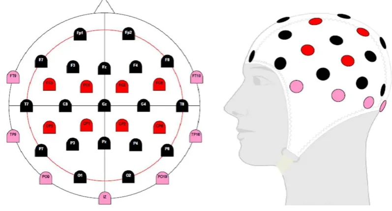
24 the nasion and inion (longitudinal line) and between the opening to each ear channel (lateral line), with each line representing 100%; and 2) the circumference of the base of the cap around the head (hat line) (see Figure 2.5, BrainProducts, 2009). Having these measures, specifically relating to the use of anatomical reference landmarks, enables replicable measures to be obtained over time, which is a valuable attribute within research, allowing examination of changes before and after an intervention over different periods of time/days/months.
Figure 2. 5: EEG Electrode System Montage (Brain Products, 2009)
25 form of stimulus (Cohen, 2011). As the focus of this literature review and thesis is related to task- and exercise-related differences in ERPs only these will be discussed further.
3.2 Event-related potentials
ERPs provide a dynamic means of examining the time course of voltage fluctuations and patterns of activity in the brain in response to a specific sensory, cognitive or motor event (stimulus) (Luck, 2005; Luck & Kappenman, 2012). These fluctuations are associated with different components which are representative of various sensory, cognitive and motor processes (Friedman et al., 2001). This type of stimulus-induced neural activity provides a picture of differences in neural responses that precede any subsequent observable behavioural response such as reaction time and are present even in the absence of a behavioural response (Kutas et al., 1977; Luck, 2005). The differences in these voltage fluctuations lead to
inferences about both the nature and location of brain function, specifically around the underlying short latency and more complex long latency components such as the N1 and P3 ERPs (Duncan et al., 2009). As ERPs are quite small (1 – 30 millionths of a volt) a number of trials are required to enable the extraction of the desired component of interest. This is
26 ERP amplitude is representative of the cognitive effort involved in the reception, integration, and processing of sensory stimuli with which subsequent goal-directed behaviour is
generated (Duncan et al., 2009; Key, Dove, & Maguire, 2005). Scalp distribution of the stimulus-driven ERP relates to the location of the neural activity (as identified by electrode scalp placement) and can provide a basis with which to determine the functional association of the pattern of voltage fluctuations in response to a given stimulus (e.g., visual or auditory) (Luck, 2005). The last characteristic is that of the experimental variable, for example the different neural responses associated with the use of an auditory compared to a visual sensory stimulus (Duncan et al., 2009). The use of an ERP methodology is multifaceted and includes the evaluation of a vast array of different neural components and is an effective and
informative means of evaluating differences in neural activity between population groups and/or interventions, such as younger and older adults and the influence of physical fitness and acute bouts of exercise. For example, Kamijo et al. (2009) found that after an acute bout of moderate intensity aerobic exercise, there was an enhanced P3 ERP amplitude in the younger (19 – 25 years) compared to older (60 – 74 years) adults, indicating an improvement in the allocation of attentional resources and working memory.
27 There are also some limitations to using ERPs to evaluate differences in neural
activity and formulating inferences of correlations between this activity and behaviour are related to: 1) the low spatial resolution as highlighted previously, specifically identifying the internal ERP generators, direction of neural activity (apical compared to transversely
orientated activity and the diffused activity measured at the scalp; 2) due to the small voltage of ERPs a large number of trials (e.g., 50-1000) are required to enable an accurate measure of the neural response to a stimulus (Luck, 2005). The need for a large number of trials relates to signal averaging, which is a process that assumes that the ERP waveform is identical in each trial, whereas extraneous noise (non-cerebral artefact) is not. Therefore, the more trials that can be performed, the clearer the resulting ERP waveform will be (Luck, 2005); and 3) Accounting for potential overlap in the ERP waveforms, which occurs when the neural response to the previous stimulus has not ended and the next stimulus is presented. This is problematic as it can cause a jittering or smearing effect of the neural data, and can lead to the data being misinterpreted. As an ERP can last several seconds the inter-stimulus interval (ISI) must account for this to minimise the risk of overlap occurring (Luck, 2005).
For the purpose of this literature review and thesis focus will be on task- and exercise-related differences in ERPs, specifically the N2 and P3 ERP component latencies and
amplitudes. The N2 ERP component is the second negative going voltage deflection after stimulus onset and occurs within a time window of 150 – 400 ms after sensory stimulus onset (Falkenstein, Hoormann, & Hohnsbein, 1999; Gajewski & Falkenstein, 2012) and is reported to be most prominent over fronto-central and posterior scalp sites (Luck, 2005; Van Veen & Carter, 2002). The N2 component is reported to be related to the process of response
28 refers to the ability to deliberately suppress a habitual response (Donkers & Van Boxtel, 2004; Yeung et al., 2004). The two main characteristics of the N2 component are latency, which is an index of the timing of information processing during visual perception, with the
peak latency representing the moment in time where sensory information is available to formulate the stimulus response decision (Schmitt et al., 2000; Thorpe et al., 1996), and
amplitude which is associated with the neural activity (i.e., degree of effort and processes) required for response monitoring and/or response inhibition (Donkers & Van Boxtel, 2004; Yeung et al., 2004).
The P3 component is a positive going waveform and includes both the P3a and P3b, which represent different phases of information processing. The P3a (or novelty P3)
subcomponent of the P3 is a large, positive deflection with a fronto-central and anterior frontal distribution and is associated with involuntary attention shifts to changes within the environment (Friedman & Simpson, 1994; Jongsma, Meeuwissen, Vos, & Maes, 2007; Spencer, Dien, & Donchin, 1999). It has a latency of approximately 250 – 350 ms and is an indicator of automated, bottom-up aspects of attention (Debener, Kranczioch, Herrmann, & Engel, 2002; Escera, Alho, Winkler, & Naatanen, 1998). This includes attentional processes (Scisco et al., 2008) and aspects of stimulus evaluation in tasks requiring some form of action (Hohnsbein, Falkenstein, & Hoormann, 1995). The P3b is a positive deflection that has a posterior-parietal distribution with a longer latency compared to that of the P3a. In tasks incorporating complex perceptual and conceptual processing the P3b latency is
29 Hohnsbein et al., 1995). The P3b is further defined into two key aspects, that of the early and late P3b, which are representative of modulation of working memory load on the encoding and retrieval phases of information processing (Brookhuis et al., 1981; Jongsma et al., 2007; Morgan et al., 2008; Scisco et al., 2008).
These subcomponents have two main characteristics, the latency which is related to the speed with which we can classify sensory stimuli and the amplitude which is related to the allocation of attentional resources and working memory (Duncan-Johnson, 1981; Kutas et al., 1977; Polich, 1987). The P3a and P3b are elicited in the performance of tasks requiring an inhibitory response, specifically related to processing of additional stimuli (such as in a dual-task context), and the subsequent updating of neural stimulus representation in working memory (see Figure 2.6).
. Figure 2. 7: “Sensory input is processed, with frontal lobe activation from attention-driven working memory changes (P3a) and temporal/parietal lobe activation from memory updating operations (P3b)” (Polich, 2003).
30 presented; the representation (red light = right button press) is accessed from working
memory to enable the performance of the correct goal-directed behaviour. In a dual-task scenario, where there are task-related differences in attention (multiple event representations in working memory), there is a reported graded effect of primary and secondary task
difficulty on the P3 amplitude (Isreal, Chesney, Wickens, & Donchin, 1980a; Isreal, Wickens, Chesney, & Donchin, 1980b). In other words there is an association between an increase in task difficulty (e.g., engaging an inhibitory response to prevent an error occurring) and a reduction in the attentional capacity resulting in a decrease in the P3 amplitude (Hahn, Wild-Wall, & Falkenstein, 2011; Kramer, Sirevaag, & Braune, 1987; Polich, 2007; Pratt et al., 2011; Strayer & Kramer, 1990).
There is strong evidence from both a behavioural and neurophysiological perspective to support the association between the increased demands on working memory and attention with more complex and cognitively demanding tasks (i.e., within a dual-task context) and a decrease in the P3 ERP amplitude (representative of the allocation of attentional resources) (Morgan et al., 2008; Pratt et al., 2011). Further, this decrease in attentional capacity is linked to fall rates in older adults (Ayers et al., 2014; Beauchet et al., 2009). The functional
repercussions of this risk may include a reduced capacity to perform activities of daily living, impaired quality of life and the resultant financial burden. Therefore, a common goal within research has been to obtain a better understanding of why these decrements occur and how we can attenuate this decline and improve overall cognitive and physical function.
4. Cognitive function and exercise
31 which include enhanced neural efficiency and allocation of attentional resources to process and manage tasks requiring interference control (Huang, Lin, Hung, Chang, & Hung, 2014). When evaluating the optimal exercise-stimulus to promote improvements in cognitive function, however, consideration must be given to the core attributes of any exercise intervention (i.e., intensity, mode, and duration).
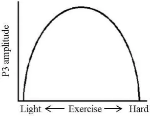
The intensity of exercise bouts includes the differential influence of continuous low, moderate and high intensity aerobic exercise on cognitive function. Evidence to date suggests that there is an inverted-U relationship between exercise intensity and cognitive performance (Kashihara, Maruyama, Murota, & Nakahara, 2009). The greatest improvement in cognitive function appears to result from moderate intensity (e.g., as defined by the anaerobic threshold; AT) compared to low and high intensity exercise (see Figure 2.7, Kashihara et al., 2009). Moderate intensity exercise has also been linked to an increase in the P3 ERP
32 Figure 2. 8: Inverted U - physiological response corresponding to cognitive function (adapted from Kashihara et al., 2009).
Figure 2. 9: Inverted U – exercise intensity corresponding to an increase in the P3 ERP amplitude (Kamijo et al., 2009).
Mode of exercise also varies between research interventions and includes aerobic and resistance-based exercise (e.g., Pesce & Audiffren, 2011; Weinberg, Hasni, Shinohara, & Duarte, 2014). Continuous aerobic exercise is the main form of exercise where exercise-induced enhancements in cognitive function have been observed, both as a result of long-term (e.g., Colcombe et al., 2006) and acute (e.g., Audiffren et al., 2008) interventions. For
33 2009). Whereas there is a wealth of research relating to the influence of aerobic exercise there is a lack of research on resistance exercise of a long-term or acute nature.
Research that has investigated the influence of resistance exercise on cognitive function, specifically the P3 ERP component, suggests that, similar to aerobic exercise, this type of exercise promotes an improvement in neural activity, namely a latency decrease and an amplitude increase in the P3 ERP component (Chang et al., 2013). When assessing these exercise modes the core attributes and differences between protocols must be considered to obtain an understanding of both behavioural and neurophysiological exercise-induced changes. For example, a basic difference between aerobic and resistance exercise being that aerobic exercise such as jogging or cycling would be classified as repetitive in nature and requires less cognitive engagement to perform the simple and rhythmic movement patterns on a stationary cycle ergometer or treadmill. Resistance exercise in contrast requires more
complex movement patterns and is dynamic by nature incorporating a higher cognitive load than aerobic exercise, thus requiring more active engagement and allocation of attentional resources which is crucial for the controlled and fluid performance of exercise (Best, 2010; Chang et al., 2013; Dai et al., 2013).
A final consideration is that of one off acute compared to long term exercise
34 allocation of attentional resources during stimulus engagement (O'Leary et al., 2011). In light of the fact that this thesis is focused on the effects of a single acute bout of exercise the following sections will discuss research relating to acute bouts of both aerobic and resistance exercise.
4.1 Acute aerobic exercise
Acute aerobic exercise has been reported to have a beneficial effect on reducing task-related deficits in performance. For example, exercise-induced enhancement in cognitive flexibility and reduction in complex switch-task costs has been observed (Pesce & Audiffren, 2011). In young, healthy populations acute exercise appears to have a positive effect on cognitive functioning including executive functioning and cognitive processing speed
(Griffin et al., 2011). Improvements in reaction times have also been reported after moderate intensity exercise (50% VO2max) compared to baseline and light intensity (30% VO2max) exercise, in younger and older adults (19 – 25 and 60 – 74 years respectively). From a neurophysiological perspective these behavioural improvements were correlated with changes in neural activity, specifically the P3 ERP latency and amplitude (Kamijo et al., 2009).
35 exhaustion), and a variation in results after a light bout of exercise (RPE 7 – 11 and 60% HRmax). Findings with regard to the P3 latency have also been equivocal with three studies not finding differences irrespective of exercise intensity (Barak et al., 2007; Kamijo et al., 2004; Kamijo et al., 2007). Kumar et al. (2012), on the other hand observed faster P3
latencies after a 20 min moderate exercise bout (60 – 80% HRmax). The ten studies that evaluated only a moderate bout of exercise on the N2 (x 4) and/or the P3 (x 9) ERP
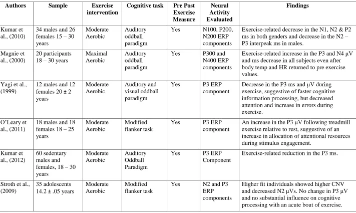
37 Table 2. 1: Articles evaluating acute exercise-related influences on the N2 andP3 ERP components
Authors Sample Exercise
intervention
Cognitive task Pre Post Exercise Measure Neural Activity Evaluated Findings Kumar et al., (2010)
34 males and 26 females 15 – 30 years Moderate Aerobic Auditory oddball paradigm
Yes N100, P200,
N200 ERP components
Exercise-related decrease in the N1, N2 & P2 ms in both genders and decrease in the N2 – P3 interpeak ms in males.
Magnie et al., (2000)
20 participants 18 – 30 years
Maximal Aerobic
Auditory oddball paradigm
Yes P300 and
N400 ERP components
Exercise-related increase in the P3 and N4 μV and ms decrease in all subjects even after body temp and HR returned to pre exercise values.
Yagi et al., (1999)
12 males and 12 females 20 ± 2 years Moderate Aerobic Auditory and visual oddball paradigm
Yes P3 ERP
component
Decrease in the P3 ms and μV during exercise, suggestive of faster cognitive information processing, but decreased attention and increase in errors during exercise.
O’Leary et al., (2011)
18 males and 18 females 18 – 25 years
Moderate Aerobic
Modified flanker task
Yes P3 ERP
component
An increase in the P3 μV following treadmill exercise relative to rest, suggestive of an increase in allocation of attentional resources during stimulus engagement.
Kumar et al., (2012)
60 sedentary males and females, 18 – 30 years Moderate Aerobic Auditory Oddball Paradigm
Yes P3 ERP
Component
Exercise-related reduction in the P3 ms.
Stroth et al., (2009)
35 adolescents 14.2 ± .05 years
Moderate Aerobic
Modified flanker task
Yes N2 and P3
ERP
components
38 Hillman et
al., (2003)
10 males (20.5 ± .05) and 9
females 20.2 ± 1.0
Graded maximal exercise test
Eriksen flanker task
Yes P3 ERP
Component
Post-exercise related increase in the P3 μV. Suggestive of an exercise effect on
neuroelectric processes underlying executive control through an increase in the allocation of neuroelectric resources, cognitive
processing and stimulus classification speed. Themanson
& Hillman (2006)
14 males and 14 females 18 – 23 years Vigorous but submaximal Aerobic Eriksen flanker task
Yes N2 ERP
component
Higher fit adults exhibited a reduced error-related negativity μV, increased error positivity μV, and increased post-error response slowing compared to lower-fit adults.
Kamijo et al., (2007)
12 males 22 – 30 years
Light and Moderate Aerobic
Modified Flanker Task
Yes P3 ERP
Component
Increase in the P3 μV across light and moderate conditions but not hard. Kamijo et
al., (2009)
24 males
12 x 60 – 74 years and
12 x 19 – 25 years
Light and Moderate Aerobic
Modified Flanker Task
Yes P3 ERP
Component
Increase in the P3 μV after moderate exercise for the younger group only. Decrease in P3 ms after both light and moderate exercise for both groups.
Kamijo et al., (2004)
12 males 22 – 33 years
Low, Medium and High Aerobic Go/No Go Reaction Time Task
Yes P3 ERP
Component
Reduced P3 μV after high-intensity exercise and an increase in the P3 μV after moderate-intensity exercise.
Drollette et al., (2014)
13 males and 27 females 9.7 ± 0.7 years
Moderate Aerobic
Modified flanker task
Yes N2 and P3
ERP
components
Exercise-related increase in the P3 μV and a reduction in the N2 μV and P3 ms, suggestive of an overall facilitation in response conflict and the speed of stimulus classification. Pontifex et
al., (2015)
21 males and 16 females
19.3 ± 0.9 years
Moderate Aerobic
Three-stimulus oddball task
Yes P3, P3a and P3b ERP component
39 Scudder et
al., (2012)
19 males and 18 females 19.7 ± 1.3 years Moderate Aerobic AX-continuous performance task
Yes N2 and P3
ERP
components
Exercise-related increase in the P3 μV within midline-parietal sites for both target and non-target trials.
Chu et al., (2015)
21 participants 19 – 24 years
Moderate Aerobic
Stop signal task Yes N1 and P3 ERP
components
Exercise-related increase in the P3 μV and ms, however no effect on the N1 component. Barak et al.,
(2007)
17 adults
21.6 ± 1.07 years
60, 75 and 90% max pulse Aerobic
Reaction time task
Yes P3 ERP
Component
40
4.2 Acute resistance exercise
Resistance exercise has been predominantly advocated in the attenuation of sarcopenia (muscle loss) and bone degeneration (osteoporosis), with resistance exercise increasing serum concentrations of bone ratio markers suggestive of increased bone turnover and bone formation (Karabulut et al., 2011). The few studies which have been conducted suggest that high intensity resistance training (100% 10 RM) improves cognitive processing speed, whereas moderate intensity resistance training (70% 10 RM) is associated with
enhanced processing speed and executive functioning immediately following exercise (Chang & Etnier, 2009b; Chang et al., 2014). These exercise-induced improvements have also been reported in relation to automatic cognitive processes, such as speed of processing in executive function tasks (e.g., Stroop colour – word) and a trend towards improvements in the
41 In summary, from the limited research that has examined the influence of acute bouts of resistance exercise on cognitive function, suggest a positive effect. However, there is no research that has evaluated the influence of an acute bout of resistance exercise on neural activity, and more specifically dual-task related neural activity.
4.3 Additional factors in assessing cognitive function and exercise
The cognitive task employed must also be considered, due to the differences in behavioural tasks available and the areas of cognitive function of interest. The modified flanker and oddball paradigms require participants to respond to a form of sensory stimuli (e.g., auditory or visual) through the execution of a button push in response to the stimulus. These paradigms are examples of cognitive tests that promote the use of executive control, specifically interference control in the evaluation of the P3 ERP component response. This response has been observed to be more prominent when performing tasks that require an inhibitory response. For example, in the use of a modified flanker task, participants are presented with a set of visual stimuli to assess their ability to suppress an incorrect response. The participant is required to identify the direction of the central target which is flanked by distractor stimuli by pressing the left or right key on a computer. The inhibitory response in an incongruent trial (e.g., central arrow facing in the opposite direction of the flanker arrows) is reported to increase the P3 latency. In other words an inhibitory response takes longer to perform, hence the increase in P3 latency (Hahn et al., 2011).