Article
C/EBPβ Is a Transcriptional Regulator of Wee1 at the
G
2/M Phase of the Cell Cycle
Ji Hae Lee †, Jeeyoung Sung †, Eun Kyung Choi †, Hyun-Kyoung Yoon, Boram Kang, Eunkyung Hong, Byunggyu Park, Yong-Nyun Kim, Seungbae Rho and Kyungsil Yoon*
Research Institute, National Cancer Center, Goyang, Gyeonggido 411-769, Korea; jhlee5@ncc.re.kr; sungjy@ncc.re.kr; museonthat@naver.com; 74670@ncc.re.kr; sirosama@hanmail.net; hongek@ncc.re.kr; bkpark@ncc.re.kr; ynk@ncc.re.kr; sbrho@ncc.re.kr
* Correspondence: kyoon@ncc.re.kr; Tel.: +1-82-31-920-2325; Fax: +1-82-31-920-2337 †These authors equally contributed as first authors.
Abstract: The CCAAT/enhancer-binding protein β (C/EBPβ) is a transcription factor that regulates cellular proliferation, differentiation, apoptosis and tumorigenesis. Although the pro-oncogenic roles of C/EBPβ have been implicated in various human cancers, how it contributes to tumorigenesis or tumor progression has not been determined. Immunohistochemistry with human non-small cell lung cancer (NSCLC) tissues revealed that higher levels of C/EBPβ protein are expressed compared to normal lung tissues. Knockdown of C/EBPβ by siRNA reduced the proliferative capacity of NSCLC cells by delaying G2/M transition of the cell cycle. In C/EBPβ-knockdown cells, a prolonged increase in phosphorylation of cyclin dependent kinase 1 at tyrosine 15 (Y15-pCDK1) was displayed with increased Wee1 and decreased Cdc25B expression, simultaneously. ChIP analysis showed that C/EBPβ bound to distal promoter regions of WEE1 and repressed WEE1 transcription through the interaction with histone deacetylase 2. Treatment of C/EBPβ-knockdown cells with a Wee1 inhibitor induced a decrease in Y15-pCDK1 and recovered cells from G2/M arrest. In the xenograft tumors, the depletion of C/EBPβ significantly reduced tumor growth. Taken together, these results indicate that Wee1 is a novel transcription target of C/EBPβ that is required for the G2/M phase of cell cycle progression, ultimately regulating proliferation of NSCLC cells.
Keywords: cell cycle; lung cancer; C/EBPβ; G2/M arrest; Wee1; Y15-pCDK1
1. Introduction
CCAAT/enhancer-binding protein β (C/EBPβ) is a member of the basic leucine zipper (bZIP) class of transcription factors and is involved in regulating cell growth, differentiation, inflammation, metabolism, survival, and tumorigenesis [1-6]. The growth-regulatory function of C/EBPβ has been reported to inhibit or promote cell proliferation, depending on the cellular context [1]. In C/EBPβ knock-out mice, hyperproliferation of epidermal keratinocytes was observed [7]. Several reports demonstrated that C/EBPβ has growth-promoting activity. In C/EBPβ knock-out mice, the number of B lymphocytes was reduced with a defect in the cell expansion, and cell proliferation in gastric mucosa was decreased [8-9]. After partial hepatectomy, C/EBPβ knock-out mice displayed reduced proliferation of hepatocytes, indicating C/EBPβ is required for liver regeneration [10]. In cancer cells, those opposing observations were also reported. Inducible expression of C/EBPβ in breakpoint cluster region-abelson murine leukemia viral oncogene homolog 1(BCR/ABL)-expressing cells inhibited cell proliferation and promotes differentiation [11]. However, in glioblastoma and gastric cancer cells, decreased expression of C/EBPβ inhibited cell growth [9, 12]. Thus, it is important to
investigate how the function of C/EBPβ is determined in the cellular context and to determine what elicits these opposite growth responses including upstream regulators and downstream effectors.
C/EBPβ was shown to be required for carcinogen-induced mouse skin tumorigenesis by regulating p53-induced apoptosis upon carcinogen treatment [13-14], even though it induced differentiation of epidermal keratinocytes in normal physiology [7]. Reduced skin-tumorigenic potential was also observed in v-Ha-Ras transgenic mice when C/EBPβ is deficient, suggesting C/EBPβ plays an oncogenic role in the downstream Ras signaling pathway [13]. The analysis of gene expression patterns of human cancers revealed that C/EBPβ is also involved in cyclin D1-induced oncogenic signature [15]. Increased expression of C/EBPβ and its oncogenic roles have been reported in breast, ovarian, colorectal, renal, and gastric cancers [16-21]. However, the detailed mechanism and function are yet to be determined.
Uncontrolled proliferation of abnormal cells is one of characteristics of cancer, implying the dysregulation of the cell cycle. Cell cycle is tightly controlled to prevent incorrect DNA replication or immature cell division that induces genomic instability, a hallmark of cancer [22]. Cell cycle progression is proceeded by cyclin-dependent kinases (CDKs) and their partner cyclins which regulate the activity of CDKs. Cyclin D-CDK4 and cyclin D-CDK6 play a role in G1 progression [23], and cyclin E-CDK2 and cycle A-CDK2 regulate G1/S transition and S phase progression, respectively [24-25]. Cyclin B-CDK1 is required for G2/M transition [26]. CDK activity is also regulated by activating phosphorylation by CDK-activating kinase [27] and inhibitory phosphorylation by Wee1 and Myt1 [28]. Dephosphorylation by Cdc25 activates CDK activity and enables cells to continue cell cycle progression. CDK inhibitors including INK4 family and Cip/Kip family control CDK activity. INK4 family consists of p15, p16, p18 and p19 and inhibits CDK4 and CDK6 [29]. p21, p27 and p57 comprising Cip/Kip family mainly inactivate G1 cyclin-CDK complexes [30-32]. In human cancers, mutation or dysregulated expression of genes involved in cell cycle regulation are frequently observed [22], which is attributed to accelerated cancer cell growth leading to more malignant progression.
Lung cancer is the leading cause of cancer death worldwide and is also most frequently diagnosed [33]. Non-small cell lung cancers (NSCLCs) account for approximately 85% of human lung cancer, and among them, lung adenocarcinoma and lung squamous cell carcinoma are most prevalent [34]. In NSCLC, altered regulation of cell cycle proteins including inactivation of p16, reduced expression of p27 and Rb, a substrate of Cdk2, and overexpression of cyclin D were also reported [22]. In addition, high expression of cyclin E and cyclin A, and low expression of Rb was correlated with unfavorable prognosis of NSCLC patients [35], implying cell cycle regulatory proteins could be important therapeutic targets or prognostic markers in lung cancer. Lung cancer is a genetically heterogeneous disease and the efforts to determine actionable mutations have led to considerable achievement in personalized molecular targeted therapy. In lung adenocarcinoma, K-RAS and epidermal growth factor receptor (EGFR) are most commonly activated by mutations [34]. Both oncogene products potentiate cell cycle progression upon mitogenic signal, resulting in more aggressive phenotypes.
As C/EBPβ has been reported to mediate several oncogenic signaling pathways, including receptor tyrosine kinases or activated Ras [13, 36], and growth-regulatory function of C/EBP in lung cancer has not been fully defined, we investigated the role of C/EBPβ in human NSCLCs. Here, we report that C/EBPβ is frequently overexpressed in lung cancer tissues compared with normal lungs tissues, and regulates cell proliferation by mediating cell cycle progression at the G2/M phase in NSCLC cells.
2. Materials and Methods
2.1. Cell Culture and Reagents
cultured in bronchial epithelial cell growth medium (BEGM) BulletKit growth media (Lonza, Walkersville, MD, USA). A549, Calu6, NCI-H1299, NCI-H1703, NCI-H23, NCI-H460, HCC2279, NCI-H358, HCC827 and BEAS2B were obtained from the American Type Culture Collection (Rockville, MD, USA). HCC95 and HCC1588 were obtained from the Korean Cell Line Bank (Seoul, Korea), and NHBE purchased from Lonza. All cell lines were maintained in media supplemented with 10% fetal bovine serum (FBS) and 1× penicillin-streptomycin, and cultured under standard conditions at 37 °C in a humidified atmosphere of 95% air and 5% CO2. Stock solutions of the Wee1 inhibitor MK-1775 (Selleck Chemicals, Houston,TX, USA) were dissolved in dimethyl sulfoxide (DMSO) and added to the media at the indicated concentrations (100 nM). Control cells were treated with vehicle alone. Doxycycline (D9891) was purchased from Sigma-Aldrich (St. Louis, MO, USA). Matrigel was purchased from Corning Inc (Corning, NY, USA).
2.2. siRNA Transfection and Generation of Conditional Knockdown Cell Lines
Cells were seeded into 6-well culture plates at a density of 2 × 10⁵ cells per well, and grown for 16 h before transfection with 20 nM of small interfering RNA (siRNA) for 48 h. The sequences of the CCAAT/enhancer binding protein (C/EBPβ) siRNA are as follows: siC/EBPβ #1, 5′-CCTCGCAGGTCAAGAGCAA-3′; siC/EBPβ #2, 5′-CCAAGAAGACCGTGGACAA-3′. SiRNA duplexes were transfected using Lipofectamine 2000 (Invitrogen, Waltham, MA, USA) following the manufacturer’s protocol. To generate doxycycline-inducible C/EBPβ-knockdown cell lines, the C/EBPβ target sequences were cloned into the Tet-pLKO-Puro plasmid (Addgene plasmid #21915,). The Tet-on-shC/EBPβ target sequence is 5′-CACCCTGCGGAACTTGTTCAA-3′. To induce C/EBPβ-knockdown, doxycycline (100 ng/mL) was added.
2.3. Cell Proliferation Assay
Cells were plated in triplicate at 10% confluence in 24 well culture plates and transfected with negative control siRNA (siNC) or C/EBPβ siRNA (siC/EBP) at final concentration of 20 nM. Cells were harvested and counted using a Coulter counter (Beckman Coulter, Brea, CA, USA) in 24 h intervals. An IncuCyte live-cell imaging system (Essen Bioscience, Ann Arbor, MI, USA) was used to measure the proliferation of Tet-on-shC/EBPβ or shNC cells using the cell confluence approach
2.4 Live/Dead cell staining
Cells were stained using LIVE/DEAD™ Viability/Cytotoxicity Kit (Thermo Scientific, Rockford, IL, USA) following manufacture’s protocol. Images were obtained by an Operetta High Content Screening (HCS) System (PerkinElmer, Waltham, MA, USA) and analysis was performed using the Harmony 3.5.2 software (PerkinElmer).
2.5. Cell Cycle Analysis
For thymidine double block [37], cells were seeded at 2 × 10⁵ cells per well in 6-well culture plates and treated with thymidine at 10 μM for 14 h. Cells were then washed, supplemented with normal media for 12 h, and treated with 10 μM thymidine for another 14 h. The cells were harvested at 2 h intervals up to 16 h and 26 h after release. Afterward, cells were harvested and fixed in 75% (v/v) cold ethanol at −20 °C for at least 2 h. The fixed cells were collected by centrifugation and resuspended in propidium iodide (PI) Staining Buffer (Sigma, St. Louis, MO, USA) to stain DNA and finally analyzed for DNA content on a flow cytometry (FACSCaliber; Becton Dickinson, Franklin Lakes, NJ, USA).
2.6. Western Blot Analysis
determined using a micro bicinchoninic acid (BCA) protein assay kit (Thermo Scientific, Rockford, IL, USA). Equal amounts of protein were separated by 6–12% Sodium dodecyl sulphate-polyacrylamide gel electrophoresis(SDS-PAGE) and transferred to a polyvinylidene difluoride (PVDF) membrane (BioRad, Hercules, CA, USA) using a wet transfer device (BioRad, Hercules, CA, USA). The following antibodies were used in this study: C/EBPβ (sc-7962), MAD2 (sc-6329), and β-actin (sc-477778) (all obtained from Santacruz Biotechnology, Santa Cruz, CA, USA); anti-Cdc2/Cdk1 (06-923), phospho-Cdk1 (Tyr15) (#9111), Cdc25B (#9525), Wee1 (#4936), and Cdc25A (#3652) (all obtained from Cell Signaling Technology Beverly, MA, USA); and anti-CyclinB1 (05-373) and Cdc25C (05-507) (all obtained from Millipore, Bedford, MA, USA ).
2.7. Quantitative Real-Time PCR
Total RNA was prepared by using Trizol (Ambion, Lifetechnologies, Carlsbad, CA, USA) and cDNA was synthesized using moloney murine leukemia virus (M-MLV) reverse transcriptase (Invitrogen, Waltham, MA, USA). Real-time polymerase chain reaction (PCR) was performed using LightCycler® 96 Real-Time PCR System (Roche, Basel, Switzerland). Each reaction was performed with 10 ng cDNA by using SYBR Green. Primer sequences used for PCR were as follows: Wee1, (F: 5′-TTCAATGAGGAGACTTGCCTG-3′ and R: 5′-ACAACAACAATCTGAGGTGCC-3′); Cdc25B, (F: 5′-GTGAGGAAGTTTCAGAACAGTCCG-3′ and R: 5′-TGGGAGGCTTGTCGCATTTG-3′), and
GAPDH, (F: 5′-TGATGACATCAAGGTGGTGAAG-3′ and R:
5′-TCCTTGGAGGCCATGTGGGCCAT-3′). PCR reactions were performed as follows: 95 °C denaturation for 5 min, followed by 40 cycles at 94 °C for 10 s, 60 °C for 10 s, 72 °C for 10 s, followed by a 9-min extension at 72 °C.
2.8. Chromatin Immunoprecipitation
The chromatin immunoprecipitation (ChIP) assay was performed using the EZ-ChIP assay (Upstate Biotechnology, Lake Placid, NY, USA) following the manufacturer’s protocol. Briefly, formaldehyde was added at a final concentration of 1% directly to cell culture media. Fixation proceeded at room temperature (RT) for 10 min and was stopped by the addition of glycine to a final concentration of 0.125 M. The cells were collected by centrifugation and rinsed in cold phosphate-buffered saline. The cell pellets were resuspended in hypotonic buffer containing 0.5 mM PMSF, protease inhibitor cocktail, and incubated on ice for 15 min. The nuclei were collected by micro-centrifugation and then resuspended in SDS lysis buffer (1% SDS, 10 mM EDTA, 50 mM Tris-HCl (pH 8.1), 0.5 mM PMSF, and protease inhibitor cocktail). The samples were sonicated to an average length of 300–500 bp with a S220 Focused-ultrasonicator (Covaris, Woburn, MA, USA). Chromatin immunoprecipitation was performed with anti-C/EBPβ (sc-150X, Santacruz Biotechnology, Santa Cruz, CA, USA) or HDAC2 (sc-9959X, Santacruz Biotechnology, Santa Cruz, CA, USA) and protein G agarose. ChIP products were eluted and DNA was recovered from reverse crosslinking and purification. C/EBPβ binding to specific sites on the Wee1 promoter was analyzed by quantitative real-time PCR (qRT-PCR). Primers for PCR analysis were follows: R1 (F, 5′-CAGTCTAGTTGTGGAGAGGCA-3′ and R, 5′-CCTGCCACTCCTGATGACAAA-3′); R2 (F, 5′-CAGTGTGTGCTTTACTCAGAGGAG-3′ and R, 5′-CTCCAGCAACCAGCACTGT-3′); R3 (F, 5′-TCAAAGTGCAAGGCTCATGT-3′ and R, 5′-TTTGCAGAATCCACATGCTT-3′); R4 (F, 5′-TGCTGATGAACATGCGGTGA-3′ and R, 5′-CTGCCTATTGGCCTCAGGAA-3′); GAPDH exon (F, 5′-TCTATAAATTGAGCCCGCAGC-3′ and R, 5′-GCGACGCAAAAGAAGATGC-3′).
2.9. Luciferase Reporter Assay
pcDNA3.1-HDAC2 as indicated using Lipofectamine 2000 (Invitrogen). After 48 h, cells were harvested. Firefly luciferase activities were determined using Luciferase assay system kit (Promega, Madison, WI, USA), as described by the manufacturer, with a luminescence plate reader (VICTOR™ X, PerkinElmer, Waltham, MA, USA). The firefly luciferase activity was normalized for transfection efficiency with protein measurement using a BCA protein assay. Data are expressed as relative luciferase activity/μg protein.
2.10. Iimmunoprecipitation
Cells were lysed in cell lysis buffer (20 mM Tris–HCl pH8.0, 150 mM NaCl, 1 mM Na2EDTA, 1 mM EGTA, 1% Triton, 2.5 mM sodium pyrophosphate, and 1 mM β-glycerophosphate). Each cell lysate (1 mg) was incubated with C/EBPβ monoclonal antibody (Santacruz Biotechnology, Santa Cruz, CA, USA) overnight at 4 °C. Following incubation, protein was immunoprecipitated using protein G agarose beads (GE Healthcare, Chicago, IL, USA) for 2 h at 4 °C with gentle rotation. The immunoprecipitates were washed three times with lysis buffer and boiled in 20 μL of 1× SDS sample buffer for 5 min at 95 °C. After centrifugation, the supernatant was analyzed using Western blot.
2.11. Xenograft Mouse Model and siRNA Delivery
A549 (5 × 10⁶) cells were suspended in 100 μL PBS and mixed with 50 μL Matrigel (Corning Inc.). The mixtures were implanted subcutaneously into 6-week-old athymic nude mice. When the tumor size reached 60 to 80 mm3, the dilute siRNA solution in sterile PBS (50 μL) was directly injected into the xenograft tumor via electroporation using NEPA21 Super Electroporator (Nepa gene Co., Chiba, Japan). The tumor size was monitored every 7 days up to 7 weeks. Tumor diameters were measured twice a week and the volume was calculated with the following formula: V (mm3) = longest diameter × shortest diameter 2/2.
2.12. Immunohistochemical Staining for Xenograft Tumor
Xenograft tumors were removed and fixed in 10% formalin, embedded in paraffin, and cut into 4-μm sections. The sections were used for immunohistochemical staining performed with the automated instrument Discovery XT (Ventana Medical Systems, Inc., Tucson, AZ, USA) using anti-C/EBβ (sc-150, Santacruz Biotechnology, Santa Cruz, CA, USA), anti-Wee1 (ab37597), Cdc25B (ab70927), phospho-Cdk1(Tyr15) (ab133463), anti-Ki67 (ab15580) (all from Abcam, Cambridge, UK), and cleaved caspase3 (#9661, Cell signaling Technology Beverly, MA, USA).
2.13. Immunohistochemical Staining for Lung Cancer Tissue Microarray
Lung cancer tissue array was obtained from Superbiochips Laboratories (Seoul, Korea), as described previously [38]. Each array contained 60 sections of 4 μm thickness obtained from 60 patients by biopsy or surgical resection. The sections were used for immunohistochemical staining performed with the Ventana BenchMark XT Staining systems (Ventana medical systems, Inc.) using C/EBPβ antibody (1:30, sc-150 Santacruz Biotechnology, Santa Cruz, CA, USA) and the UltraView Universal DAB detection kit (Ventana Medical Systems, Inc.). Parallel sections incubated with normal IgG antibody instead of primary antibodies were used for negative controls. The stainings were scored from 0 to 4 based on the intensity and proportion of positive staining in a tissue field. Stained tissue array was reviewed by two experienced medical pathologists. To obtain representative images, slides were scanned by the Aperio ScanScope scanner (Aperio Technologies, Vista, CA, USA) and images were captured using Aperio ImageScope software.
2.14. Statistical Analysis
defined as a p value <0.05. For survival analysis, Kaplan-Meier plotter [39] was used to investigate the association of C/EBPβ mRNA expression in overall survival and post-progression survival of lung cancer patients.
3. Results
3.1.Levels of C/EBPβ Protein in Human Lung Cancer Tissues Were Elevated
Figure 1. CCAAT/enhancer-binding protein β (C/EBPβ) expression in human lung cancer tissues. (A) Patients-derived lung cancer tissue array was examined for C/EBPβ expression using the immunoperoxidase method. Staining results were graded according to the intensity and proportion of positive area. Images were captured at a magnification of 200X by using the Aperio ImageScope software. Scale bars: 200 μm. (B) The histogram represents the percentage of the immunohistochemistry (IHC) score for C/EBPβ in 68 normal tissues, 95 primary, and 9 metastatic tumor tissues. The statistical significance was determined using the t-test, p < 0.05.(C) The association between C/EBPβ mRNA expression and overall survival of adenocarcinoma (whole dataset) and squamous cell carcinoma patients (GSE37745) was analyzed using the Kaplan–Meier Plotter. Hazard ratio (HR) significance was found with log-rank tests.
Table 1. Immunohistochemistry (IHC) scoring of C/EBPβ
score Normal
n (%)
Tumor n (%)
Metastasis n (%)
0 47 (69.1) 31 (32.6) 1 (11.1)
1 20 (29.4) 36 (37.9) 3 (33.3)
2 1 (1.5) 10 (10.5) 2 (22.2)
3 0 (0) 13 (13.7) 3 (33.3)
4 0 (0) 5 (5.3) 0 (0)
Total 68 (100) 95 (100) 9 (100)
Table 2. Summary of C/EBPβ expression and clinicopathological feature in the lung cancer patients.
C/EBPβ expression
n (%) n (%) n (%) Sex
Male 23 (31) 52 (69) 75 (100)
0.4343
Female 8 (40) 12 (60) 20 (100)
Age
≤57 11 (31) 24 (69) 35 (100)
0.8505
>57 20 (33) 40 (67) 60 (100)
Clinical Stage
Ⅰ 10 (27) 27 (73) 37 (100)
0.2305
Ⅱ 12 (32) 25 (68) 37 (100)
Ⅲ 9 (43) 12 (57) 21 (100)
N classification
0 15 (28) 38 (72) 53 (100)
0.2204
1 8 (33) 16 (67) 24 (100)
2 8 (44) 10 (66) 18 (100)
T classification
1 1 (13) 7 (87) 8 (100)
0.3039
2 23 (33) 47 (67) 70 (100)
3 5 (46) 6 (54) 11 (100)
4 2 (33) 4 (67) 17 (100)
Histology
Adenocarcinoma 13 (46) 15 (54) 28 (100) 0.0180* 0.0256†
Squamous carcinoma 15 (31) 34 (69) 49 (100) 0.2941#
Other NSCLC 3 (17) 15 (83) 18 (100)
* Adenocarcinoma vs. squamous cell carcinoma, † Adenocarcinoma vs. other NSCLC, # Squamous cell carcinoma vs. other NSCLC, * † significantly different between two groups, p < 0.05.
3.2. Knockdown of C/EBPβ Inhibits Cell Proliferation Rates of NSCLC Cells
We measured the levels of C/EBPβ protein in normal human bronchiolar epithelial cells (NHBE), immortalized human bronchial epithelial cells (BEAS-2B), and various NSCLC cell lines. NHBE cells expressed C/EBPβ protein in marginal amounts compared with the BEAS-2B cells and NSCLC cell lines tested (Figure 2A). To examine the function of C/EBP in lung cancer, we knocked down C/EBPβ using two different siRNAs in NSCLC cell lines. Transfection of each siC/EBPβ dramatically suppressed proliferation of A549 cells with C/EBP down-regulation (Figure 2B). We also employed doxycycline-inducible shRNA targeting another sequence of C/EBP and observed that doxycycline decreased cell proliferation (Figure 2C).
Figure 2. C/EBPβ promotes cell proliferation of various lung cancer cell lines. (A) C/EBPβ protein levels in normal human bronchial epithelial cells (NHBE), immortalized human bronchial epithelial cells, BEAS-2B and various NSCLC cell lines were determined by Western blot analysis. (B) Live cells of A549 transfected with si-Negative Control (siNC), siC/EBPβ #1, or siC/EBPβ #2 were counted with tryphan blue staining at indicated times after transfection. Data are presented as fold increase. (C) Doxycycline-inducible shC/EBPβ cells using A549 were generated and treated with or without doxycycline (100 ng/mL). Using the IncuCyte live cell imaging system, proliferation cells was monitored and quantified by the percentage of cell confluence. (D) Cell number of lung cancer cell lines transfected with siNC or siC/EBPβ (#1+#2) was counted using Coulter counter at intervals of 24 h up to 120 h after transfection of siRNA. (E) The protein levels of C/EBPβ were detected by Western blotting to check C/EBPβ-knockdown in each cell line. Data are presented as mean ± standard deviation (SD). The statistical significance was determined using the t-test, * p < 0.05, ** p < 0.01.
3.3. C/EBPβ-Knockdown Delayed G2/M Phase of Cell Cycle Progression with Elevated Inhibitory Phosphorylation of CDK1
3C). The expression of Cdc25A and Cdc25C remained unchanged between the groups. C/EBPβ-knockdown did not appeared to affect protein levels of Myt1, Cyclin B1, and mitotic arrest deficeint 2 (Mad2), mitotic spindle checkpoint component [46]. Taken together, these data indicate that C/EBPβ-knockdown cells are arrested at the G2/M phase of the cell cycle, displaying elevated Y15-pCDK1 possibly due to increased Wee1 and decreased Cdc25B.
Figure 3. C/EBPβ-knockdown inhibits cell cycle progression. (A) A549 cells were transfectected with siNC or siC/EBPβ for 24 and 48 h. Live cells were stained by green calcein AM, while dead cells were stained by red ethidium homodimer-1 (EthD-1). Cell images were taken at a magnification of 100X using Operetta High Content Screening (HCS) System. Scale bar: 200 μm. (B) A549 cells were transfected with control siRNA or C/EBPβ siRNA for 48 h. The cell cycle was analyzed by fluorescence-activated cell sorting (FACS) after DNA staining with propidium iodide (PI). M1: subG0/G1 M2: G0/G1, M3: S, and M4: G2/M phase. Percentage of cells in each cell cycle phase is shown as a bar graph. (C) Whole cell lysates were prepared 48 h after transfection and the levels of the G2/M cell cycle-related proteins in control or C/EBPβ-knockdown cells were analyzed by Western blotting. β-actin was used as a loading control. Data are presented as mean ± SD. The statistical significance was determined using the t-test, * p < 0.01.
levels corerlated with a decrease in Y15-pCDK1, which is an important event for mitotic entry. However, Y15-pCDK1 stayed higher levels for a longer time with an increase in Wee1 and a delayed and attenuated Cdc25B induction in the C/EBPβ-knockdown cells compared with control cells (Figure 4C). Taken together, our data demonstrate that C/EBPβ is an important mediator at the G2/M phase of the cell cycle progression by regulating the expression of Wee1 and Cdc25B, which critically modulates Y15-pCDK1.
Figure 4. C/EBPβ knockdown delays G2/M-cell cycle transition. (A) The time course study of cell cycle analysis was performed in control or C/EBPβ-knockdown A549 cells. Cells were released from thymidine double block-induced G1/S synchronization; and 0, 2, 4, 6, 8, 10, 12, 14, 16, and 26 h after releasing, cell were collected and stained with PI for measuring DNA content using FACS. (B) The data are expressed as the percentage of cells in the subG0/G1, G0/G1, S, and G2/M phase at the indicated time points. (C) Whole cell lysates were prepared at the indicated times and Western blot analysis was performed for expression of proteins associated with the G2/M transition (Y15-pCDK1, Wee1, Cdc25B and Cyclin B1). SE; short exposure, LE; long exposure
3.4. C/EBPβ Regulates Wee1 Expression at the Transcription Levels
C/EBPβ has been reported to interact with histone deacetylase1 (HDAC1) and repress the activation of C/EBP promoter [48]. Our data showed that C/EBPβ interacts with HDAC2, not with HDAC1 in A549 cells (Figure 5D). In addition, the HDAC2-CHIP assay revealed that HDAC2 also bound to the R2 and R3 regions, suggesting that C/EBPβ recruits HDAC2 at the WEE1 distal promoter. To determine if C/EBPβ in conjunction with HDAC2 repress WEE1 promoter activity, we constructed a promoter-reporter vector containing either R2 or R3 region of distal WEE1 promoter. Enforced expression of C/EBP or HDAC2 alone reduced WEE1 promoter-reporter activity and C/EBP along with HDAC2 further decreased transcriptional activity of WEE1 promoter, indicating that C/EBP recruits HDAC2 to WEE1 promoter, negatively regulating WEE1 transcription (Figure 5F). Taken together, we identified Wee1, a mitosis inhibitor, as a novel transcription target gene of C/EBP
luciferase activity/ug protein standardized by control pGL3-promoter vector. Data are presented as mean ± SD. The statistical significance was determined using the t-test, * p < 0.05, ** p < 0.01.
3.5. C/EBPβ Knockdown Cells Treated with MK1775 Were Recovered from G2/M Arrest
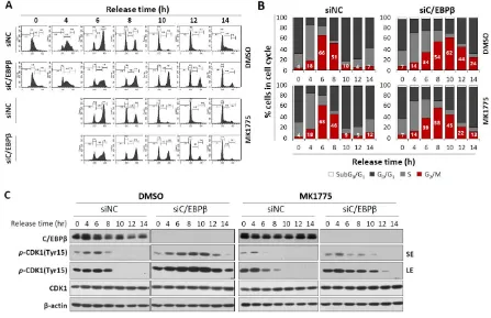
To confirm Wee1 plays an important role in the G2/M cell cycle arrest shown in C/EBP deficiency, we examined whether the inhibition of Wee1 can release C/EBPβ-knockdown cells from the G2/M arrest. Four hours after release into the cell cycle at the completion of the thymidine double block, cells were treated with a Wee1 inhibitor, MK1775, or DMSO. MK1775 treatment of control cells had little effect on the cell cycle progression (Figures 6A,B). However, Wee1 inhibition of C/EBPβ-knockdown cells diminished accumulation of cell population in the G2/M phase induced by C/EBPβ-deficiency (Figures 6A,B). More specifically, C/EBP-knockdown cells were accumulated in the G2/M phase up to 10 h (62.2%) because of the G2/M arrest and decreased to 43. 6% at 12 h after release. However, upon MK-1775 treatment of C/EBP-knockdown cells, cell population in the G2/M phase reached maximally up to 57.8% at 8 h, further progressed into the G0/G1 and S phase, and then decreased to 44.9% and 22% at 10 and 12 h after release, respectively. Consistent with the results in Figure 3 and 4, compared with control cells, Y15-pCDK1 increased in the C/EBPβ-knockdown cells (Figure 6C). Treatment with MK1775 induced a rapid decrease in Y15-pCDK1 with little changes in both total levels of CDK1. As MK1775 treatment rescued C/EBP-knockdown cells from the G2/M arrest, we argue that increased Y15-pCDK1 by Wee1 is responsible for the G2/M arrest with defective C/EBPβ function.
Figure 6. C/EBPβ knockdown cells treated with MK1775 were recovered from G2/M arrest. (A,B) A549 cells transfected with siNC or siC/EBPβ were treated with either DMSO or MK1775 4 h after being released from thymidine double block. Cells were harvested for cell cycle analysis at different time points from release, and DNA contents with PI staining were analyzed using FACS. (C) Expression of cell cycle-associated proteins was analyzed in DMSO- or MK1775-treated control cells and C/EBPβ-knockdown cells. Percentage of cells in each cell cycle phase is shown as a bar graph. SE; short exposure, LE; long exposure
Since C/EBPβ-knockdown inhibits cell proliferation inducing G2/M cell cycle arrest, we tested if C/EBPβ-knockdown reduces the growth of xenograft tumors in vivo. Tumors were produced by injecting A549 cells subcutaneously into dorsal area of athymic nude mice, and siNC or siC/EBPβ RNA was delivered into the tumors via electroporation. Tumor growth was monitored for seven weeks. As shown in Figure 7A, siC/EBPβ treatment markedly suppressed tumor growth by at least 50% compared with the siNC treatment. Consistent with in vitro results, immunohistochemistry analysis revealed that treatment with siC/EBPβ increases the expression of Y15-pCDK1 and Wee1 while decreasing Cdc25B expression (Figure 7B). C/EBPβ-knockdown tumors displayed lower Ki67 and higher cleaved caspase-3 expression compared with the control. These data indicate that C/EBPβ is important for tumor growth in vivo.
Figure 7. C/EBPβ-knockdown inhibits tumor growth. (A) A549 cells (5 × 10⁶) were implanted subcutaneously into athymic nude mice. When tumor size reached 60 to 80 mm3, siNC or siC/EBP were delivered into tumors via electroporation once a week for seven weeks. Tumors were measured at the indicated time and tumor volume was calculated as described in Section 2. Photos from siNC- or siC/EBPβ-treated tumors are shown. Similar results were observed in three independent experiments. Data are presented as mean ± SD. * p < 0.05, ** p < 0.01, significantly different from siC/EBP-treated tumor volume. (B) Immunohistochemical staining for C/EBPβ, Y15-pCDK1, Wee1, Cdc25B, Ki67, and cleaved caspase-3 (CC-3) was conducted with paraformaldehyde-fixed, paraffin-embedded xenograft tumors. Images were captured at a magnification of 400X by using the Aperio ImageScope software. Scale bars: 100 μm.
4. Discussion
transcriptional activation and is yet to be determined. C/EBPβ bound to two WEE1 distal promoter regions, −4.7 to −4.9 kB and −4.4 to −4.5 kB, upstream of transcription start site and repressed transcription through recruiting HDAC2. These findings are summarized as a schematic diagram in Figure 8. To the best of our knowledge, this is the first report that Wee1, a key regulator of G2/M progression, is a transcriptional target of C/EBPβ. Finally, we showed that a Wee1 inhibitor, MK-1775, significantly recovers C/EBPβ-knockdown cells from the G2/M arrest. Taken together, these results indicate that C/EBPβ is a transcriptional regulator of Wee1, mitosis inhibitor protein, ultimately regulating the G2/M phase of the cell cycle progression.
Cell cycle proteins need to be tightly controlled temporally and spatially. Wee1 increases during the S and G2 phase to block immature mitotic entry and then decreases during M phase of the cell cycle [50]. Regulation of Wee1 protein has been extensively studied and several mechanisms on its kinase activity and protein levels have been demonstrated [51]. For example, Wee1 kinase activity is regulated by Akt-mediated phosphorylation on Ser642 and phosphorylated Wee1 binds to 14–3-3θ and translocates to the cytoplasm, resulting in G2/M cell cycle progression. [52]. In late G2 phase, phosphorylation of Wee1 by CDK1 and polo-like kinase 1 creates a phosphodegron which targets for SCF–β-TrCP ubiquitin ligase-mediated proteasomal degradation [53]. However, transcriptional regulation of WEE1 is not well known. WEE1 transcription is repressed by direct binding of kruppel-like factor 2 (KLF2) and chromodomain helicase DNA binding protein 5 (CHD5) and activated by c-Fos/activator protein-1 (AP-1) [54-56]. Our results also showed that Wee1 is downregulated by C/EBP via direct binding to the distal promoter and it is required for the G2/M cell cycle progression in lung cancer cells.
In our study, control cells after release from the thymidine double block progressed to the S phase rapidly reaching the peak levels in 2 h, and majority of them moved into the G2/M phase in another 4 hours (Figures 4A, B, 6A, B). However, in C/EBP-knockdown cells, G1/S transition was delayed and significant population of cells remained in the G0/G1 phase (Figures 4A, B), suggesting that G1/S cell cycle arrest is also induced in the absence of C/EBP. Consistent with our observation, C/EBP-deficient human endometrial stromal cells underwent G1/S arrest shown by the reduction of BrdU incorporation after release from thymidine double block. In those cells, cyclin E was downregulated and thereby cyclin E-CDK2 was not functional for G1/S phase progression [57]. C/EBP has been shown to cooperate with E2F in activating transcription of E2F target genes involved in the G1/S transition by recruiting CBP/p300 coactivator [58].
C/EBPs are expressed in human lung epithelium and they play roles in lung development and differentiation [59]. C/EBPα-deficient mice displayed hyper-proliferative alveolar type II cells and a defect in alveolar architecture and suffered from respiratory distress [60-61], whereas lung phenotypes of C/EBPβ knockout mice were not emphasized [59]. Even if C/EBPβ is involved in the lung-specific gene expression in lung epithelial cells [62-63], it does not seem to be essential in basal lung development or differentiation, possibly due to functional redundancy with other C/EBP family members, as described in liver and mouse skin [64-65]. Rather acute lung injury is induced by lipopolysaccharide (LPS)-induced C/EBPβ mRNA [66], suggesting C/EBPβ might play a distinct role in pathological status. C/EBPβ positively regulated the proliferation of various NSCLC cells, whereas it did not have a substantial effect on immortalized lung epithelial cells. Even if we identified Wee1 and Cdc25B as downstream players of C/EBPβ in the cell cycle regulatory role, the upstream cue, possibly altered in NSCLC, remains to be determined.
that the function of C/EBPβ is not vital in the lung, C/EBPβ could be an attractive target for cancer therapy alone or in combination.
Figure 8. Schematic representation of the potential role of C/EBPβ in the G2/M phase of cell cycle progression. (A) C/EBPβ represses WEE1 transcription by directly binding to WEE1 distal promoter regions and recruiting HDAC2. (B) Wee1 and Cdc25B are key regulators of phosphorylation of tyrosine-15 residue of CDK1, which blocks mitotic entry. C/EBPβ activates Cdc25B expression via unknown mechanism but inhibits Wee1 expression. In the absence of C/EBP, cells undergo G2/M arrest displaying increased CDK1 phsophorylation along with increased Wee1 and decreased Cdc25B levels.
Supplementary Materials: The following are available online, Figure S1: Expression and gene alteration of C/EBPβ in human lung cancers, Figure S2: Overall survival (OS) and post-progression survival (PPS) of lung cancer patients.
Author Contributions: Conceptualization, E.K.C. and K.Y.; Methodology, E.K.C., B.K., S.R. and K.Y.; Validation, J.L., E.K.C. and H.Y.; Formal analysis, J.L., J.S. and E.H.; Investigation, J.L., J.S., E.K.C., H.Y. and B.K.; resources, B.P., Y.K. and S.R.; writing—original draft preparation, J.L., J.S., E.K.C. and K.Y.; writing—Review & Editing, J.L., J.S., Y.K. and K.Y.; Supervision &Funding Acquisition, K.Y.
Funding: This research was supported by the Basic Science Research Program through the National Research Foundation (NRF) funded by the Korean government [NRF-2016R1A2B4016618] and National Cancer Center Research Grant (1710310). And the APC was funded by [NRF-2016R1A2B4016618].
Acknowledgments: We would like to thank Geon Kook Lee, medical pathologist and Dongwan Hong, National Cancer Center for their comments on analysis of immunohistochemistry and public database.
References
1. BibliographySebastian, T.; Johnson, P. F., Stop and go: anti-proliferative and mitogenic functions of the
transcription factor C/EBPbeta. Cell Cycle 2006, 5 (9), 953-957.
2. BibliographyCao, Z.; Umek, R. M.; McKnight, S. L., Regulated expression of three C/EBP isoforms during
adipose conversion of 3T3-L1 cells. Genes Dev 1991, 5, 1538-1552.
3. BibliographyPoli, V., The role of C/EBP isoforms in the control of inflammatory and native immunity
functions. J Biol Chem 1998, 273, 29279-29282.
4. BibliographyZahnow, C. A., CCAAT/enhancer binding proteins in normal mammary development and breast
cancer. Breast Cancer Res 2002, 4, 113-121.
5. BibliographyDiehl, A. M., Roles of CCAAT/enhancer-binding proteins in regulation of liver regenerative
growth. J Biol Chem 1998, 273, 30843-30846.
6. BibliographyRamji, D. P.; Foka, P., CCAAT/enhancer-binding proteins: structure, function and regulation.
Biochem J 2002, 365, 561-575.
7. BibliographyZhu, S.; Oh, H. S.; Shim, M.; Sterneck, E.; Johnson, P. F.; Smart, R. C., C/EBPbeta modulates the
early events of keratinocyte differentiation involving growth arrest and keratin 1 and keratin 10 expression.
Mol Cell Biol 1999, 19 (10), 7181-7190.
8. BibliographyChen, X.; Liu, W.; Ambrosino, C.; Ruocco, M. R.; Poli, V.; Romani, L.; Quinto, I.; Barbieri, S.;
Holmes, K. L.; Venuta, S.; Scala, G., Impaired generation of bone marrow B lymphocytes in mice deficient in
C/EBPbeta. Blood 1997, 90 (1), 156-164.
9. BibliographyRegalo, G.; Forster, S.; Resende, C.; Bauer, B.; Fleige, B.; Kemmner, W.; Schlag, P. M.; Meyer, T. F.;
Machado, J. C.; Leutz, A., C/EBPbeta regulates homeostatic and oncogenic gastric cell proliferation. J Mol Med
(Berl) 2016, 94 (12), 1385-1395.
10. BibliographyGreenbaum, L. E.; Li, W.; Cressman, D. E.; Peng, Y.; Ciliberto, G.; Poli, V.; Taub, R., CCAAT
enhancer- binding protein beta is required for normal hepatocyte proliferation in mice after partial
hepatectomy. J Clin Invest 1998, 102 (5), 996-1007.
11. BibliographyGuerzoni, C.; Bardini, M.; Mariani, S. A.; Ferrari-Amorotti, G.; Neviani, P.; Panno, M. L.; Zhang,
Y.; Martinez, R.; Perrotti, D.; Calabretta, B., Inducible activation of CEBPB, a gene negatively regulated by
BCR/ABL, inhibits proliferation and promotes differentiation of BCR/ABL-expressing cells. Blood 2006, 107
(10), 4080-4089.
12. BibliographyAguilar-Morante, D.; Cortes-Canteli, M.; Sanz-Sancristobal, M.; Santos, A.; Perez-Castillo, A.,
Decreased CCAAT/enhancer binding protein beta expression inhibits the growth of glioblastoma cells.
Neuroscience 2011, 176, 110-119.
13. BibliographyZhu, S.; Yoon, K.; Sterneck, E.; Johnson, P. F.; Smart, R. C., CCAAT/enhancer binding
protein-beta is a mediator of keratinocyte survival and skin tumorigenesis involving oncogenic Ras signaling.
Proc Natl Acad Sci U S A 2002, 99 (1), 207-212.
14. BibliographyYoon, K.; Zhu, S.; Ewing, S. J.; Smart, R. C., Decreased survival of C/EBP beta-deficient
keratinocytes is due to aberrant regulation of p53 levels and function. Oncogene 2007, 26 (3), 360-367.
15. BibliographyLamb, J.; Ramaswamy, S.; Ford, H. L.; Contreras, B.; Martinez, R. V.; Kittrell, F. S.; Zahnow, C. A.;
Patterson, N.; Golub, T. R.; Ewen, M. E., A mechanism of cyclin D1 action encoded in the patterns of gene
expression in human cancer. Cell 2003, 114 (3), 323-334.
16. BibliographyZahnow, C. A.; Younes, P.; Laucirica, R.; Rosen, J. M., Overexpression of C/EBPbeta-LIP, a
naturally occurring, dominant-negative transcription factor, in human breast cancer. J Natl Cancer Inst 1997, 89
17. BibliographyRask, K.; Thorn, M.; Ponten, F.; Kraaz, W.; Sundfeldt, K.; Hedin, L.; Enerback, S., Increased
expression of the transcription factors CCAAT-enhancer binding protein-beta (C/EBBeta) and C/EBzeta
(CHOP) correlate with invasiveness of human colorectal cancer. Int J Cancer 2000, 86 (3), 337-343.
18. BibliographySundfeldt, K.; Ivarsson, K.; Carlsson, M.; Enerback, S.; Janson, P. O.; Brannstrom, M.; Hedin, L.,
The expression of CCAAT/enhancer binding protein (C/EBP) in the human ovary in vivo: specific increase in
C/EBPbeta during epithelial tumour progression. Br J Cancer 1999, 79 (7-8), 1240-1248.
19. BibliographySankpal, N. V.; Moskaluk, C. A.; Hampton, G. M.; Powell, S. M., Overexpression of CEBPbeta
correlates with decreased TFF1 in gastric cancer. Oncogene 2006, 25 (4), 643-649.
20. BibliographyOya, M.; Horiguchi, A.; Mizuno, R.; Marumo, K.; Murai, M., Increased activation of
CCAAT/enhancer binding protein-beta correlates with the invasiveness of renal cell carcinoma. Clin Cancer
Res 2003, 9 (3), 1021-1027.
21. BibliographyRegalo, G.; Canedo, P.; Suriano, G.; Resende, C.; Campos, M. L.; Oliveira, M. J.; Figueiredo, C.;
Rodrigues-Pereira, P.; Blin, N.; Seruca, R.; Carneiro, F.; Machado, J. C., C/EBPbeta is over-expressed in gastric
carcinogenesis and is associated with COX-2 expression. J Pathol 2006, 210 (4), 398-404.
22. BibliographyVermeulen, K.; Van Bockstaele, D. R.; Berneman, Z. N., The cell cycle: a review of regulation,
deregulation and therapeutic targets in cancer. Cell Prolif 2003, 36 (3), 131-149.
23. BibliographySherr, C. J., G1 phase progression: cycling on cue. Cell 1994, 79 (4), 551-555.
24. BibliographyGirard, F.; Strausfeld, U.; Fernandez, A.; Lamb, N. J., Cyclin A is required for the onset of DNA
replication in mammalian fibroblasts. Cell 1991, 67 (6), 1169-1179.
25. BibliographyOhtsubo, M.; Theodoras, A. M.; Schumacher, J.; Roberts, J. M.; Pagano, M., Human cyclin E, a
nuclear protein essential for the G1-to-S phase transition. Mol Cell Biol 1995, 15 (5), 2612-2624.
26. BibliographyKing, R. W.; Jackson, P. K.; Kirschner, M. W., Mitosis in transition. Cell 1994, 79 (4), 563-571.
27. BibliographyJeffrey, P. D.; Russo, A. A.; Polyak, K.; Gibbs, E.; Hurwitz, J.; Massague, J.; Pavletich, N. P.,
Mechanism of CDK activation revealed by the structure of a cyclinA-CDK2 complex. Nature 1995, 376 (6538),
313-320.
28. BibliographyLew, D. J.; Kornbluth, S., Regulatory roles of cyclin dependent kinase phosphorylation in cell
cycle control. Curr Opin Cell Biol 1996, 8 (6), 795-804.
29. BibliographyCarnero, A.; Hannon, G. J., The INK4 family of CDK inhibitors. Curr Top Microbiol Immunol 1998,
227, 43-55.
30. BibliographyLee, M. H.; Reynisdottir, I.; Massague, J., Cloning of p57KIP2, a cyclin-dependent kinase
inhibitor with unique domain structure and tissue distribution. Genes Dev 1995, 9 (6), 639-649.
31. BibliographyHarper, J. W.; Elledge, S. J.; Keyomarsi, K.; Dynlacht, B.; Tsai, L. H.; Zhang, P.; Dobrowolski, S.;
Bai, C.; Connell-Crowley, L.; Swindell, E.; et al., Inhibition of cyclin-dependent kinases by p21. Mol Biol Cell
1995, 6 (4), 387-400.
32. BibliographyPolyak, K.; Lee, M. H.; Erdjument-Bromage, H.; Koff, A.; Roberts, J. M.; Tempst, P.; Massague, J.,
Cloning of p27Kip1, a cyclin-dependent kinase inhibitor and a potential mediator of extracellular
antimitogenic signals. Cell 1994, 78 (1), 59-66.
33. BibliographyBray, F.; Ferlay, J.; Soerjomataram, I.; Siegel, R. L.; Torre, L. A.; Jemal, A., Global cancer statistics
2018: GLOBOCAN estimates of incidence and mortality worldwide for 36 cancers in 185 countries. CA Cancer
J Clin 2018, 68 (6), 394-424.
34. BibliographyHerbst, R. S.; Morgensztern, D.; Boshoff, C., The biology and management of non-small cell lung
35. BibliographySinghal, S.; Vachani, A.; Antin-Ozerkis, D.; Kaiser, L. R.; Albelda, S. M., Prognostic implications
of cell cycle, apoptosis, and angiogenesis biomarkers in non-small cell lung cancer: a review. Clin Cancer Res
2005, 11 (11), 3974-3986.
36. BibliographySelagea, L.; Mishra, A.; Anand, M.; Ross, J.; Tucker-Burden, C.; Kong, J.; Brat, D. J., EGFR and
C/EBP-beta oncogenic signaling is bidirectional in human glioma and varies with the C/EBP-beta isoform.
Faseb j 2016, 30 (12), 4098-4108.
37. BibliographyGavvovidis, I.; Rost, I.; Trimborn, M.; Kaiser, F. J.; Purps, J.; Wiek, C.; Hanenberg, H.; Neitzel, H.;
Schindler, D., A novel MCPH1 isoform complements the defective chromosome condensation of human
MCPH1-deficient cells. PLoS One 2012, 7 (8), e40387.
38. BibliographyLee, H. S.; Lee, H. K.; Kim, H. S.; Yang, H. K.; Kim, W. H., Tumour suppressor gene expression
correlates with gastric cancer prognosis. J Pathol 2003, 200 (1), 39-46.
39. BibliographyGyorffy, B.; Surowiak, P.; Budczies, J.; Lanczky, A., Online survival analysis software to assess
the prognostic value of biomarkers using transcriptomic data in non-small-cell lung cancer. PLoS One 2013, 8
(12), e82241.
40. BibliographyObaya, A. J.; Sedivy, J. M., Regulation of cyclin-Cdk activity in mammalian cells. Cell Mol Life Sci
2002, 59 (1), 126-142.
41. BibliographyO'Farrell, P. H., Triggering the all-or-nothing switch into mitosis. Trends Cell Biol 2001, 11 (12),
512-519.
42. BibliographyParker, L. L.; Piwnica-Worms, H., Inactivation of the p34cdc2-cyclin B complex by the human
WEE1 tyrosine kinase. Science 1992, 257 (5078), 1955-1957.
43. BibliographyMueller, P. R.; Coleman, T. R.; Kumagai, A.; Dunphy, W. G., Myt1: a membrane-associated
inhibitory kinase that phosphorylates Cdc2 on both threonine-14 and tyrosine-15. Science 1995, 270 (5233),
86-90.
44. BibliographyDonzelli, M.; Draetta, G. F., Regulating mammalian checkpoints through Cdc25 inactivation.
EMBO Rep 2003, 4 (7), 671-677.
45. BibliographyDunphy, W. G.; Kumagai, A., The cdc25 protein contains an intrinsic phosphatase activity. Cell
1991, 67 (1), 189-196.
46. BibliographyGlotzer, M., Mitosis: don't get mad, get even. Curr Biol 1996, 6 (12), 1592-1594.
47. BibliographyKent, W. J.; Sugnet, C. W.; Furey, T. S.; Roskin, K. M.; Pringle, T. H.; Zahler, A. M.; Haussler, D.,
The human genome browser at UCSC. Genome Res 2002, 12 (6), 996-1006.
48. BibliographyWang, G. L.; Salisbury, E.; Shi, X.; Timchenko, L.; Medrano, E. E.; Timchenko, N. A., HDAC1
promotes liver proliferation in young mice via interactions with C/EBPbeta. J Biol Chem 2008, 283 (38),
26179-26187.
49. BibliographyCastedo, M.; Perfettini, J. L.; Roumier, T.; Kroemer, G., Cyclin-dependent kinase-1: linking
apoptosis to cell cycle and mitotic catastrophe. Cell Death Differ 2002, 9 (12), 1287-1293.
50. BibliographyMcGowan, C. H.; Russell, P., Cell cycle regulation of human WEE1. Embo j 1995, 14 (10),
2166-2175.
51. BibliographyPerry, J. A.; Kornbluth, S., Cdc25 and Wee1: analogous opposites? Cell Div 2007, 2, 12.
52. BibliographyKatayama, K.; Fujita, N.; Tsuruo, T., Akt/protein kinase B-dependent phosphorylation and
inactivation of WEE1Hu promote cell cycle progression at G2/M transition. Mol Cell Biol 2005, 25 (13),
53. BibliographyWatanabe, N.; Arai, H.; Nishihara, Y.; Taniguchi, M.; Watanabe, N.; Hunter, T.; Osada, H.,
M-phase kinases induce phospho-dependent ubiquitination of somatic Wee1 by SCFbeta-TrCP. Proc Natl Acad
Sci U S A 2004, 101 (13), 4419-4424.
54. BibliographyWang, F.; Zhu, Y.; Huang, Y.; McAvoy, S.; Johnson, W. B.; Cheung, T. H.; Chung, T. K.; Lo, K. W.;
Yim, S. F.; Yu, M. M.; Ngan, H. Y.; Wong, Y. F.; Smith, D. I., Transcriptional repression of WEE1 by
Kruppel-like factor 2 is involved in DNA damage-induced apoptosis. Oncogene 2005, 24 (24), 3875-3885.
55. BibliographyKawasaki, H.; Komai, K.; Ouyang, Z.; Murata, M.; Hikasa, M.; Ohgiri, M.; Shiozawa, S.,
c-Fos/activator protein-1 transactivates wee1 kinase at G(1)/S to inhibit premature mitosis in antigen-specific
Th1 cells. Embo j 2001, 20 (16), 4618-4627.
56. BibliographyQuan, J.; Adelmant, G.; Marto, J. A.; Look, A. T.; Yusufzai, T., The chromatin remodeling factor
CHD5 is a transcriptional repressor of WEE1. PLoS One 2014, 9 (9), e108066.
57. BibliographyWang, W.; Taylor, R. N.; Bagchi, I. C.; Bagchi, M. K., Regulation of human endometrial stromal
proliferation and differentiation by C/EBPbeta involves cyclin E-cdk2 and STAT3. Mol Endocrinol 2012, 26 (12),
2016-2030.
58. BibliographyWang, H.; Larris, B.; Peiris, T. H.; Zhang, L.; Le Lay, J.; Gao, Y.; Greenbaum, L. E., C/EBPbeta
activates E2F-regulated genes in vivo via recruitment of the coactivator CREB-binding protein/P300. J Biol
Chem 2007, 282 (34), 24679-24688.
59. BibliographyCassel, T. N.; Nord, M., C/EBP transcription factors in the lung epithelium. Am J Physiol Lung Cell
Mol Physiol 2003, 285 (4), L773-781.
60. BibliographySugahara, K.; Iyama, K. I.; Kimura, T.; Sano, K.; Darlington, G. J.; Akiba, T.; Takiguchi, M., Mice
lacking CCAAt/enhancer-binding protein-alpha show hyperproliferation of alveolar type II cells and
increased surfactant protein mRNAs. Cell Tissue Res 2001, 306 (1), 57-63.
61. BibliographyFlodby, P.; Barlow, C.; Kylefjord, H.; Ahrlund-Richter, L.; Xanthopoulos, K. G., Increased hepatic
cell proliferation and lung abnormalities in mice deficient in CCAAT/enhancer binding protein alpha. J Biol
Chem 1996, 271 (40), 24753-24760.
62. BibliographyPark, Y.; Kemper, B., The CYP2B1 proximal promoter contains a functional C/EBP regulatory
element. DNA Cell Biol 1996, 15 (8), 693-701.
63. BibliographyHe, Y.; Crouch, E., Surfactant protein D gene regulation. Interactions among the conserved
CCAAT/enhancer-binding protein elements. J Biol Chem 2002, 277 (22), 19530-19537.
64. BibliographyChen, S. S.; Chen, J. F.; Johnson, P. F.; Muppala, V.; Lee, Y. H., C/EBPbeta, when expressed from
the C/ebpalpha gene locus, can functionally replace C/EBPalpha in liver but not in adipose tissue. Mol Cell Biol
2000, 20 (19), 7292-7299.
65. BibliographyHouse, J. S.; Zhu, S.; Ranjan, R.; Linder, K.; Smart, R. C., C/EBPalpha and C/EBPbeta are required
for Sebocyte differentiation and stratified squamous differentiation in adult mouse skin. PLoS One 2010, 5 (3),
e9837.
66. BibliographySugahara, K.; Sadohara, T.; Sugita, M.; Iyama, K.; Takiguchi, M., Differential expression of
CCAAT enhancer binding protein family in rat alveolar epithelial cell proliferation and in acute lung injury.
Cell Tissue Res 1999, 297 (2), 261-270.
67. BibliographyGombart, A. F.; Hofmann, W. K.; Kawano, S.; Takeuchi, S.; Krug, U.; Kwok, S. H.; Larsen, R. J.;
Asou, H.; Miller, C. W.; Hoelzer, D.; Koeffler, H. P., Mutations in the gene encoding the transcription factor
CCAAT/enhancer binding protein alpha in myelodysplastic syndromes and acute myeloid leukemias. Blood
68. BibliographyLourenco, A. R.; Coffer, P. J., A tumor suppressor role for C/EBPalpha in solid tumors: more than
fat and blood. Oncogene 2017, 36 (37), 5221-5230.
69. BibliographyHalmos, B.; Huettner, C. S.; Kocher, O.; Ferenczi, K.; Karp, D. D.; Tenen, D. G., Down-regulation
and antiproliferative role of C/EBPalpha in lung cancer. Cancer Res 2002, 62 (2), 528-534.
70. BibliographyTada, Y.; Brena, R. M.; Hackanson, B.; Morrison, C.; Otterson, G. A.; Plass, C., Epigenetic
modulation of tumor suppressor CCAAT/enhancer binding protein alpha activity in lung cancer. J Natl Cancer