ISSN: 2319-8753
I
nternational
J
ournal of
I
nnovative
R
esearch in
S
cience,
E
ngineering and
T
echnology
(An ISO 3297: 2007 Certified Organization)
Vol. 2, Issue 9, September 2013
Characterization and Study of Turkish Perlite
Sakshi Kabra
1, Stuti Katara
2, Ashu Rani
3*
Research Scholar, Department of Pure and Applied Chemistry, University of Kota, Kota, Rajasthan, India1
Research Scholar, Department of Pure and Applied Chemistry, University of Kota, Kota, Rajasthan, India2
Head, Department of Pure and Applied Chemistry, University of Kota, Kota, Rajasthan, India3*
Abstract: An examination of the structural, morphological and mineralogical features of Turkish perlite sample was
carried out by FT-IR, SEM, SEM-EDX, XRD, TGA and N2-adsorption-desorption analytical techniques. Perlite is a
natural, silica enriched igneous rock. This article features the primary step of study of basic characterization of perlite to investigate its utilization in different areas. Higher silica and alumina content was shown by EDX analysis. Both XRD and FT-IR studies reveal the presence of amorphous silica in perlite. TGA and LOI analysis illustrate the presence of water in the sample. The irregular morphology was exposed through SEM images. It was evident from the results that silica-enriched perlite could be developed further into a potential material in several area including catalyst synthesis by using various activation techniques.
Keywords: perlite, characterization, XRD, FT-IR.
I. INTRODUCTION
The basic objective of this paper is to provide the comprehensive characterization of Turkish perlite and suggest its suitability towards several fields of utilization. Perlite is a hydrated, widely occurring amorphous igneous rock formed
by cooling of volcanic eruptions. Its unique structure consists of numerous concentric layers having SiO2, Al2O3, K2O
and Na2O as major constituents while TiO2, CaO, MgO, Fe2O3 and hydrated water as well as unburned carbon remain
present in varying quantities [1]. On heating the perlite to its softening range, i.e., above 850°C, crystalline water molecules vaporize and escape resulting in unusual expansion of perlite up to 7-16 times of its original volume,
creating inert, non-toxic, lightweight particles with specific surface area of about 1.22 m2g-1 [2], density in the range of
0.6 - 2.30 gml-1 [3] and particle size in range of 0.2-4 mm. [4]. Physio-chemical properties of perlite need to be
examined prior to evaluation of its possible utilization in various fields. As far as applications of perlite are concerned, it is mainly consumed as fillers, filter aids, in producing building construction materials [5], [6], adsorptive materials, precursor for geo-polymer formation [7], removal of heavy metal ions and other pollutants from atmosphere [8], in thermal insulation [9], removal of dyes [10], [11], in horticulture [12], sorption of oil [4] etc. Perlite is a naturally occurring waste, estimating about 700 million tons worldwide reserves. Turkey has rich resources of perlite, approximately 160 tons [13], since, domestic demand is very limited, so, most of the produced perlite is exported to various countries including India. Thus its effective, conducive and eco-friendly utilization has always been a challenge for scientific community. Above applications could thrive up to only some level in utilizing the huge reserves of perlite. However, the search of new applications of the perlite as either catalyst or catalyst support material is still enduring. Literature accounts only for the few applications of perlite as photo catalyst [14], pyrolysis catalyst [15] and catalyst used for different chemical reactions such as, immobilization [16], methacrolein production [17], zeolite synthesis [18], [19] etc.
ISSN: 2319-8753
I
nternational
J
ournal of
I
nnovative
R
esearch in
S
cience,
E
ngineering and
T
echnology
(An ISO 3297: 2007 Certified Organization)
Vol. 2, Issue 9, September 2013
characteristics of perlite and their study using spectroscopic and microscopic analysis through FT-IR, SEM,
SEM-EDX, XRD, TGA and N2-adsorption-desorption techniques.
II. EXPERIMENTAL
A. Materials
Perlite sample was imported from Turkey and supplied to us by Indica Chem. Ind. Pvt. Ltd., India.
B. Procedure
Perlite sample was taken as such and thermally treated in a muffle furnace under static conditions at 800°C for 3 h and
named as TAP.
C. Characterization
Physiochemical properties of perlite and TAP were studied by FT-IR, SEM and SEM-EDX, XRD, TGA, and N2
adsorption-desorption studies.
The FT-IR study of the sample was done by Bruker FT-IR Spectrophotometer (TENSOR 27) in DRS mode by mixing
samples with KBr in 1:20 weight ratio. The spectra were recorded in the range 550-4000 cm-1 with a resolution of
4 cm-1. SEM (Model-JEOL-JSM 5600) was used to investigate morphology and surface texture of the sample while
elemental analysis was studied by SEM-EDX analysis (Model-INCA Oxford). ray diffraction study was done by
X-ray powder diffractometer (Bruker D8 Advance) using Cu Kα radiation (λ = 1.5406A˚). The samples were scanned in
2θ range of 5-65˚ at a scanning rate of 0.04˚s-1
. TGA of the sample was carried out using Mettler Toledo thermal analyser (TGA/DSC1 SF/752), by heating the sample in the range of 50–850 °C with a heating rate of 10 °C/min under
nitrogen flow (50 cm3/min). BET surface area of the samples was measured at liquid nitrogen temperature (77 K) using
Quantachrome NOVA 1000e surface area analyser. The samples were degassed under vacuum at 120 °C for 2 h, prior to measurement.
III. RESULTS AND DISCUSSION
Perlite is white or light-gray coloured material which turns to light pinkish on thermal treatment. FT-IR spectrum of
perlite and TAP (Fig. 1) shows a broad band between 3600-3300 cm-1, which is attributed to surface –OH groups of –
Si-OH and water molecules adsorbed on the surface. The broadness of band indicates the presence of strong hydrogen bonding in the sample [20]. The hydroxyl groups exist in higher degree of association with each other which results in extensive hydrogen bonding, while in FT-IR spectrum of TAP, the intensity and broadness of band is decreased which
confirms the loss of water (Fig. 1b). The strong band at 1178 cm-1 is due to the structural siloxane framework, which is
the vibrational frequency of the Si-O-Si bond. The peak is shifted to higher wave number, i.e., 1192 cm-1 after thermal
treatment, which is normally observed in amorphous silica samples [21]. An intense peak at 1633 cm-1 in the spectrum
of perlite is attributed to bending mode (δO-H) of water molecule [22], which is again highly decreased in case of TAP.
The region around 805 cm-1 is characteristic of Si-O-Si symmetric stretching modes [23], [24]. Amorphous silica
exhibited a relatively strong peak at about 800 cm–1 and it can be distinguished from the band of crystalline silicate
ISSN: 2319-8753
I
nternational
J
ournal of
I
nnovative
R
esearch in
S
cience,
E
ngineering and
T
echnology
(An ISO 3297: 2007 Certified Organization)
Vol. 2, Issue 9, September 2013
Fig 1. FT-IR spectrum of (a) perlite and (b) TAP
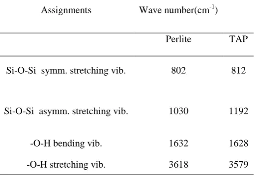
Table I. The Observed Frequencies of IR Bands of Perlite, TAP and their Possible Assignments
Assignments Wave number(cm-1)
Perlite TAP
Si-O-Si symm. stretching vib. 802 812
Si-O-Si asymm. stretching vib. 1030 1192
-O-H bending vib.
H-O-H bending vib.
1632 1628
-O-H stretching vib.
-O-H stretching vib.
3618 3579
SEM micrograph of perlite and its magnified view (Fig. 2 a and b) revealed the irregular morphology of perlite particles with broken or ragged edges. Similar pattern was observed in other reported micrographs of perlite [7]. SEM images of TAP and its magnified view (Fig. 3 a and b) are mainly fragmatic and random as a result of thermal activation [4]. But here, the morphology is less irregular which confirms the evaporation of water from the perlite sample on heating at high temperature.
D:\FTIR DATA\New Folder\BS\EXSTRA\BASE-RAW FA JAMESHEDPUR.0 BASE-RAW FA JAMESHEDPUR DRS
D:\FTIR DATA\New Folder\BS\EXSTRA\BASE-MAFA JAMESHEDPUR.0 BASE-MAFA JAMESHEDPUR DRS
17/03/2012 17/03/2012 34 33 .4 3 19 83 .5 4 18 71 .9 8 17 92 .8 8 16 14 .4 5 13 09 .2 3 10 37 .1 7 75 2. 86 70 8. 07 63 5. 66 59 4. 70 1000 1500 2000 2500 3000 3500 Wavenumber cm-1 -2 0 -0 20 40 60 80 1 0 0 T ra n sm itta n ce [% ] Page 1/1 (a) (b) 3618 3579 1632 1628 1030 802 1192 812
D:\FTIR DATA\New Folder\BS\EXSTRA\BASE-RAW FA JAMESHEDPUR.0 BASE-RAW FA JAMESHEDPUR DRS
D:\FTIR DATA\New Folder\BS\EXSTRA\BASE-MAFA JAMESHEDPUR.0 BASE-MAFA JAMESHEDPUR DRS
17/03/2012 17/03/2012 34 33 .4 3 19 83 .5 4 18 71 .9 8 17 92 .8 8 16 14 .4 5 13 09 .2 3 10 37 .1 7 75 2. 86 70 8. 07 63 5. 66 59 4. 70 1000 1500 2000 2500 3000 3500 Wavenumber cm-1 -2 0 -0 20 40 60 80 1 0 0 T ra n sm itta n ce [% ] Page 1/1
D:\FTIR DATA\sakshi\VA NEW.0 VA NEW DRS
D:\FTIR DATA\sakshi\TAVA-800.3HUNWASH.1 TAVA-800.3HUNWASH DRS
ISSN: 2319-8753
I
nternational
J
ournal of
I
nnovative
R
esearch in
S
cience,
E
ngineering and
T
echnology
(An ISO 3297: 2007 Certified Organization)
Vol. 2, Issue 9, September 2013
(a) (b)
Fig 2. SEM micrograph of (a) perlite and (b) its magnified view
(a) (b)
Fig 3. SEM micrograph of (a) TAP and (b) its magnified view
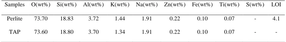
The chemical composition of perlite and TAP were determined by SEM-EDX technique which is shown in Table 2.
Table II. EDX Analysis of Perlite and TAP
Samples O(wt%) Si(wt%) Al(wt%) K(wt%) Na(wt%) Zn(wt%) Fe(wt%) Ti(wt%) S(wt%) LOI
Perlite 73.70 18.83 3.72 1.44 1.91 0.22 0.10 0.07 - 4.1
TAP 73.60 18.80 3.70 1.34 1.91 0.22 0.10 0.07 - -
ISSN: 2319-8753
I
nternational
J
ournal of
I
nnovative
R
esearch in
S
cience,
E
ngineering and
T
echnology
(An ISO 3297: 2007 Certified Organization)
Vol. 2, Issue 9, September 2013
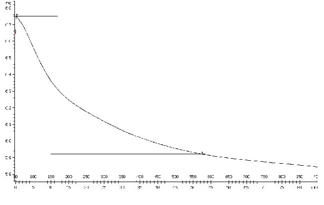
900°C. Weight loss in lower temperature range relates to the removal of moisture content of the sample together with some volatile materials. While, the weight loss of sample within range of 550-900˚C would correspond to the burning of carbonaceous materials that were firmly adsorbed on the surface of the solid materials remaining or volatilization of some trace metal oxides [27].
Fig 4. TGA curve of perlite
BET surface area of perlite and TAP samples were found to be 2.6 and 2.3 m2/g respectively.
The broad powder X-ray diffraction pattern of perlite (Fig. 5a), confirmed the absence of any ordered crystalline structure [28] which is typical for amorphous solids. However, heating of perlite at temperature over 800˚C for 3 h could convert less ordered structure to a more highly ordered structure and a single crystalline peak appears at 2θ = 27.642˚ (Fig. 5b) which shows presence of quartz in the sample [29], along with a broad peak at 2θ = 22-23˚ confirming amorphous nature of silica [24], [30].
ISSN: 2319-8753
I
nternational
J
ournal of
I
nnovative
R
esearch in
S
cience,
E
ngineering and
T
echnology
(An ISO 3297: 2007 Certified Organization)
Vol. 2, Issue 9, September 2013
IV. CONCLUSIONS
Results obtained in the present investigation confirm silica and alumina as major constituents of perlite. It is also observed that the nature of silica in perlite is mainly amorphous with irregular morphology and only a single crystalline peak appears on thermal treatment. On the basis of analysis, it can also be said that high number of hydroxyl groups and Si-O-Si network is also present in perlite. Moreover, comparison of perlite with TAP shows the effects of thermal treatment on structure, mineralogy, colour, surface area and morphology of perlite. The contemporary report aims at study of fundamental characteristics of perlite for its further applications in novel fields. The results like presence of amorphous silica network and surface hydroxyl groups in perlite indicate towards the potential capability of perlite as a support material in catalyst synthesis.
Fig 5. X-ray diffraction pattern of (a) Perlite and (b) TAP
(a) (b)
In
ten
si
ty
(c
o
u
2 ThetaDegree
n
ts
)
ISSN: 2319-8753
I
nternational
J
ournal of
I
nnovative
R
esearch in
S
cience,
E
ngineering and
T
echnology
(An ISO 3297: 2007 Certified Organization)
Vol. 2, Issue 9, September 2013
ACKNOWLEDGEMENT
The authors are thankful to Dr. Mukul Gupta, Dr. D.M. Phase, Er. V.K. Ahiray from UGC-DAE CSR Lab, Indore, India for XRD and SEM, SEM-EDX respectively. The financial support was provided by Fly Ash Mission, DST, New Delhi, India. The authors are also grateful to UGC, New Delhi, India for their Junior Research Fellowship scheme.
REFERENCES
[1] Dogan, M., Alkan, M., Cakir, U., “Electrokinetic Properties of Perlite”, J. Colloid Interface Sci. Vol. 192, pp.114-18, 1997. [2] Dogan, M., Alkan, M., “Adsorption kinetics of methyl violet onto perlite”, Chemosphere, Vol.50, pp.517-28, 2003.
[3] Roulia, M., Chassapis, K.., Kapoutsis, J.A.., Kamitsos, E.I., Savvidis, T., “Influence of thermal treatment on the water release and the glassy structure of perlite”,J. Mater. Sci., Vol.41, pp.5870-81, 2006.
[4] Bastani, D., Safekordi, A.A., Alihosseini, A., Taghikhani, V., “Study of oil sorption by expanded perlite at 298.15 K”, Sep. Purif. Technol., Vol.52, pp.295-300, 2006.
[5] Morsy, M.S., Shebl, S.S., Abd El Gawad Saif, M., “Development of perlite-gypsum-slag-Lime sludge-composite system for building application”,Building Research Journal, Vol.56, pp.49-58, 2008.
[6] Aglan, H., Morsy, M., Allie, A., Fouad, F., “Evaluation of fiber reinforced nanostuctured perlite-cementitious surface compounds for building skin applications”, Construction and Building Materials, Vol.23 (1), pp.138-45, 2009.
[7] Vance, E.R., Perera, D.S., Imperia, P., Cassidy, D.J., Davis, J., Gourley, J.T., “Perlite Waste as a Precursor for Geopolymer Formation”,
Journal ofthe Australian Ceramic Society, Vol.45 (1), pp.44-49, 2009.
[8] Mostaedi, M.T., Ghassabzadeh, H., Maragheh, M.G., Ahmadi, S.J., Taheri, H., “Removal of cadmium and nickel from aqueous solution using expanded perlite”, Brazilian Journal of Chemical Engineering, Vol.27 (2), pp. 299-308, 2010.
[9] Vaou, V., Panias, D., “Thermal insulating foamy geopolymers from perlite”, Miner. Eng., Vol.23 (14), pp.1146-51, 2010.
[10] Vijaykumar, G., Dharmendirakumar, M., Renganathan, S., Sivanesan, S., Baskar, G., Elango, K.P., “Removal of Congo Red from Aqueous Solutions by Perlite”, Clean- Soil, Air, Water, Vol.37 (4-5), pp.355-64, 2009.
[11] Dogan, M., Alkan, M., “Removal of methyl violet from aqueous solution by perlite”, J. Colloid Interface Sci., Vol.267 (1), pp. 32-41, 2003. [12] Silber, A., Bar-Yosef, B., Levkovitch, I., Kautzky, L., Minz, D., “Kinetics and mechanisms of pH-dependent Mn (II) reactions in plant-growth
medium”, Soil Biol. Biochem., Vol. 40, pp. 2787-95, 2008.
[13] Celik, A.G., Kilic, A.M., Cakal, G.O., “Expanded perlite aggregate characterization for use as a lightweight construction raw material”, Physicochem. Probl. Miner. Process. Vol. 49 (2), pp. 689-700, 2013.
[14] Hosseini, S.N., Borghei, S.M., Vossoughi, M., Taghavinia, N., “Immobilization of TiO2 on perlite granules for photocatalytic degradation of
phenol”, Appl. Catal., B, Vol. 74 (1-2), pp. 53-62, 2007.
[15] Balat, M., “Diesel-like Fuel Obtained by Catalytic Pyrolysis of Waste Engine Oil”, Energy, Exploration and Exploitation, Vol. 26 (3), pp. 197-208, 2008.
[16] Jafarzadeh, N.K., Sharifnia, S., Hosseini, S.N., Rahimpour, F., “Statistical optimization of process conditions for photocatalytic degradation of phenol with immobilization of nano TiO2 on perlite granules”, Korean J. Chem. Eng., Vol. 28 (2), pp. 531-38, 2011.
[17] Schlaefer, Francis, W., Hansen, A., “Methacrolein production utilizing novel Catalyst”, George, US Patent, 05/428, pp. 150, 1978.
[18] Kongkachuichay, P., Lohsoontorn, P., “Phase Diagram of Zeolite Synthesized from Perlite and Rice Husk Ash”,Science Asia, Vol. 32, pp. 13-16, 2006.
[19] Wang, P., Shen, B., Shen, D., Peng, T., Gao, “Synthesis of ZSM-5 zeolite from expanded perlite/kaolin and its catalytic performance for FCC naphtha aromatization”, J. Catal. Commun., Vol. 8 (10), pp. 1452-56, 2007.
[20] Silverstein, R.M., Webster, F.X. SpectrometricIdentification of Organic Compounds, Sixth ed., John Wiley Pub., Wiley India Pvt. Ltd., pp. 88., 2006.
[21] Adam, F., Balakrishnan, S., Wong, P.L., “Rice Husk Ash Silica as a Support Material for Ruthenium based Heterogenous Catalyst”, Journal
of Physical Science, Vol. 17 (2), pp. 1-13, 2006.
[22] Khatri, C., Rani, A., “Synthesis of a nano-crystalline solid acid catalyst from fly ash and its catalytic performance”, Fuel, Vol. 87, pp. 2886-92, 2008.
[23] Kabra, S., Sharma, A., Katara, S., Hada, R., Rani, A., „DRIFT- spectroscopic study of modification of surface morphology of perlite during thermal activation”, Indian Journal of Applied Research, Vol. 3 (4), pp. 40-42, 2013.
[24] Javed, S.H., Naveed, S., Feroze, N., Zafar, M., Shafaq, M., “Crystal and Amorphous Silica from KMnO4 treated and untreated Rice Husk
Ash”, Journal of Quality and Technology Management, Vol. 6 (1), pp. 81-90, 2010.
[25] Ojima, J., “Determining of Crystalline Silica in Respirable Dust Samples by Infrared Spectrophotometry in the Presence of Interferences”, J.
Occup. Health, Vol. 45, pp. 94-103, 2003.
[26] Kordatos, K., Gavela, S., Ntziouni, A., Pistiolas, K.N., Kyritsi, A., Kasselouri-Rigopoulou, V., “Synthesis of highly siliceous ZSM-5 zeolite using silica from rice husk ash”, Microporous Mesoporous Mater., Vol. 115 (1-2), pp. 189-96, 2008.
[27] Sekkina, M.M.A., Issa, R.M., El-Deen, A., Bastawisy, M., El-Helece, W.A., “Characterization and Evaluation of Thermodynamic Parameters for Egyptian Heap Fired Rice Straw Ash (RSA”, Int. J.Chem., Vol. 2 (1), pp. 81-88, 2010.
[28] Kalapathy, U., Proctor, A., Shultz, J., “
A
simple method for production of pure silica from rice hull ash”, Bioresour. Technol., Vol. 73, pp. 257-62, 2000.[29] Jain, D., Khatri, C., Rani, A., “Synthesis and characterization of novel solid base catalyst from fly ash”, Fuel, Vol. 90, pp.2083-88, 2011. [30] Amutha, K., Ravibaskar, R., Sivakumar, G., “Extraction, Synthesis and Characterization of Nanosilica from Rice Husk Ash”, International
ISSN: 2319-8753
I
nternational
J
ournal of
I
nnovative
R
esearch in
S
cience,
E
ngineering and
T
echnology
(An ISO 3297: 2007 Certified Organization)
Vol. 2, Issue 9, September 2013
BIOGRAPHY
Mrs. Sakshi Kabra is pursuing Ph.D. from Department of Pure and Applied Chemistry, University of Kota, Kota (Rajasthan). She secured All India Rank 385 in National Eligibility Test (June-2009) and acquired JRF. She is currently working as UGC-SRF in the department. Her research interests mainly include synthesis of heterogeneous acid catalysts and their various applications, solid waste management, material synthesis etc.
Ms. Stuti Katara is doing Ph.D. from Department of Pure and Applied Chemistry, University of Kota, Kota (Rajasthan). She got all India Rank 353 in National Eligibility Test (June-2009) and attained UGC-JRF. She is associated with solid base catalysis, green catalysis etc. and presently working as UGC-SRF in the department.
Prof. Ashu Rani is Head, Department of Pure and Applied Chemistry, Dean of post graduate departments and Convenor of various boards at University of Kota, Kota (Rajasthan). Several research scholars are pursuing M. Phil and Ph. D. under her guidance. Her wide research interests involve Heterogeneous Catalysis, Waste management, climate change, Lithospheric salt transport kinetics, Nanotechnology, Material Synthesis. She is associated with several National and International Collaborations and is a Principle Investigator of several projects funded by central and state governments.