HoxGene Function and Cell
Identity In
Drosophila
by
Philip Ronald Elstob
Thesis submitted to the University of London, for the
degree of Doctor of Philosophy
ProQuest Number: U643752
All rights reserved
INFORMATION TO ALL USERS
The quality of this reproduction is dependent upon the quality of the copy submitted.
In the unlikely event that the author did not send a complete manuscript
and there are missing pages, these will be noted. Also, if material had to be removed, a note will indicate the deletion.
uest.
ProQuest U643752
Published by ProQuest LLC(2016). Copyright of the Dissertation is held by the Author.
All rights reserved.
This work is protected against unauthorized copying under Title 17, United States Code. Microform Edition © ProQuest LLC.
ProQuest LLC
789 East Eisenhower Parkway P.O. Box 1346
‘A fool can ask more questions than a wise man can answer’
ACKNOWLEDGEMENTS
Firstly I would like to thank Alex Gould, my supervisor, for taking me on as his first
PhD student and for his guidance and continual enthusiasm over the past four
years. I am hugely indebted to Patricia Serpente and Bruno Bello, who were the
only lab members for much of my PhD. Patricia provided support and humour from
day one when I arrived in an essentially empty lab. I am very grateful to Patricia for
efficiently establishing and running the lab. Patricia was an invaluable source of
conversation and advice, whether the topic was the intricacies of molecular biology
or how our respective football teams were performing. I thank Bruno for teaching
me all that he knows about Drosophila and how to be “Lord of the Flies”. I do not think Bruno has ever answered one of my questions with a simple yes or no,
however this lead to many stimulating discussions during which I learnt a great
deal. I often found myself in Room 101 during my PhD; pushing flies, listening to
CDs and teaching Bruno the finer points of the English language - Northern style. I
am very grateful to Bruno for all his guidance and companionship. I owe a big
thank you to Véronique Brodu who joined the lab approximately 6 months before
my departure and unselfishly helped with experiments for the Development paper. I
express my thanks to JP Vincent and Cyrille Alexandre for sharing their knowledge
and reagents, and to other members of Mammalian Development for their advice. I
would like to thank Frank Johnson and Lesley MacNiell for help with the cartoons
presented in this thesis.
The late Rosa Beddington was a scientist for whom I had the utmost respect, she
was a great inspiration and I feel privileged to have known her.
I would like to say a huge thank you to all of my PhD friends for the forest treks,
snooker matches, the kicking I received in TaeKwondo and football, and for making
life at Mill Hill as fun as it could be. Finally I am indebted to Claire, my parents and
PREFACE
The research reported in this thesis was carried out in the Division of Mammalian
Development at the National Institute for Medical Research (Mill Hill, London)
under the supervision of Dr. Alex Gould,
This thesis describes my own original work with the exception of Figure 4.1 A, D-F
and Figure 5.3B. These panels show the preliminary work on oenocytes carried out
by Dr. Alex Gould and were included to provide the necessary background
ABSTRACT
The /-/ox/Homeotic genes pattern the anteroposterior axis of animal embryos.
However, the mechanisms by which these conserved transcription factors generate
morphological diversity remain largely unknown. Here I describe both a molecular
and a cellular study of Hox gene function. In the molecular approach, a model Hox
target enhancer, the late neural enhancer (LNE) of the mouse Hoxb4 gene, was
dissected in Drosophila. Individual analysis of two essential HOX binding sites
(HS1 and HS2) revealed that each site has a different Hox specificity and
modulates responses to more than one Hox input. Activation of the LNE requires
the group 4-6 H o x genes and is largely dependent on the H o x cofactor
Extradenticle. Furthermore, three conserved regions, remote to HS1/2, were found
to influence LNE activity along the anteroposterior and dorsoventral axes. In
summary, the LNE contains at least five regulatory modules required for correct
enhancer expression.
In a cellular approach, I have investigated how the Hox gene abdominal A
micromanages segment identity in Drosophila by studying its role in specifying a single cell identity: the larval oenocyte. An initial study of this cell type revealed
three stages of morphogenesis: 1) induction, 2) anterior movement out of the
posterior compartment and 3) ventral migration. Induction occurs in response to
EGFR signalling from primary chordotonal sensory organ precursors. Ectodermal
cells are primed to become oenocytes by virtue of a genetic prepattern, one
component of which is spall SPALT also suppresses EGFR-mediated induction of
an alternative cell fate, the chordotonal organ. Given that both abdominal A and
EGFR ligand can induce ectopic oenocytes, I propose that abdominal A might
specify oenocytes non-cell autonomously, through regulating local EGFR ligand
TABLE OF CONTENTS
Page 3 4 Acknowledgements PrefaceAbstract ^
List of Figures
Abbreviations
Chapter One: General Introduction
1.1 Hox gene function and segment identity in Drosophila 17
1.2 Establishing the Drosophila body plan 17
1.3 The HoA/Homeotic genes 21
1.4 Systematic screens for Hox target identification 25
1.5 Focussed candidate approaches to Hox target identification 27
1.6 Hox target genes - a summary 34
1.7 Hox genes and the micromanagement of segment identity 35
1.8 Co-operative HOX-cofactor interactions in HOX target gene 37
regulation
1.9 Two models for achieving HOX functional specificity: binding site 40
selection versus activity regulation
1.10 A composite binding site selection-activity regulation model 46
1.11 Co-operative interactions with EXD do not account for all Hox- 48
regulated morphological events
1.12 Synopsis 49
Chapter Two: Materials and Methods
2.1 Drosophila stocks and genetic manipulations 52 2.2 Drosophila transformation 54
2.3 Generation of LNE cis mutations and transformation constructs 57
2.4 cloning strategy 59
Chapter Three: Molecular Mechanisms of Hox Gene Function
3.1 Introduction 64
3.2 Results
3.2.1 LNE expression in the maxillary segment is associated with 69
the common salivary duct
3.2.2 A role for De/brmecy during salivary duct development? 75
3.2.3 HS1 and HS2 are targets for regulation by multiple Hox 76
genes
3.2.4 Remote sequences and the restriction of the HOX response 82
3.2.5 EXD is required for the in vivo activation of the LNE 86
3.3 Discussion
3.3.1 MSI and HS2 have different Hox specificities in Drosophiia 94
3.3.2 Conserved sequences remote to MSI and HS2 modulate 101
Hox activation of the LNE
3.3.3 A major role for cofactors in Hox activation of the LNE 102
3.3.4 Perspectives 103
C h apter Four: The oenocyte: a model fo r studying c ellu la r
mechanisms of Hox gene function
4.1 Introduction 106
4.2 Results
4.2.1 A screen for new oenocyte genes 109
4.2.2 The embryonic development of oenocytes 109
4.2.3 Ventral migration of oenocytes; a role for seven up 117
4.2.4 Anterior movement of oenocyte precursors 128
4.2.5 A requirement for engrailed buX not wingless 137
4.2.6 Is a lateral break in the WG stripe required for anterior
movement?
142
Discussion
4.3.1 Oenocyte development - whorls, strings and clusters 151
4.3.2 Escape from the posterior compartment 152
Chapter Five: Oenocyte Induction by abdA and the EGFR Pathway
5.1 Introduction 159
5.2 Results
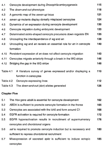
5.2.1 abdA is necessary and sufficient for oenocyte formation 161
5.2.2 Oenocyte precursors are associated with the most dorsal 167
primary COP
5.2.3 Oenocyte induction is regulated by EGFR activation 171
5.2.4 The degree of EGFR signalling controls cell-number but not 175
cell-type
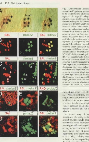
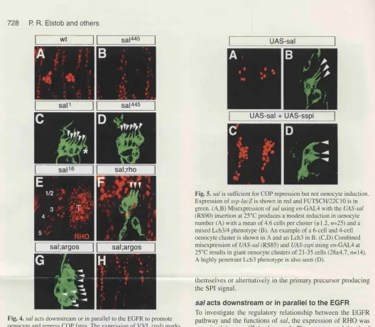
5.2.5 sal is sufficient to suppress COP recruitment and is required 179
for oenocyte induction
5.2.6 Misexpression of the SPI inductive signal is sufficient to 183
produce ectopic oenocytes in the thorax
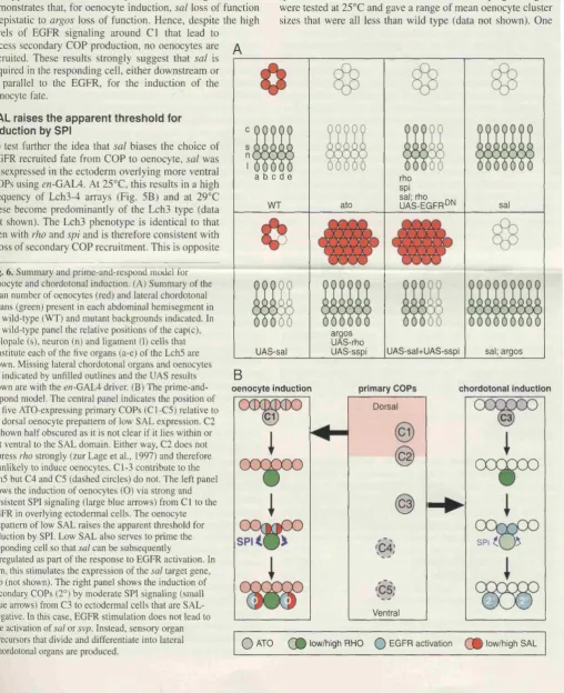
5.3 Discussion
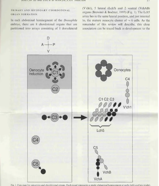
5.3.1 Oenocytes and secondary COPs are recruited by the same 187
inducer
5.3.2 Fate mapping oenocyte and secondary COP induction 189
5.3.3 A prime-and-respond model 194
5.3.4 abcW and oenocyte specification 198
Chapter Six: General Discussion
6.1 From Hox genes to morphogenesis 202
6.2 The LNE is a target for multiple HOX proteins 202
6.3 Larval oenocytes - a new model system to study Hox specification 204
at the single cell level
6.4 Oenocyte movement can be separated into AP and DV phases 205
6.5 Do oenocytes have serial homologues in other segments? 206
6.6 Multiple responses from one receptor: the role of prepatterns 207
6.7 Keeping an eye on oenocytes and COPs 208
6.8 What defines the oenocyte prepattern? 209
6.9 Does abdA function in the 01 COP, in the responding ectoderm or 210
in both?
Bibliography 216
Appendices 243
Publications
Elstob, P. R., Brodu, V. and Gould, A. P. (2001). spa/f-dependent switching Back
between two cell fates that are induced by the Drosophila EG F receptor. sleeve
Development'IIQ, 723-732.
Gould, A. P., Elstob, P. R. and Brodu, V. (2001). Insect oenocytes: a model Back
LIST OF FIGURES
Chapter One
Page
1.1 The genetic hierarchy for anteroposterior patterning in the 19
Drosophila embryo
1.2 Hox genes and their expression in the Drosophila embryo 23
1.3 The regulation of midgut morphogenesis by Hox target genes 28
1.4 Repression of wing genes by Ubx in the haltere 32
1.5 Binding site selection and activity regulation models for HOX 41
specificity
Chapter Two
2.1 P-element excision 55
Chapter Three
3.1 Hox activation and repression of the LNE in Drosophila 66
3.2 LNE-positive cells invaginate with the common salivary duct 70
3.3 Cell movements during salivary gland development in Drosophila 72
3.4 LNE sequence conservation and transgenesis constructs 77
3.5 Reduced LNE activation resulting from mutation of MSI or HS2 79
3.6 Conserved regions outside of HS1/HS2 have a role in LNE 84
function
3.7 The LNE is almost fully exd-dependent 88
3.8 LNE activity is increased in a weak hth allele but reduced in a 92
strong allele
3.9 A summary of LNE cis mutations and cofactor mutant backgrounds 95
Chapter Four
4.1 Oenocyte development during DrosopA7//a embryogenesis 115
4.2 The ûfoivn-anûf-oi/f phenotype 118
4.3 A genomic map of the seven up locus 121
4.4 seven up mutants display dorsally misplaced oenocytes 124
4.5 Dynamics of e/7 expression during oenocyte development 130
4.6 Oenocyte migration during embryonic development 133
4.7 Delaminated sickle-shaped oenocyte precursors down-regulate EN 135
4.8 Uncoupling the interdependence of wg and en 138
4.9 Uncoupling wg and en reveals an essential role for en in oenocyte 140
formation
4.10 Persistent expression of en does not affect oenocyte migration 143
4.11 Oenocytes migrate anteriorly through a break in the WG stripe 146
4.12 Bridging the gap in the WG stripe 148
Table 4.1 A literature survey of genes expressed and/or displaying a 110
function in oenocytes
Table 4.2 Oenocyte expressing lines 112
Table 4.3 The down-and-out {don) alleles generated 126
Chapter Five
5.1 The Hox gene abdA is essential for oenocyte development 162
5.2 ABDA is sufficient to promote oenocyte formation in the thorax 165
5.3 Oenocytes are associated with the Ich5 and form around 01 168
5.4 EGFR activation is required for oenocyte formation 172
5.5 EGFR hyperactivation results in recruitment of supernumerary 176
oenocytes and chordotonal organs
5.6 s a /is required to promote oenocyte induction but is necessary and 180
sufficient to repress chordotonal recruitment
5.7 Misexpression of secreted spitz is sufficient to induce ectopic 185
5.8 A fate map for oenocytes and abdominal chordotonal organs 191
5.9 The prime-and-respond model 195
Chapter Six
6.1 A model for oenocyte induction by abdA and the EGFR pathway 213
Appendices
Appendix I Expression levels of LN E-/acZ constructs in stage 13 244
embryos
Appendix II The only abdominal chordotonal organs to derive from the 246
ABBREVIATIONS
A A1-A8 abdA AbdB Aldh-lll ANT-C Antp AP arm ato P-gal bed bp BX-C C ci cnn con CR3 C-terminal dap Dfd Dll DNA don dpp DV EGFR EGTA en AdenineAbdominal segments 1 to 8
abdominal A Abdominal B
Aldehyde dehydrogenase type III
Antennapedia complex Antennapedia Anteroposterior armadillo atonal P-galactosidase bicold base-pair Bithorax complex Cytosine cubitus Interruptus centrosomln connectin
Conserved region 3 of the mouse Hoxb4 gene
Carboxy terminal dacapo Deformed DIstalless deoxyribonucleic acid down-and-out decapentaplegic Dorsoventral
Epidermal growth factor receptor
ethyleneglycol-bis-(B-amino-ethyl ether) N,N^-tetranic acid
esg escargot exd extradenticle fkh forkhead ftz fushi tarazu
G Guanine
hh hedgehog
hnf4 hepatocyte nuclear factor 4
HRP Horse radish peroxidase
hth homothorax
IPCR Inverse polymerase chain reaction
lab labial
lacZ The gene encoding p-galactosidase
LNE Late neural enhancer of the mouse i
Meis1 myeloid ecotropic insertion ^ite 1
mRNA messenger ribonucleic acid
mrr mirror
NES Nuclear export signal
NLS Nuclear localisation sequence
N-terminal Amino terminal
pb proboscipedia
PBS Phosphate buffered saline
PCR Polymerase chain reaction
PIPES piperazine-N, N^-bis[2-ethane-sulphi
prd paired
Prep1 Pbx regulating protein 1
ps parasegment
r6/7 rhombomere 6/7 boundary
rho rhomboid sal spalt sea scabrous
Scr Sex combs reduced svp seven up
T1-T3 Thoracic segments 1 to 3
TALE Three amino acid loop extension
TGFP Transforming growth factor p
tsh teashirt Ubx Ultrabithorax
VM Visceral mesoderm
w l ventral veinless also known as drifter {dff) wg wingless
CHAPTER ONE
1.1 Hox gene function and segment identity in Drosophila
The main body axis in all animals is that which runs from head-to-tail, or anterior to
posterior. In the Drosophila embryo this anteroposterior (AP) axis is divided into a series of repeated units or segments. A great deal is known about how the
segmented body plan of Drosophila is established, including the genetic hierarchy that controls these events. The /-/ox/Homeotic genes lie at the bottom of this
hierarchy and function to give each segment its individual identity and morphology.
However, relatively little is known about the molecular mechanisms that link Hox
genes to morphogenetic events. This gap in our knowledge of AP patterning is the
focus of the work presented in this thesis.
1.2 Establishing the Drosophila body plan
The mechanisms by which the AP axis is set up in the Drosophila embryo have
been described in great detail. It is only appropriate here to give a brief summary of
these events (for excellent reviews see Wolpert et al., 1998; Carroll et al., 2001;
Slack, 2001). Segmentation, and thus the basic D ro so p h ila body plan, is
established by the sequential expression of genes in overlapping domains along
the AP axis. The first embryonic co-ordinates are set up by maternal mRNAs that
are deposited into the egg by the mother. Several such mRNAs are localised to
distinct regions of the embryo, and upon fertilisation they are translated. As the
early embryo is a syncytium in which nuclei are present in a common cytoplasm,
protein gradients can form along the AP axis. For example, bicold {bod} mRNA is localised to the anterior pole of the embryo, meanwhile BCD protein diffuses to
factors, distributed in overlapping patterns, switch on the first zygotic genes, the
gap genes (Figure 1.1). These genes are expressed in distinct broad regions in the
embryo and encode transcription factors. Thus the maternal and gap genes divide
the AP axis of the Drosophila embryo into regions containing different
combinations of transcription factors.
The first sign of molecular segmentation in the Drosophila embryo occurs
when the aperiodic patterns of maternal and gap proteins establish the periodic
expression of pair-rule genes in the ectoderm (Figure 1.1). Also encoding
transcription factors, the pair-rule genes control the initial expression of segment
polarity genes (Figure 1,1). The segment polarity genes encode a mixed bag of
proteins including transcription factors, secreted signalling molecules and
membrane receptors, which serve to define 14 parasegmental units
(Martinez-Arias, 1993). Although the mature Drosophila embryo is composed of segments,
the parasegment has been identified as an earlier developmental unit of the same
period but out-of-phase with the segments (Lawrence, 1992). More specifically, a
parasegment is made up of the posterior compartment of one segment and the
anterior compartment of the more caudal segment. A compartment comprises a
non-intermingling set of lineage-restricted cells (Garcia-Bellido et al., 1973), and
thus cell lineages in the anterior compartment are kept segregated from those in
the posterior compartment. This results in a straight boundary between these two
cell populations that is termed the compartment boundary (reviewed in Dahmann
and Basler, 1999).
Genes of the maternal, gap, pair-rule and segment polarity classes
Figure 1.1 The genetic hierarchy for anteroposterior patterning in the
Maternal genes
Gap genes
Pair-rule genes
Segment Polarity
genes
Hox genes
domains of two or more parasegments. As discussed below, the Hox genes encode transcription factors that function to give each segment its identity.
1.3 The Hox/Homeotic genes
First described by Bateson in 1894, homeotic mutations cause the transformation
of one body region into the “likeness of another". In the Drosophila embryo/larva these mutations are generally characterised with respect to the patterning of
denticle belts found in the ventral epidermis. Each segment has a characteristic
pattern of denticles, or hairs, which serves to identify it. Loss-of-function mutations
in Hox genes result in homeotic transformations of these denticle belts (Lewis,
1978; Wakimoto and Kaufman, 1981). In the adult fly, viable mutations altering Hox
gene function produce more spectacular homeotic transformations. The
gain-of-function mutations in the Antennapedia (Antp) gene provide a dramatic example
where the antennal appendages of the head are transformed into legs (Kaufman et
al., 1990). Another famous homeotic transformation is displayed in the four-winged
fly, the result of loss-of-function mutations at the Ultrabithorax (Ubx) locus (Lewis, 1963). In this exquisite creature, the halteres, a pair of balancing appendages
found on the third thoracic segment, are transformed into a pair of wings, usually
associated with the second thoracic segment. The apparent complete segment
transformations observed with Drosophiia Hox mutations indicate a role for these
genes in directing segment morphogenesis.
The Hox genes encode a conserved family of transcription factors that
specify morphological differences along the AP axis in animals as diverse as
vertebrates and arthropods. These genes are organised into clusters in the
Drosophila genome contains a single Hox complex that is split into two clusters on the third chromosome (Figure 1.2), the Antennapedia complex (ANT-C) and the
Bithorax complex (BX-C). In contrast, vertebrates including humans, mice (Figure
1.2) and chickens have four Hox complexes due to large-scale duplications of
chromosomal segments, or even of entire genomes (tetraploidization), during
evolution (for an excellent review see Carroll et al., 2001). One of the most
intriguing features of all Hox gene complexes is colinearity, whereby the position of a gene in the complex correlates with its domain of expression along the AP axis
(Figure 1.2 and Lewis, 1978; Duboule and Dolle, 1989; Graham et al., 1989).
However, a discussion of colinearity is beyond the scope of this thesis.
In 1975 Garcia-Bellido proposed the “selector gene hypothesis”, whereby
the expression of a Hox gene within a compartment directs the fate of all cells within that metamere (Garcia-Bellido, 1975). The model treated the compartment
as an independent unit that is uniformly instructed by a single Hox gene to adopt a
specific segment identity. It also suggested that continuous Hox expression is
required to direct cells in this unit along a defined developmental pathway. In
addition, Garcia-Bellido correctly predicted that, rather than directly specifying
morphological differences along the AP axis, the Hox “selector” genes control a
battery of target “realizator” genes. These target genes more directly promote
segment-specific cellular properties such as growth, mitosis, adhesion and cell
differentiation (Garcia-Bellido, 1975). Thus, according to this model, the
complexities of segment-specific morphogenesis lay entirely downstream of the
Figure 1.2 Hox genes and their expression in the Drosophila embryo
(A) A schematic representation of Hox gene expression in the ectoderm of a
Drosophila embryo at the extended germ-band stage. This figure depicts the
general trend of Hox expression within any particular segment/parasegment as
high (large block) or moderate (small block). However it should be noted that, due
to intrasegmental modulation of Hox expression, levels within a block are not
necessarily uniform.
(B) The single split Hox gene cluster of Drosophila comprises the ANT-C and BX-C complexes. Colours are as in panel (A) to show the relationship between the
position of a Hox gene in the cluster and its domain of expression along the
anteroposterior axis (termed colinearity).
(C) The mouse genome contains four different clusters of Hox genes (A, B, C and
D) located on different chromosomes. Hox genes are coloured by paralogue group
Gnathal Thoracic Abdominal
T
Segments
lab
pb
Ubx
AbdB
Pro Mn Mx La 11 12 13 A1 A2 A3 A4 A5 A6 A7 A8 Telson
B
ANT-C lab 5 pb BX-C
3'
3'
-3'
A3
B3
- A3 - A4 - A5 — A6
- B4 - | B5 - B6
I A7 I--- 1 A9 |- [ Â Ï Ô - | A l l I---1 A 13|— 5'
17 - B8 - [ B9 I--- B13 — 5
C4 — C5 — C6 C8 W C9 M C10H C11 c n k - 5'
1.4 Systematic screens for Hox target identification
The Drosophila Hox genes are expressed in precise segmental domains (Figure 1.2), suggesting that their downstream targets would also be differentially
expressed between segments. Based on this assumption, two studies were
undertaken to look for genes specifically activated or repressed in response to
ectopic expression of a Hox gene. In the first of these, the expression of enhancer
traps was analysed in the developing antenna following misexpression of Antp
(Wagner-Bernholz et al., 1991). Enhancer trap lines contain a single copy of a
transposon that carries a fusion between lacZ and a basal promoter, which comes
under the regulation of enhancer elements close to its random insertion site in the
genomic DNA. Thus reporter expression generally reflects the whole, or part of the
expression pattern of a neighbouring gene. Wagner-Bernholz and colleagues
(1991) used enhancer traps to identify regulatory elements and the corresponding
genes under the control of ANTP during the antenna-to-leg homeotic
transformation. In a second approach, Feinstein et al. (1995) used subtractive
hybridisation to enrich for genes transcribed following ectopic expression of UBX,
and thus only isolated genes potentially activated by this HOX protein.
The above approaches do not distinguish between direct and indirect
targets. To circumvent this problem, two molecular approaches have been used to
isolate direct HOX targets based on HOX-DNA interactions. The first strategy used
In vivo immunopurification to isolate UBX-bound chromatin fragments from embryonic nuclei (Gould et al., 1990). The second used a yeast one-hybrid
screening assay that relied on UBX-mediated activation of a reporter gene,
(Mastick et al., 1995). Although both approaches have the advantage of isolating
potential direct targets of UBX, immunopurification has been the most successful.
This strategy benefited from more stringent conditions, as targets were isolated
from wild type D ro s o p h ila embryos. Therefore unlike the yeast assay,
immunopurification combined the presence of physiological UBX levels with the
proteins involved in co-operative DNA binding with UBX (see Section 1.8). Gould
and colleagues isolated several targets, including connectin {con), a gene involved in hemophilic cell-cell adhesion (Gould and White, 1992; Nose et al., 1992). Using
a similar immunopurification approach, Heuer et al. (1995) isolated the ANTP
target centrosomin (cnn), which encodes an essential centrosomal protein. As
discussed in the next section, cnn has been implicated in the cell shape changes that occur during development of the second constriction of the midgut. In a third,
closely related screen, this time incorporating DNA-protein cross-linking, scabrous
(sea), a gene encoding a secreted protein involved in cellular communication
during neurogenesis, was identified as a putative direct target of UBX (Graba et al.,
1992).
The above studies suggested that the number of Hox target genes is large, and that these genes are highly varied in nature and do not fall into any one
particular family. Despite the identification of several Hox targets using such
approaches, it has been very difficult to relate these to segment-specific
morphological events. Thus we know very little about how Hox genes instruct
1.5 Focussed candidate approaches to Hox target identification
Many of the Drosophila Hox target genes have been identified from previously
characterised genes, known to be involved in morphogenetic processes, that are
under Hox control. Studies on the formation of the central region of the midgut,
during Drosophila embryogenesis, have provided one of the best examples of how
Hox genes direct morphogenetic processes (reviewed in Graba et al., 1997). Ubx
and abdominal A (abdA) expression in the visceral mesoderm (VM) controls the formation of the second midgut constriction and also cell differentiation events in
the underlying endoderm (refer to Figure 1.3). Ubx is expressed in the VM adjacent to parasegment 7 (ps 7) where it controls the expression of decapentaplegic {dpp,
Capovilla et al., 1994), a signalling molecule belonging to the TGF-|3 family. VM
cells in parasegment 8 (ps 8) express abdA, which activates the transcription of
wingless {wg, Reuter et al., 1990), a gene encoding a secreted factor of the WNT class. The combinatorial action of DPP and WG controls the expression of teashirt {tsh), a transcription factor required for formation of the second midgut constriction (Mathies et al., 1994). Previous studies revealed that the VM imposes the
constriction on the underlying endoderm, and that VM cells close to the inner limits
of the constriction contained dense bundles of microtubules (Reuter and Scott,
1990). centrosomin, isolated in one of the target gene screens described above, is involved in microtubule-dependent processes and has been implicated in these
mechanical events. Isolated as a putative direct target of ANTP, cnn is positively
regulated by U b x in the VM, and mutants for cnn lack the second midgut
Figure 1.3 The regulation of midgut morphogenesis by Hox target genes
A summary of the genetic cascade initiated by Ubx and abdA that produces a
central constriction of the visceral mesoderm and also endodermal cell
differentiation. Note that by regulating target genes encoding secreted signalling
molecules {dpp and wg), a Hox gene expressed in one parasegment can mediate
e
an Xffect in an adjacent parasegment. See text for details. This figure was
Parasegment 7 ! Parasegment 8
abdA
n
Constriction
Visceral
mesoderm
DPP
VVG
lab
Endoderm
WG
High level
lab
Copper cells
1
Large flat cells
its role in microtubule organisation, CNN participates directly in the cell shape
changes that occur during midgut constriction.
H o x gene expression in the VM of parasegments 7 and 8
non-autonomously patterns the underlying endoderm through the target genes dpp and
wg (refer to Figure 1.3). The integrated action of these signalling molecules results in expression of the Hox gene labial {lab) in the endodermal cells of ps 7. The closer endodermal cells are to the ps 8 WG source, the greater the transcription of
lab. Hence a gradient of LAB is observed, with high levels underlying posterior ps 7 and increasingly lower levels towards the anterior of this parasegment. lab is both necessary and sufficient for the specification of copper cells in the midgut
endoderm (Hoppler and Bienz, 1994). Interestingly, the level of LAB determines
the degree of differentiation of these highly specialised cuprophilic cells. Thus the
largest and most distinct copper cells are observed in the most posterior region of
ps 7 while poorly differentiated cells are found more anteriorly (Hoppler and Bienz,
1994). Although WG acts in ps 7 to promote lab expression, the higher degree of
wg signal received by the ps 8 endodermal cells results in the repression of lab and hence no copper cells (Hoppler and Bienz, 1995). Instead, these endodermal cells
differentiate into the so-called “large flat cells”.
To summarise, in the developing midgut Ubx and abdA lie at the top of genetic hierarchies that result in the specification of structures in ps 7 and ps 8.
However, cross talk occurs between the downstream targets of Ubx and abdA, and
this is essential for morphogenetic events in both parasegments. The identification
of intercompartmental signalling molecules as HOX targets is in direct contrast to
the “selector gene hypothesis”, in which the compartment was considered an
As alluded to in section 1.3, it is remarkable that by altering the expression
of a single Hox gene, the identity of a whole segment can be affected. Originally, based on the classical “selector gene” model proposed by Garcia-Bellido, it was
though that a Hox gene acted to instruct all cells in a compartment to adopt a particular segmental identity. However, the results presented in recent studies
strongly suggest that the Hox genes act at many levels in genetic hierarchies,
independently regulating selected genes (Weatherbee et al., 1998). One good
example is the development of wings and halteres, two serially homologous
structures found on adjacent adult thoracic segments. Like most adult structures
these develop from imaginai discs, the monolayer epithelial sacs set aside during
embryogenesis. These proliferate during larval life and differentiate during
metamorphosis to form the adult structures. Normally, no Hox genes are required
or expressed in the developing wing-blade primordium of the wing disc while the
haltere imaginai disc develops under the influence of Ubx. Removal of Ubx function from the developing haltere results in selection of the wing developmental pathway,
and thus formation of a four-winged fly. Meanwhile, misexpression of Ubx can
transform the identity of a structure as complex as a wing into that of a haltere.
Weatherbee and colleagues (1998) dissected the function of UBX in the
developing haltere through the examination of genes already known to be involved
in wing patterning. They demonstrated that Ubx acts in the haltere imaginai disc to repress genes involved in wing development (Figure 1.4). Although direct
Figure 1.4 Repression of wing genes by Ubx in the haltere
The haltere and wing are serial homologues. The wing develops in the absence of any Hox input, whilst
Ubx expression promotes haltere development. Shown here is the wing regulatory hierarchy and in red are
those genes under the control of Ubx in the developing haltere. Ubx appears to repress genes at many
w levels in the wing genetic hierarchy. However, it should be noted that direct regulation has not been
Anteroposterior
Dorsoventral
en
\
\
vein
positioning
wing cell
growth and
ap
POST
Ser
wg P ^ T
dpp
sal
omb
DSRF
AS-C
cut
\
1.6 Hox target genes - a summary
From the studies to date, Hox target genes encode a diverse range of molecules
including transcription factors, growth factor-like molecules and membrane
receptors (reviewed in Graba et al., 1997). Interestingly, most of these target genes
have more than one HOX regulator and many have functions, often earlier in
development, that are independent of Hox genes.
Based on expression patterns alone, thousands of genes are predicted to
lie directly or indirectly downstream of the Hox genes (Bellen et al., 1989; Bier et al., 1989; Biggin and McGinnis, 1997). Given this number, it is surprising that less
than 30 Hox target genes have been identified in Drosophila to date (reviewed in Pradel and White, 1998). In most instances, the direct nature of the target has not
been demonstrated due to the laborious strategy that is necessary to prove this
type of regulation in vivo (Schier and Gehring, 1992). In one case however,
Capovilla et al. (1994) showed that UBX interacts directly with DNA binding sites in
the dpp303 enhancer to regulate dpp expression in ps 7 of the VM. This was
achieved by making corresponding changes to the binding specificity of UBX, and
in its DNA binding sites within a dpp303-/acZ construct. LacZ expression was only observed from the mutant construct in ps 7 of the VM when the mutant UBX
protein was also expressed in this tissue. This second site suppression strategy
demonstrated that in the endogenous situation, UBX binds directly to the dpp303
1.7 Hox genes and the micromanagement of segment identity
The Hox genes are initially transcribed in broad domains consisting of two or more
parasegments. However, later in development Hox gene expression becomes
more complex, and at any one time each segment may contain a heterogeneous
population of cells expressing and not expressing a specific Hox gene. This
intrasegmental spatial and temporal regulation of Hox gene expression can have
important effects on the final morphology of a segment. This was demonstrated for
Ubx in preventing leg development in the first abdominal (A1) but not in the third thoracic (T3) segment, despite being expressed in both segments in the relevant
cells (Castelli-Gair and Akam, 1995). These two distinct morphological outcomes
result from the differential regulation of Distalless (Dll), a UBX target gene required for leg development. In T3, Dll is activated in the leg primordia through an early enhancer (Vachon et al., 1992), and expression is maintained through an
autoregulatory loop involving a late enhancer element (Vachon et al., 1992;
Castelli-Gair and Akam, 1995). The early enhancer is sensitive to UBX-mediated
repression but is activated in T3 before Ubx expression is initiated. Meanwhile, Ubx
is expressed earlier in the presumptive leg precursor cells of A1. Therefore, in this
segment UBX can bind to the early enhancer, block the initial expression of Dll,
and thus prevent the feedback loop and leg formation (Castelli-Gair and Akam,
1995), It has been proposed that although Ubx is expressed in the T3 leg following earlier specification, this Hox gene serves to modulate the structure of the leg, making it different from those on T1 and T2 (Castelli-Gair et al., 1994). Therefore
patterns of Hox expression has been extended to the non-segmented nematode
Caenorhabditis elegans, where they are also critical for regulating several diverse developmental processes (Salser and Kenyon, 1996).
The work of Gair and Akam (reviewed in Akam, 1998a;
Castelli-Gair, 1998), together with that of Weatherbee et al. (1998), strongly suggests that,
through the fine-grained spatio-temporal pattern of HOX target regulation,
numerous cell-fate decisions are made on a cell-by-cell basis within a segment as
a whole. This contrasts sharply with the original selector gene hypothesis, which
predicted a uniform and continual requirement for Hox expression within a
segment. Based on these studies, the present hypothesis is that Hox genes
micromanage at the cellular level many diverse cell fates and behaviours, and it is
the summation of all these cell fates that defines the identity of a segment (Akam,
1998b). These theories have led some researchers to change tack and begin to
address how Hox genes specify the fate of a subset of cells within a segment, such as those in an organ. This is an altogether much simpler task than studying the
segment as a whole unit as fewer cell types are involved.
Two excellent Drosophila organogenesis models that have been exploited
recently are the salivary glands and the posterior spiracles. Contrary to haltere
development, both of these structures are promoted rather than repressed by Hox
genes. Sex combs reduced {Sci) is both necessary and sufficient to induce salivary
gland development (Panzer et al., 1992), acting with the Hox cofactor Extradenticle
(see next section) to specify both duct and gland cell fates in the salivary primordia
(Henderson and Andrew, 2000). Input from the Epidermal Growth Factor Receptor
(EGFR) receptor pathway makes the distinction between duct and gland
on duct cell genes, whilst in those that do not see EGFR ligand, Scr modulates the
expression of gland specific genes (Isaac and Andrew, 1996; Andrew, 1998;
Andrew et al., 2000).
The posterior spiracles in Drosophila are distinct structures that form the posterior opening for the larval tracheal system. Similar to Scr-mediated
specification of salivary glands. Abdominal B {AbdB) is both necessary and
sufficient to direct posterior spiracle morphogenesis (Kuziora, 1993). Hu and
Castelli-Gair (1999) showed that ABDB activates the transcription of different
genes expressed in at least four distinct cell types that contribute to the posterior
spiracle. However, even in such organ-based approaches, the exact number of
distinct cell states and the mechanisms by which any single fate is specified by a
Hox gene have not been elucidated. To address Hox gene function at the level of a single cell, I introduce a model cell type, the larval oenocyte, which is specified by
a single Hox gene, abdA.
1.8 Co-operative HOX-cofactor interactions in HOX target gene regulation
The Hox protein products all contain a highly conserved 60 amino acid DNA
binding domain termed the homeodomain, which is encoded by the homeobox.
There is very little extended sequence conservation between the HOX proteins
outside of the homeodomain. One notable exception is the hexapeptide (or YPWM)
motif that is found at varied distances N-terminal to the HOX homeodomain
(Acampora et al., 1989). The homeodomain was first described in genes of the
ANT-C and BX-C complexes (McGinnis et al., 1984), and has subsequently been
(Desplan et al., 1988; Hoey and Levine, 1988; Kalionis and O'Farrell, 1993).
However, this does not appear to reflect the situation in vivo, where HOX proteins have distinct biological activities. The promiscuous binding of HOX proteins to DNA
in vitro raises the question of how these transcription factors discriminate between their targets in vivo.
Through co-operative binding, Hox cofactors play a significant role in
modulating the activity, affinity and specificity of the HOX proteins for their DNA
target sites. The best characterised Hox cofactor in Drosophila is extradenticle {exd), a member of the PBC family that includes the vertebrate Pbx genes (Burglin and Ruvkun, 1992). This homeobox gene was isolated in a screen for mutations
that disrupt cuticle patterning in the Drosophila larva (Wieschaus et al., 1984). Reduced levels of EXD result in homeotic transformations of the larval cuticle, a
phenotype that is normally associated with the altered expression of Hox genes.
However, Peifer and Wieschaus (1990) showed that this was not so in ex d
mutants, as most Hox genes were expressed normally. This suggested that EXD is
not involved in regulating Hox expression but instead modulates HOX protein
function. Based on these results, Peifer and Wieschaus proposed that EXD forms
a complex with the homeotic gene products and alters their in vivo specificity of binding. More recent biochemical and genetic experiments have highlighted such a
role and, as discussed in the following section, this forms the basis for the binding
site selection model of HOX target recognition.
homeotic phenotype in the absence of alterations in the expression patterns of the
trunk Hox genes (Rieckhof et a!., 1997). These observations, together with genetic interaction studies (Rieckhof et a!., 1997), suggested that exd and hth act in the same pathway and that both genes are required for correct Hox gene activity. As
described below, it has subsequently been shown that hth plays two roles; it
controls the activity of EXD and also acts in a complex containing HOX/EXD
proteins.
The functional activity of EXD is regulated at the subcellular level (Mann
and Abu-Shaar, 1996; Aspland and White, 1997). In Drosophila, HTH is necessary
for the nuclear localisation and thus the activity of EXD (Rieckhof et al., 1997;
Kurant et al., 1998; Pai et al., 1998). In the absence of HTH, EXD is exported from
the nucleus due to the presence of a nuclear export signal (NES, Abu-Shaar et al.,
1999; Berthelsen et al., 1999). It has been proposed that on binding, HTH induces
a conformational change in EXD that unmasks a nuclear localisation sequence
(NLS) identified in the EXD homeodomain (Abu-Shaar et al., 1999; Berthelsen et
al., 1999). Thus, in the absence of HTH the EXD NES dominates, while interaction
with HTH alters the targeting activity in favour of the NLS. The protein product of
the vertebrate homologue of exd, Pbx1, is similarly either nuclear or cytoplasmic in the mouse embryo (Gonzalez-Crespo et al., 1998). The nuclear translocation of
PBX1 is presumably mediated via a similar mechanism, involving Meis1, the
Once in the nucleus, PBC family members co-operatively bind to DNA with
the HOX proteins . Structural analysis of HOX-PBG-DNA complexes shows that the
HOX YPWM motif sits inside a hydrophobic pocket in the EXD/PBX homeodomain
(Passner et al., 1999; Piper et al., 1999). This pocket is formed in part by a three
amino acid loop extension (TALE) between helix 1 and helix 2 in the
homeodomain. Recently HTH, also a TALE family member (Burglin, 1998), was
shown to bind to DNA as part of a trimeric complex with LAB and EXD (Ryoo et al.,
1999). Most importantly, this HOX protein complex was shown to be essential for
the activation of a natural in vivo HOX target (Ryoo et al., 1999). A similar trimeric
binding site has also been identified in a murine Hox target element and was
shown to be essential for its in vivo activity (Jacobs et al., 1999; Ferretti et al., 2000).
1.9 Two models for achieving HOX functional specificity: binding site
selection versus activity regulation
Following the dissection of a handful of Hox target genes, two models have been proposed to account for HOX specificity in vivo: the activity regulation model (also referred to as the widespread binding model) and the binding site selection model
(see Figure 1.5A-B and Mann and Morata, 2000). According to the binding site
selection model, through their interaction with cofactors, HOX proteins recognise
and bind to specific DNA sequences. Therefore, a single HOX protein will control a
specific subset of genes that contain the appropriate binding sites. In contrast, the
activity regulation model predicts that different HOX-cofactor heterodimers can
Figure 1.5 Binding site selection and activity regulation models for HOX
specificity
(A) According to the binding site selection model all HOX monomers have a weak
but observable affinity (dashed arrows) for most HOX binding sites (top). The
bottom panel shows that EXD would raise the affinity (thin arrows) moderately for
all HOX proteins, but would selectively raise the affinity even further (thick arrow)
for one particular HOX/EXD heterodimer (in this example DFD/EXD).
(B) In the activity regulation model, multiple HOX/EXD heterodimers (in this
example DFD/EXD and UBX/EXD) bind to the same bipartite site. Functional
specificity is achieved post-binding through the abilities of a HOX/EXD dimer to
Binding Site Selection
-E X D
DFD
EXD
+ EXD
(a n t i
SCR UBX DFD
DFD
EXD
B
Activity Regulation
Able to recruit correct co-activators (or co-repressors)
then conferred after binding by the ability to recruit the correct co-activators and/or
co-repressors.
The binding site selection model suggests that HOX proteins distinguish
their target sites from other closely related sequences through co-operative binding
with cofactors. Thus, in the presence of a cofactor, each HOX protein acts
specifically on its battery of in v/Vo target genes (Figure 1.5A). The characterisation
of both Drosophila and vertebrate Hox target elements has increased our
understanding of how PBC family members might function to increase HOX
specificity (Bergson and McGinnis, 1990; TremmI and Bienz, 1992; Zeng et al.,
1994; Lou et al., 1995; Popped et al., 1995; Gould et al., 1997; Ryoo et al., 1999).
HOX and EXD/PBX bind to DNA as a heterodimer through their respective
homeodomains. The bipartite consensus binding site is ^’N N A TNNATCA^. where
the HOX site is in bold and the overlapping EXD/PBX site is underlined. Although
trimeric HTH/MEIS1-H0X-EXD/PBX1 complexes can regulate Hox targets in vivo
(Jacobs et al., 1999; Ryoo et al., 1999), as yet there is no evidence for HTH/Meisi
directly modulating HOX sequence specific binding. Therefore, focussing on
HOX/EXD interactions, how does a single cofactor confer specificity upon
numerous Hox genes in Drosophila? It has been suggested that co-operative
binding with EXD might induce conformational changes in HOX proteins, which
could reveal latent specificity in the homeodomain (Mann and Chan, 1996; Mann
and Morata, 2000). One region of the homeodomain already known to contribute to
the specificity of HOX proteins is the N-terminal arm (Chang et al., 1996). This
region, lying immediately N-terminal to helix 1 in the homeodomain, is less highly
conserved than the rest of the homeodomain (Laughon, 1991). Interestingly, the
located in their N-terminal arms (Gibson et al., 1990). SCR-ANTP protein chimeras
have been used to show that the N-terminal arms have a major influence on the
functional specificity of these proteins in vivo (Furukubo-Tokunaga et al., 1993; Zeng et al., 1993). It has been predicted that HOX homeodomain specificity
imparted by the N-terminal arm is through an interaction with the two variable
central bases (NN) in the HOX/PBC recognition site (see above and Chan and
Mann, 1996; Mann and Chan, 1996). Chan et al. (1997) demonstrated that
changing these central nucleotides from CC to TA switched the in vivo specificity of
a 20bp Hox-responsive element from ia b to Deformed {Dfd). In another
experiment, mutations were introduced in the two central basepairs of a HOX/EXD
site found in an enhancer of the forkhead gene that is exclusively targeted by SCR (Ryoo and Mann, 1999). This simple change resulted in reduced specificity, such
that this 37bp element was now activated by SCR, ANTP and UBX, and repressed
by ABDA.
In summary, both the N-terminal arms and the central base-pairs in the
bipartite site appear to contribute to the specificity of HOX target recognition.
Crystal structures determined for two different HOX-PBC heterodimers bound to
DNA reveal only a single non-specific contact between the HOX homeodomain
N-terminal arm and either of the two variable central basepairs (Passner et al., 1999;
Piper et al., 1999). However, more interactions are likely, as most of the HOX
N-terminal arm was not visible in these structures (Mann and Morata, 2000). There
also remains the possibility that factors other than EXD are required in vivo to uncover further cryptic DNA binding specificities in the HOX homeodomain. A good
candidate for such a factor is HTH/MEISI, the EXD/PBX1 cofactor that is known to
Ryoo et al., 1999). Prep1 is another candidate factor that could contribute to HOX specificity in vertebrates. In vitro studies have shown that PREP1 binds together with HOX and PBX1 proteins to MEIS1-H0X-PBX1 binding sites (Berthelsen et al.,
1998a; Jacobs et al., 1999; Ferretti et al., 2000). Although a MEIS1/PREP1 binding
site is necessary for activation of at least one HOX target element in vivo, it remains to be determined which protein (MEIS1 or PREP1) naturally forms a
ternary complex with HOX and PBX1 on such sites.
Considering the affinity regulation (or widespread binding) model (Figure
1.5B), UV crosslinking studies have been used to demonstrate the widespread
DNA binding of homeodomain proteins in vivo (Walter et al., 1994; Walter and
Biggin, 1996). However, Li et al. (1999a) have shown that for at least one HOX
protein, DFD, binding to a HOX recognition site is not sufficient for the
transcriptional activation of reporter genes in vivo. This suggests that many of the homeoprotein-DNA interactions observed by UV crosslinking may be low affinity
and/or functionally insignificant. Also, given the promiscuous in vitro binding of homeodomain monomers, it is not surprising that widespread binding is observed
in vivo.
Bipartite HOX/PBC sites, rather than simple HOX recognition sequences,
have been identified in most of the HOX target elements studied to date. According
to the binding site selection model, HOX-EXD heterodimers bind to defined
bipartite sites. In contrast, the activity regulation model suggests that a single
HOX/EXD site can be occupied by most HOX-EXD heterodimers in vivo (Biggin
and McGinnis, 1997; Mann and Morata, 2000). HOX functional specificity is then
achieved post-binding by the subsequent recruitment of different