O R I G I N A L R E S E A R C H
Prognostic and Clinicopathological Signi
fi
cance of
EphB3 and Dysadherin Expression in Extrahepatic
Cholangiocarcinoma
This article was published in the following Dove Press journal: Cancer Management and Research
Zhengchun Wu1
Rushi Liu2 Li Xiong1
Xiongying Miao1
Daiqiang Li3
Qiong Zou4
Yuan Yuan4
Zhulin Yang 1
1Department of General Surgery, Second Xiangya Hospital, Central South University, Changsha, Hunan 410011, People’s Republic of China;2Laboratory of Medical Molecular and Immunological Diagnostics, School of Medicine, Hunan Normal University, Changsha, Hunan 410013, People’s Republic of China; 3Department of Pathology, Second Xiangya Hospital, Central South University, Changsha, Hunan 410011, People’s Republic of China;4Department of Pathology, Third Xiangya Hospital, Central South University, Changsha, Hunan 410013, People’s Republic of China
Aim: EphB3 and dysadherin are involved in tumorigenesis and progression of many neoplasms. However, the roles of EphB3 and dysadherin in extrahepatic cholangiocarcinoma (ECC) remain to be revealed. In this study, we aimed to evaluate the expression of EphB3 and dysadherin, and investigate their clinicopathological significance in ECC.
Methods: We examined EphB3 and dysadherin expression in 100 ECC, 30 peritumoral
tissues, 10 adenoma and 15 normal biliary tract tissues using EnVision immunohistochem-istry. The relationship between EphB3 or dysadherin expression and clinicopathological features was evaluated using theχ2test or Fisher’s exact test. The overall survival of ECC patients was analyzed using Kaplan-Meier univariate survival analysis and Log rank tests.
Results: We found that EphB3 expression was significantly down-regulated and dysadherin
expression was significantly up-regulated in ECC tissues compared with normal tissues (P< 0.01). EphB3 expression was negatively correlated with dysadherin expression in ECC (P< 0.01). The positive rate of EphB3 expression and negative rate of dysadherin expression was significantly higher in patients with well-differentiated type, no lymph node metastasis, no surrounding tissues and organs invasion, early TNM stages (I + II) and radical resection (P< 0.01). The survival of ECC patients with positive EphB3 or negative dysadherin expression was significantly longer than patients with negative EphB3 or positive dysadherin expression (P< 0.01). Cox multivariate analysis demonstrated that negative EphB3 or positive dysadherin expression were independent poor prognostic factors in ECC patients. The ROC curves suggested that EphB3 and dysadherin combined diagnostic efficacy (AUC=0.688, 95%CI: 0.603-0.772) was significantly higher EphB3 diagnostic efficacy (AUC=0.654, 95%CI: 0.564-0.743) or dysadherin diagnostic efficacy (AUC=0.648, 95%CI: 0.558-0.737) alone.
Conclusion:EphB3 and dysadherin are involved in the carcinogenesis and progression of ECC,
and ECC patients with negative EphB3 or positive dysadherin expression have a poor prognosis.
Keywords:extrahepatic cholangiocarcinoma, EphB3, dysadherin, prognosis, clinicopathological
significance
Introduction
Cholangiocarcinoma (CCA), a malignant neoplasm arising from epithelial cells of the biliary tract, occurs at any location along the biliary tree.1 CCA is an aggressive malignancy and has a poor prognosis with an only 10% 5-year survival rate.2There are a number of established risk factors associated with CCA tumorigenesis, such as primary sclerosing cholangitis, congenital hepaticfibrosis, Caroli disease, choledochal cysts, biliary stone disease, chronic infection with liver flukes.3 Based on arising
Correspondence: Zhulin Yang
Department of General Surgery, Second Xiangya Hospital, Central South University, 139 Renmin Road, Changsha, Hunan 410011, People’s Republic of China Email yangzhulin8@csu.edu.cn
Cancer Management and Research
Dove
press
open access to scientific and medical research
Open Access Full Text Article
Cancer Management and Research downloaded from https://www.dovepress.com/ by 118.70.13.36 on 24-Aug-2020
anatomical location of the tumor, CCA is classified into intrahepatic cholangiocarcinoma (ICC) and extrahepatic cholangiocarcinoma (ECC). ECC is the most common CCA and accounts for approximately 90% of CCA.1,4The typical clinical symptom of ECC is features of biliary obstruction including jaundice, pale stool, dark urine and pruritus.5–7The diagnosis of ECC is based on clinical man-ifestation, blood test, imaging, and histology and cytology.5 Imaging is the main diagnostic method for ECC, including ultrasonography, computed tomography (CT), magnetic resonance imaging (MRI), endoscopic retrograde cholangio-pancreatography (ERCP).6Current treatment strategies for ECC include surgery, systemic chemotherapy and targeted radiation.8Surgical resection is the only curative treatment approach for ECC. Chemotherapy and radiotherapy have no definitive therapeutic effect for unresectable ECC. Due to the lack of early clinical manifestations and reliable diagnostic biomarkers, most of ECC patients are diagnosed at an advanced stage and lost the opportunity to receive radical surgery so that these patients have a poor clinical outcome.9 Therefore, it is imperative to discover new specific diagnostic biomarkers for the early diagnosis of ECC.
The Ephrin (Eph) receptors are the largest subfamily of receptor tyrosine kinase superfamily in humans. According to their structure and their affinity for the corresponding ephrin ligands, these receptors are divided into A- and B-types, consisting of EphA1-8, EphA10, EphB1-4 and EphB6.10All Eph receptors belong to single transmembrane protein with intrinsic tyrosine activity. The Eph receptors play an important role in regulating angiogenesis, tumorigen-esis, cell attachment, shape and motility.11As a member of the Eph family, EphB3 is also involved in many physiologi-cal and pathological processes in different organ systems.12,13EphB3 expresses in a variety of organ systems, including lung, liver, kidney, intestine muscle, heart, and brain. Recently, several studies have demonstrated that EphB3 is associated with tumorigenesis of various types of human cancers, such as colorectal cancer, gastric cancer, head and neck tumor, non-small-cell lung cancer (NSCLC), ovarian cancer and prostate cancer.14–20 These studies revealed that EphB3 is closely related to pathogenesis, pro-gression and biological behaviors of above malignant lesions. Nevertheless, there has been no study regarding the role of EphB3 in ECC.
Dysadherin, also known to FXYD5 that interacts with Na, K-ATPase and regulates its properties, is a cancer-associated membrane glycoprotein composed of 178 amino acids.21,22 Dysadherin expresses in a limited number of normal cell
types, including lymphocytes, endothelial cells, and basal cells of stratified squamous epithelium.23 Dysadherin is involved in modulating ion transport and its expression is found in kidney, duodenum, spleen, and lung.21Dysadherin expression is up-regulated in a variety of human cancer cells, and dysadherin can promote cancer metastasis and progres-sion via down-regulating E-cadherin mediated cell-cell adhe-sion and up-regulating vimentin.22,24–28Previous studies have revealed that overexpression of dysadherin is related to metas-tasis and poor clinical outcome of many human different cancer types.22,24–28However, the relationship between dys-adherin expression and ECC is never reported.
Therefore, we examined EphB3 and dysadherin expression in ECC using immunohistochemistry and ana-lyzed their clinicopathological significance and prognostic values in this study.
Materials and Methods
Case Selection
This study was approved by the Ethics Committee for Human Research, Central South University, and was performed according to the Declaration of Helsinki. This study is exempt from informed consent since it is a retrospective study and the data collection and analysis were carried out without disclos-ing patients’identities. Tissue specimens were collected from the Second and Third Xiangya Hospitals, Central South University from January 2001 to December 2013. These specimens included 100 ECC, 30 peritumoral tissues, 10 bile tract adenoma, and 15 normal bile tract tissues. The 15 normal bile tract tissues were obtained from contributors of liver transplantation who were voluntary civilian organ donors. All specimens were histologically confirmed by two patholo-gists. Tumors were restaged based on the 7th TNM Classification of Malignant Tumors and classified following the World Health Organization tumor classification system.29 Tumor differentiation degrees were defined based on the World Health Organization criteria. We collected the survival information of the 100 patients with ECC via letter or tele-phone interviews. The follow-up time was 30 months, and patients who survived over 30 months were included in the analysis as censored cases.30
Main Reagents
Rabbit human EphB3 and dysadherin polyclonal anti-bodies were purchased from Dako Corporation (Carpentaria, CA, USA). EnVisionTM Detection Kit was purchased from Dako Laboratories (CA, USA).
Cancer Management and Research downloaded from https://www.dovepress.com/ by 118.70.13.36 on 24-Aug-2020
Immunohistochemistry
EnVision immunohistochemistry was conducted in accor-dance with the user manual. Briefly, 4 μm-thick paraffin slices were cut and then dewaxed. The slices were treated with 3% H2O2 for 15 min. Next, Heat-induced epitope
retrieval was performed with sodium citrate buffer at 96°C for 30 min. The slices were soaked in PBS for 3 × 5 min and then incubated with the primary antibody (1:100 dilution) at 37°C for 2 hrs. Then, the slices were incubated with solu-tion A for 30 min, followed by DAB staining and haema-toxylin counter-staining. Finally, the slices were dehydrated, soaked in xylene, and mounted with neutral balsam. Two observers independently examinedfive hun-dred cells from ten random fields of per section, and the percentage of positive cells was counted. The staining eva-luation was based on an average percentage of positive cells from these two observers. Cases with an average percentage of positive cells≥25% were classed as positive expression, while other cases were classed as negative expression.30–32
Statistical Analysis
We analyzed data with the SPSS 17.0 (statistical package for the Social Sciences, Version 17.0). We used the Chi-squared test or Fisher’s exact test to evaluate the relationships between EphB3 and dysadherin and clinicopathological fac-tors. Kaplan-Meier univariate survival analysis and Log rank tests were used to analyze the overall survival of ECC patients. Univariate analysis and multivariate analysis with the Cox proportional hazards model were applied, and the 95% confidence interval was calculated. A probability level ofP< 0.05 was considered statistically significant.
Results
Characteristics of Patients
As shown in Table 1, the 100 ECC patients included 61 men and 39 women, and their ages varied from 35 to 80 (58.8 ±10.2) years. Histologically, the 100 ECCs consisted of 31 well-differentiated tumors (31.0%), 34 moderately differentiated tumors (34.0%) and 35 poorly differentiated tumors (35.0%). Among the 100 patients with ECC, 67% patients occurred invasion of region tissues and/or organs; 38.0% patients presented regional lymph node metastasis; and 31.0% patients had bile stone. Based on TNM staging, 35 ECC patients were classified as stage I + II, 38 ECC patients were classified as stage III and 27 ECC patients were classified as stage IV. Among the 100 ECC patients, 54 patients (54%) received radical resection; 36 patients
(36%) received palliative resection; and 10 patients (10%) only received a biopsy.
Thirty peritumoral tissues were obtained from 20 male patients (66.6%) and 10 female patients (33.3%), their ages varied from 35 to 72 (48.5 ± 9.2) years. Histologically, among the 30 peritumoral tissues, 12 were normal tissues, 8 presented mild dysplasia, 6 presented moderately dyspla-sia and 4 presented severe dyspladyspla-sia. Ten bile tract adenoma tissues were obtained from 6 male patients (60.0%) and 4 female patients (40.0%) whose ages varied from 33 to 70 (46.7 ± 10.2) years. Histologically, among the 10 bile tract adenoma tissues, 6 were simple adenoma tissues, 2 pre-sented mild dysplasia and 2 prepre-sented moderate to severe dysplasia. Fifteen normal biliary tract tissues were obtained from contributors of liver transplantation and were all nor-mal biliary tract tissues based on pathological examination.
EphB3 and Dysadherin Protein
Expression in ECC, Peritumoral Tissues,
Adenoma, and Normal Tissues
To study the expression of EphB3 and dysadherin in ECC, peritumoral tissues, adenoma, and normal tissues, EnVision immunohistochemistry was performed. As shown in
Figures 1 and 2, immunohistochemical staining revealed that positive EphB3 expression was observed at the cyto-plasm and positive dysadherin expression was observed at the cytomembrane and cytoplasm. Among the 100 cases of ECC, EphB3 and dysadherin were positively expressed in 42 (42%) cases and 55 (55%) cases, respectively (Table 2). Among the 10 cases of adenomas, EphB3 and dysadherin were positively expressed in 8 (80%) cases and 3 (30%) cases, respectively. All 15 normal tissues showed EphB3 positive expression and dysadherin negative expression. As presented inTable 2, ECC tissues exhibited a significantly lower positive rate of EphB3 expression and higher positive rate of dysadherin expression compared with normal tissues (P < 0.01). Moreover, Peritumoral tissues and adenoma with negative EphB3 and/or positive dysadherin expression exhibited moderate to severe dysplasia.
We further analyzed the relationship between EphB3 expression and dysadherin expression in ECC byχ2test. Among the 42 cases with positive EphB3 expression, 10 cases showed positive dysadherin expression. Among the 58 cases with negative EphB3 expression, 13 cases exhib-ited negative dysadherin expression. EphB3 expression was negatively correlated with dysadherin expression in ECC (Table 3,P< 0.01).
Cancer Management and Research downloaded from https://www.dovepress.com/ by 118.70.13.36 on 24-Aug-2020
Association of EphB3 and Dysadherin
Expression with Clinicopathological
Features in ECC
We further evaluated the potential correlation between EphB3 or dysadherin expression and clinicopathological parameters of the 100 patients with ECC. EphB3-positive expression was significantly correlated to well-differentiated type, the negativity of lymph node metastasis, the negativity of
surrounding tissues and organs invasion and early TNM stage (I + II) (P< 0.01). The patients received radical resection showed a higher positive rate of EphB3 expression than the patients underwent no resection (biopsy only) (P < 0.01). Inversely, dysadherin-positive expression was significantly correlated to poorly differentiated type, the positivity of lymph node metastasis, the positivity of surrounding tissues and organs invasion, and advanced TNM stage (III or IV)
Table 1Correlations of EphB3 and Dysadherin Protein Expression with the Clinicopathological Characteristics of ECC
CPC Number of Patients(n) EphB3 Dysadherin
Pos No. (%) χ2 P Pos No. (%) χ2 P
Age (year)
≤45 years 17 10 (58.8) 2.380 0.123 7 (41.2) 1.581 0.209
>45 years 83 32 (38.6) 48 (57.8)
Gender
Male 61 24 (39.3) 0.453 0.501 34(55.7) 0.034 0.853
Female 39 18 (46.2) 21 (53.8)
Differentiation
Well 31 21 (67.7) 17.373 0.000 9(29.0) 15.390 0.000
Moderately 34 15 (44.1) 19 (55.9)
Poorly 35 6 (17.1) 27 (77.1)
Tumor size
≤3cm 62 27 (43.5) 0.161 0.689 33 (53.2) 0.208 0.649
>3cm 38 15 (39.5) 22 (57.9)
Tumor location
Hilar site 27 9 (33.3) 1.850 0.397 21 (77.8) 9.135 0.010
Hepatic duct 4 1 (25.0) 3 (75.0)
Distal duct 69 32 (46.4) 31 (44.9)
Bile stone
Absent 69 31 (44.9) 0.783 0.376 36 (52.2) 0.718 0.397
Present 31 11 (35.5) 19 (61.3)
Lymph node metastasis
Negative 62 37 (59.7) 20.930 0.000 24 (38.7) 17.494 0.000
Positive 38 5 (13.2) 31 (81.6)
Invasion
Negative 33 21 (63.6) 9.465 0.002 9 (27.3) 15.300 0.000
Positive 67 21 (31.3) 46 (68.7)
TNM stage
I + II 35 23 (65.7) 21.460 0.000 8 (22.9) 27.608 0.000
III 38 17 (44.7) 23 (60.5)
IV 27 2 (7.4) 24 (88.9)
Surgery
Radical 54 32 (59.3) 14.371 0.001 20 (37.0) 15.383 0.000
Palliative 36 8 (22.2) 27 (75.0)
Biopsy 10 2 (20.0) 8 (80.0)
Abbreviations:CPC, Clinicopathological characteristics; Pos No., Positive Number.
Cancer Management and Research downloaded from https://www.dovepress.com/ by 118.70.13.36 on 24-Aug-2020
(P < 0.01). The patients received radical resection showed a lower positive rate of dysadherin expression than the patients underwent no resection (biopsy only) (P< 0.01). However, there was no significant correlation between expression of EphB3 or dysadherin and other clinicopathological parameters including gender, age, tumor size, and the existence of biliary stone (P> 0.05) (Table 1).
EphB3 and Dysadherin Protein
Expression Correlated with Overall
Survival in Patients with ECC
Overall survival was analyzed in the 100 patients with ECC. Among the 100 patients, 59 patients survived no longer than 12 months, 24 patients survived no longer than 24 months, 9 patients survived no longer than 30 months, and 8 patients who survived over 30 months were included in the analysis as censored cases. As shown inTable 4, the average overall survival time of ECC patients was closely related to several
clinicopathological factors, including tumor differentiation, lymph node metastasis, invasion of surrounding tissues and organs, TNM stage and surgical procedure (P < 0.01) (Table 4). Kaplan-Meier survival curves showed that the overall survival time of patients with EphB3 positive or dysadherin negative expression was significantly longer than patients with negative EphB3 or positive dysadherin expression (P< 0.01) (Table 4,Figure 3). Furthermore, we defined four groups by the expression of EphB3 and dysad-herin; positive expression of both EphB3 and dysadherin (+/+), positive and negative (+/−); negative and positive (−/+), and both negative (−/−). Kaplan-Meier survival curves revealed that the group with EphB3 positive and dysadherin negative expression had longest overall survival time than other groups, and the group with EphB3 negative and dys-adherin positive expression had shortest overall survival time than other groups (Table 4,Figure 3).
According to univariate analysis and multivariate ana-lysis using Cox’s proportional hazards model, this study
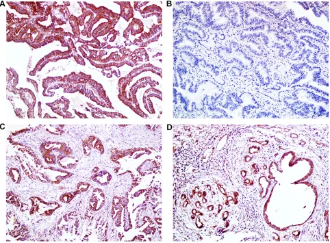
Figure 1Immunohistochemical staining of EphB3, ×200. (A) Positive expression of EphB3, well differentiated ECC. (B) Negative expression of EphB3, moderately-differentiated ECC. (C) Positive expression of EphB3, peritumoral tissues. (D) Positive expression of EphB3, adenoma.
Cancer Management and Research downloaded from https://www.dovepress.com/ by 118.70.13.36 on 24-Aug-2020
found that several clinicopathological parameters nega-tively correlated with overall survival and posinega-tively cor-related with mortality, including poorly differentiated type, the positivity of lymph node metastasis, the positivity of surrounding tissues and organs invasion, and advanced TNM stages (III or IV), which are risk factors and inde-pendent prognostic predictors (Tables 5 and 6). Negative
EphB3 expression or positive dysadherin expression nega-tively correlated with overall survival and posinega-tively cor-related with mortality, which are risk factors and independent prognostic predictors (Tables 5and6).
Lastly, we depicted the receiver operating characteris-tic (ROC) curves to evaluate the diagnoscharacteris-tic efficacy of EphB3 expression, dysadherin expression, and EphB3 and dysadherin expression, respectively. The AUC of EphB3 was 0.654 (95% CI: 0.564–0.743), the AUC of dysadherin expression was 0.648 (95% CI: 0.558–0.737), and the AUC of EphB3 and dysadherin expression was 0.688 (95% CI:0.603–0.772) (Figure 4). Our results sug-gested that EphB3 and dysadherin combined diagnostic efficacy was significantly higher EphB3 diagnostic effi -cacy or dysadherin diagnostic efficacy alone.
Discussion
ECC is an aggressive malignancy and has a poor prognosis. In this study, our data showed that the average survival time
Figure 2Immunohistochemical staining of dysadherin, ×200. (A) Positive expression of dysadherin, moderately differentiated ECC. (B) Negative expression of dysadherin, well differentiated ECC. (C) Positive expression of dysadherin, peritumoral tissues. (D) Positive expression of dysadherin, adenoma.
Table 2 Comparison of EphB3 and Dysadherin Expression in
Normal Tissue, Adenoma, Peritumoral Tissue and ECC
Tissue Type Number of Patients (N)
EphB3 Positive (%)
Dysadherin Positive (%)
ECC 100 42 (42.0) 55 (55.0)
Peritumoral tissues
30 17(56.7) 11 (36.7)
adenoma 10 8 (80.0)* 3(30.0)
Normal tissues 15 15 (100.0)** 0 (0.0)**
Notes:Compared to ECC: *P< 0.05; **P< 0.01.
Abbreviation:ECC, extrahepatic cholangiocarcinoma.
Cancer Management and Research downloaded from https://www.dovepress.com/ by 118.70.13.36 on 24-Aug-2020
of patients with early TNM stages (I + II) is significantly longer than patients with advanced stages (III or IV). Additionally, the average survival time of patients received radical surgery are significantly longer than patients received a biopsy. These results demonstrated that early diagnosis is essential to improve the clinical prognosis of ECC. However, most patients with ECC are diagnosed at an advanced stage due to the lack of specific clinical manifestations and diag-nostic biomarkers in the early stage. Hence, it is very urgent tofind new specific diagnostic markers for early diagnosis of ECC.
Although EphB3 and dysadherin are related to the pro-gression and prognosis of various human cancers, no studies have investigated their expression and biological roles in ECC. Thus, we assessed EphB3 and dysadherin expression in ECC and non-tumor tissues using immunohistochemistry and evaluated their correlations with clinicopathological parameters and survival. Significantly decreased EphB3 expression and significantly increased dysadherin expression was observed in ECC, compared with normal tissues. Additionally, negative EphB3 expression and positive dys-adherin expression were closely associated with poorly dif-ferentiated type, the positivity of surrounding tissues and organs invasion, the positivity of lymph node metastasis, advanced TNM stages, and poor prognosis in ECC. To our knowledge, this is the first study to report the correlation between EphB3 or dysadherin expression and clinicopatho-logic characteristics and survival in ECC patients.
Many Eph receptors and ephrin ligands are associated with the development and progression of several human cancers. As one of Eph receptors family, EphB3 is also involved in tumorigenesis and progression of certain human cancers, such as colorectal cancer, ovarian serous carcinomas, prostate can-cer, papillary thyroid cancan-cer, head and neck squamous cell carcinoma, and NSCLC.15–20It has been reported that EphB3 is over-expressed in normal prostate cell lines compared with prostate tumor cell lines.33Previously studies demonstrated
Table 4 Correlations of Clinicopathological Characteristics,
EphB3 and Dysadherin Expression with the Mean Survival in Patients with ECC
Group Number of
Patients (n) Median Survival (Month) Log-Rank χ2 P Sex
Male 61 12.67 (3–30) 0.001 0.980
Female 39 12.59 (4–30)
Age (year)
≤45 17 13.82 (3–30) 0.667 0.414
>45 83 12.10 (3–30)
Differentiation
Well 31 18.46 (5–30) 27.655 0.000
Moderately 34 11.41 (3–30)
Poorly 35 7.97 (3–30)
Tumor size
≤3cm 62 12.62 (3–30) 0.235 0.628
>3cm 38 12.03 (5–30)
TNM stage
I + II 35 18.57 (7–30)
III 38 11.05 (3–30) 57.569 0.000
IV 27 6.26 (3–13)
Lymph node metastasis
No 62 15.52 (4–30) 39.001 0.000
Yes 38 7.18 (3–25)
Invasion
No 33 17.52 (4–30) 17.399 0.000
Yes 67 9.87 (3–30)
Surgery
Radical 54 16.62 (3–30) 48.388 0.000 Palliative 36 7.58 (4–24)
Biopsy 10 6.90 (3–14)
EphB3
− 58 8.35 (3–25) 37.806 0.000
+ 42 17.88 (7–30)
Dysadherin
− 45 17.11 (5–30) 32.224 0.000
+ 55 8.46 (3–25)
EphB3 and Dysadherin
EphB3(−) and Dysadherin (−)
13 10.92(5–20) 48.278 0.000
EphB3(+) and Dysadherin (−)
32 19.63(7–30)
EphB3(−) and Dysadherin (+)
45 7.60(3–25)
EphB3(+) and Dysadherin (+)
10 12.30(7–24)
Abbreviations:−, negative expression; +, positive expression.
Table 3 The Association Between EphB3 Expression and
Dysadherin Expression in ECC
EphB3 Dysadherin Total
− +
− 13 45 58
+ 32 10 42
Total 45 55 100
Notes:χ2
=28.464,P= 0.000.
Abbreviations:−, negative expression; +, positive expression.
Cancer Management and Research downloaded from https://www.dovepress.com/ by 118.70.13.36 on 24-Aug-2020
that EphB3 expression is down-regulated in ovarian serous carcinoma and colorectal cancer samples.15,18Consistent with these previous studies, our study showed that EphB3 were significant down-expression in ECC compared with normal tissues, suggesting that EphB3 may be involved in tumorigen-esis of ECC. Moreover, several studies have revealed that EphB3 is related to clinical prognosis and clinicopathological
characteristics of several human cancers. EphB3 acts as a tumor suppressor in colorectal cancer and ovarian serous carcinomas.15,18Chiu reported that overexpression of EphB3 in HT-29 colorectal cancer cells inhibits tumor growth and EphB3 expression levels is significantly decreased in advanced Dukes’stage of human colorectal cancer.20Xuan found that the EphB3 expression level was negatively
Figure 3Kaplan-Meier curves for ECC. (A) Positive and negative expression of EphB3 in ECC. (B) Positive and negative expression of dysadherin in ECC. (C) EphB3 and dysadherin expression in ECC.
Table 5Univariate Cox Regression Analysis of Survival Rate in Patients with ECC and EphB3 and Dysadherin Expression
Groups Factors B SE Wald P HR 95% CI
Lower Upper
Differentiated degree Well/moderately/poorly 0.659 0.136 23.466 0.000 1.933 1.480 2.523
Tumor size ≤3cm/>3cm 0.099 0.214 0.212 0.645 1.104 0.725 1.680
Lymph node metastasis No/Yes 1.285 0.228 31.705 0.000 3.615 2.311 5.655
Invasion No/Yes 0.912 0.237 14.841 0.000 2.489 1.565 3.957
TNM stage I/II/III/IV 1.023 0.158 41.673 0.000 2.782 2.039 3.795
Surgery Radical/Palliative/Biopsy 0.883 0.149 35.301 0.000 2.417 1.807 3.234
EphB3 −/+ −1.323 0.239 30.720 0.000 0.266 0.167 0.425
Dysadherin −/+ 1.181 0.229 26.595 0.000 3.257 2.079 5.102
Abbreviations:−, negative expression; +, positive expression; HR, hazard ratio; CI, confidence interval.
Cancer Management and Research downloaded from https://www.dovepress.com/ by 118.70.13.36 on 24-Aug-2020
correlated to the depth of tumor invasion, lymph node metas-tasis, TNM stage and differentiation of colorectal cancer, and the overall survival of patients with high EphB3 expression is
significantly longer than patients with negative or weak EphB3 expression.15Gao also reported that positive EphB3 expression is negatively related to histological grade and
Table 6Multivariate Cox Regression Analysis of Survival Rate in Patients with ECC and EphB3 and Dysadherin Expression
Groups Factors B SE Wald P HR 95% CI
Lower Upper
Differentiated degree Well/moderately/poorly 0.615 0.155 15.724 0.000 1.849 1.364 2.505
Tumor size ≤3cm/>3cm 0.202 0.233 0.756 0.385 1.224 0.776 1.932
Lymph node metastasis No/Yes 1.181 0.277 18.216 0.000 3.258 1.894 5.604
Invasion No/Yes 0.829 0.349 5.659 0.017 2.292 1.157 4.539
TNM stage I/II/III/IV 0.721 0.244 8.695 0.003 2.056 1.273 3.318
Surgery Radical/Palliative/Biopsy 0.495 0.182 7.395 0.007 1.640 1.148 2.343
EphB3 −/+ −0.826 0.290 8.131 0.004 0.438 0.248 0.772
Dysadherin −/+ 0.739 0.264 7.824 0.005 2.093 1.247 3.513
Abbreviations:−, negative expression; +, positive expression; HR, hazard ratio; CI, confidence interval.
Figure 4ROC of diagonal segments. (A) ROC of diagonal segments is produced by ties of EphB3 in ECC. (B) ROC of diagonal segments is produced by ties of dysadherin in ECC. (C) ROC of diagonal segments is produced by ties of EphB3 and dysadherin in ECC.
Cancer Management and Research downloaded from https://www.dovepress.com/ by 118.70.13.36 on 24-Aug-2020
FIGO stage of ovarian serous carcinomas.18 Similarly, our study found that positive rates of EphB3 expression were significantly higher in cases with well-differentiation, no sur-rounding tissues and organs invasion, no lymph node metas-tasis and early TNM stages (I + II), and the patients with positive EphB3 expression exhibited longer survival time than patients with negative EphB3 expression. Thus, EphB3 may function as a tumor suppressor in ECC, which needs further study to identify its potential mechanism. However, EphB3 expression is significantly elevated in NSCLC and papillary thyroid cancer, and EphB3 can promote cell migration and metastasis of papillary thyroid cancer and lung cancer, which contradictory to our results.17,19This may be due to the organ specificity. Therefore, the effect of EphB3 on development and progression of cancer is divergent and dependent on cancer types.
Dysadherin is a protein that is involved in tumorigenesis by down-regulating cell-cell adhesion and up-regulating che-mokine production.34Dysadherin expression has been studied in various human cancer types and is a mark of poor prognosis of these cancers.22,26,28,35In most human cancers studies, the increased dysadherin expression is associated with decreased E-cadherin expression and reflects tumor aggressiveness.34,35 Several studies showed that dysadherin was frequently expressed in cancer cells, but not expressed in the cells of corresponding normal tissues.22,26,36In agreement with pre-vious reports, we found that positive dysadherin expression in ECC was higher compared to normal tissues and dysadherin was not detected in normal biliary tract tissue, indicating that dysadherin may be involved in the oncogenesis of ECC. To further identify the role of dysadherin in ECC, we analyzed the relationship between dysadherin expression and several clinicopathological features including tumor differentiation, lymph node metastasis, invasion, TNM stage, and surgical procedure. Previous studies have shown that dysadherin can promote cancer metastasis and progression of several human cancers, such as colorectal cancer, ovarian carcinomas, gastric cancer.22,24,25,34Likewise, our data demonstrated that dysad-herin positive expression was higher in the cases with poor differentiation, lymph node metastasis, invasion, advanced TNM stages, suggesting that dysadherin may be contributed to metastasis and development of ECC. Furthermore, the current study also revealed that dysadherin positive expression had a significant effect on ECC patient survival and was significantly correlated to poor prognosis of ECC patient, which is consistent with previous reports.22,26,28,35Thus, dys-adherin may play an unneglected role in carcinogenesis and
progression of ECC, which needs further in vivo and in vitro study to explore its underlying mechanism.
In this study, we found that EphB3 expression was significantly down-regulated and dysadherin expression was significantly up-regulated in ECC. In peritumoral tis-sues and adenoma tistis-sues, negative EphB3 or positive dys-adherin expression was positively related to dysplasia of biliary tract epithelia. Thus, down-expression of EphB3 or overexpression of dysadherin may be involved in the pro-cesses that benign lesions evolve into ECC. Furthermore, EphB3 negative expression or dysadherin positive expres-sion was closely associated with several clinicopathological features of ECC, which could reflect aggressiveness and malignant degree of the tumor. The survival of patients with positive expression of EphB3 was significantly longer than patients with negative expression of EphB3, which was contrary to the relationship between dysadherin expression and patient survival. Cox multivariate analysis further demonstrated that negative EphB3 or positive dysadherin expression was an independent predictor for poor prognosis in patients with ECC. The AUC of EphB3 and dysadherin suggested that the expression of EphB3 and dysadherin may have potential clinicopathological diagnostic value. These results showed that EphB3 and dysadherin may function an important role in the tumorigenesis and progression of ECC. Therefore, detection of EphB3 or dysadherin expres-sion in biliary duct tissues may have important clinical significance in the prevention or earlyfinding of ECC.
Conclusions
EphB3 and dysadherin are involved in the carcinogenesis and progression of ECC, and ECC patients with negative EphB3 or positive dysadherin expression have a poor prognosis.
Abbreviations
ECC, extrahepatic cholangiocarcinoma; ICC, intrahepatic cholangiocarcinoma; TNM, tumor-node-metastasis; PSC, Primary Sclerosing Cholangitis; CT, ultrasonography, com-puted tomography; MRI, magnetic resonance imaging; ERCP, endoscopic retrograde cholangiopancreatography; NSCLC, non-small-cell lung cancer; CPC, Clinicopathological charac-teristics; Pos No., Positive Number; RR, relative risk; CI, confidence interval.
Author Contributions
Zhengchun Wu, Rushi Liu, and Li Xiong carried out studies and wrote the paper; Zhulin Yang designed the
Cancer Management and Research downloaded from https://www.dovepress.com/ by 118.70.13.36 on 24-Aug-2020
study and revised the paper; Zhulin Yang and Xiongying Miao performed the statistical analysis; Daiqiang Li, Yuan Yuan, and Qiong Zou collected specimens and experimental materials. All authors contributed to data analysis, drafting or revising the article, gave final approval of the version to be published, and agree to be accountable for all aspects of the work.
Funding
This work was funded by the National Natural Science Foundation of China (grant number 81472738); Natural Science Foundation of Hunan Province, China (2019JJ10002); and Hunan Provincial Key Research and Development Program (2019SK2042).
Disclosure
The authors declare that they have no conflicts of interest regarding this work.
References
1. Esnaola NF, Meyer JE, Karachristos A, et al. Evaluation and manage-ment of intrahepatic and extrahepatic cholangiocarcinoma.Cancer.
2016;122(9):1349–1369. doi:10.1002/cncr.29692
2. Rizvi S, Gores GJ. Pathogenesis, diagnosis, and management of cholangiocarcinoma. Gastroenterology. 2013;145(6):1215–1229. doi:10.1053/j.gastro.2013.10.013
3. Labib PL, Goodchild G, Pereira SP. Molecular pathogenesis of cholangiocarcinoma. BMC Cancer. 2019;19(1):185. doi:10.1186/ s12885-019-5391-0
4. Razumilava N, Gores GJ. Cholangiocarcinoma. Lancet. 2014;383 (9935):2168–2179. doi:10.1016/S0140-6736(13)61903-0
5. Khan SA, Davidson BR, Goldin RD, et al. Guidelines for the diag-nosis and treatment of cholangiocarcinoma: an update.Gut.2012;61 (12):1657–1669. doi:10.1136/gutjnl-2011-301748
6. Alsaleh M, Leftley Z, Barbera TA, et al. Cholangiocarcinoma: a guide for the nonspecialist. Int J Gen Med. 2019;12:13–23. doi:10.2147/IJGM.S186854
7. Khan SA, Davidson BR, Goldin R, et al. Guidelines for the diagnosis and treatment of cholangiocarcinoma: consensus document. Gut.
2002;51(Suppl 6):I1–I9. doi:10.1136/gut.51.suppl_6.vi1
8. Doherty B, Nambudiri VE, Palmer WC. Update on the diagnosis and treatment of cholangiocarcinoma. Curr Gastroenterol Rep.2017;19 (1):2. doi:10.1007/s11894-017-0542-4
9. Liang Z, Liu X, Zhang Q, et al. Diagnostic value of microRNAs as biomarkers for cholangiocarcinoma. Dig Liver Dis. 2016;48 (10):1227–1232. doi:10.1016/j.dld.2016.07.006
10. Aasheim HC, Patzke S, Hjorthaug HS, et al. Characterization of a novel Eph receptor tyrosine kinase, EphA10, expressed in testis. Biochim Biophys Acta. 2005;1723(1–3):1–7. doi:10.1016/ j.bbagen.2005.01.011
11. Surawska H, Ma PC, Salgia R. The role of ephrins and Eph receptors in cancer. Cytokine Growth Factor Rev. 2004;15(6):419–433. doi:10.1016/j.cytogfr.2004.09.002
12. Baker RK, Vanderboom AK, Bell GW, et al. Expression of the receptor tyrosine kinase gene EphB3 during early stages of chick embryo development.Mech Dev.2001;104(1–2):129–132. doi:10.1016/S0925-4773(01)00363-X
13. Adams RH, Wilkinson GA, Weiss C, et al. Roles of ephrinB ligands and EphB receptors in cardiovascular development: demarcation of arterial/venous domains, vascular morphogenesis, and sprouting angiogenesis.Genes Dev.1999;13(3):295–306. doi:10.1101/gad.13.3. 295
14. Lee SY, Na YJ, Jeong YA, et al. Upregulation of EphB3 in gastric cancer with acquired resistance to a FGFR inhibitor.
Int J Biochem Cell Biol. 2018;102:128–137. doi:10.1016/j. biocel.2018.07.008
15. Xuan Z, Huang J, Gao L, et al. Receptor tyrosine kinase EphB3: a prognostic indicator in colorectal carcinoma.Pathol Oncol Res.
2018. doi:10.1007/s12253-018-0562-x
16. Bhatia S, Griego A, Lennon S, et al. Role of EphB3 receptor in mediating head and neck tumor growth, cell migration, and response to PI3K inhibitor.Mol Cancer Ther.2018;17(9):2049–2059. doi:10. 1158/1535-7163.MCT-17-1163
17. Li JJ, Sun ZJ, Yuan YM, et al. EphB3 stimulates cell migration and metastasis in a kinase-dependent manner through Vav2-Rho GTPase axis in papillary thyroid cancer.J Biol Chem.2017;292(3):1112–1121. doi:10.1074/jbc.M116.750349
18. Gao W, Zhang Q, Wang Y, et al. EphB3 protein is associated with histological grade and FIGO stage in ovarian serous carcinomas.
APMIS.2017;125(2):122–127. doi:10.1111/apm.12646
19. Ji XD, Li G, Feng YX, et al. EphB3 is overexpressed in non-small-cell lung cancer and promotes tumor metastasis by enhancing non-small-cell survival and migration. Cancer Res. 2011;71(3):1156–1166. doi:10.1158/0008-5472.CAN-10-0717
20. Chiu ST, Chang KJ, Ting CH, et al. Over-expression of EphB3 enhances cell-cell contacts and suppresses tumor growth in HT-29 human colon cancer cells. Carcinogenesis. 2009;30(9):1475–1486. doi:10.1093/carcin/bgp133
21. Lubarski I, Pihakaski-Maunsbach K, Karlish SJ, et al. Interaction with the Na,K-ATPase and tissue distribution of FXYD5 (related to ion channel).J Biol Chem.2005;280(45):37717–37724. doi:10.1074/ jbc.M506397200
22. Aoki S, Shimamura T, Shibata T, et al. Prognostic significance of dysadherin expression in advanced colorectal carcinoma. Br J Cancer.2003;88(5):726–732. doi:10.1038/sj.bjc.6600778 23. Ino Y, Gotoh M, Sakamoto M, et al. Dysadherin, a cancer-associated
cell membrane glycoprotein, down-regulates E-cadherin and pro-motes metastasis.Proc Natl Acad Sci U S A.2002;99(1):365–370. doi:10.1073/pnas.012425299
24. Raman P, Purwin T, Pestell R, et al. FXYD5 is a marker for poor prognosis and a potential driver for metastasis in ovarian carcinomas.Cancer Inform.2015;14:113–119. doi:10.4137/CIN.S30 565
25. Shimada Y, Yamasaki S, Hashimoto Y, et al. Clinical significance of dysadherin expression in gastric cancer patients.Clin Cancer Res.
2004;10(8):2818–2823. doi:10.1158/1078-0432.CCR-0633-03 26. Shimamura T, Sakamoto M, Ino Y, et al. Dysadherin overexpression
in pancreatic ductal adenocarcinoma reflects tumor aggressiveness: relationship to e-cadherin expression. J Clin Oncol. 2003;21 (4):659–667. doi:10.1200/JCO.2003.06.179
27. Kyzas PA, Stefanou D, Batistatou A, et al. Dysadherin expression in head and neck squamous cell carcinoma: association with lymphan-giogenesis and prognostic significance.Am J Surg Pathol.2006;30 (2):185–193. doi:10.1097/01.pas.0000178090.54147.f8
28. Sato H, Ino Y, Miura A, et al. Dysadherin: expression and clinical significance in thyroid carcinoma.J Clin Endocrinol Metab.2003;88 (9):4407–4412. doi:10.1210/jc.2002-021757
29. FT B, C F, RH H, et al.World Health Organization Classification of Tumours of the Digestive System. Lyon: IARC Press;2010. 30. Liu R, Yang Z, Huang S, et al. The expressions of HMGA2 and Thy1 in
extrahepatic cholangiocarcinoma and their clinicopathological significances. Surg Oncol. 2019;29:41–47. doi:10.1016/j.suronc.2019. 01.013
Cancer Management and Research downloaded from https://www.dovepress.com/ by 118.70.13.36 on 24-Aug-2020
31. He J, Yang Z, Wu Z, et al. Expression of FOXP1 and FOXO3a in extrahepatic cholangiocarcinoma and the implications in clinico-pathological significance and prognosis. Onco Targets Ther.
2019;12:2955–2965. doi:10.2147/OTT.S197001
32. Wu ZC, Xiong L, Wang LX, et al. Comparative study of ROR2 and WNT5a expression in squamous/adenosquamous carcinoma and ade-nocarcinoma of the gallbladder. World J Gastroenterol. 2017;23 (14):2601–2612. doi:10.3748/wjg.v23.i14.2601
33. Fox BP, Tabone CJ, Kandpal RP. Potential clinical relevance of Eph receptors and ephrin ligands expressed in prostate carcinoma cell lines. Biochem Biophys Res Commun. 2006;342(4):1263–1272. doi:10.1016/j.bbrc.2006.02.099
34. Nam JS, Hirohashi S, Wakefield LM. Dysadherin: a new player in cancer progression.Cancer Lett.2007;255(2):161–169. doi:10.1016/ j.canlet.2007.02.018
35. Tamura M, Ohta Y, Tsunezuka Y, et al. Prognostic significance of dysadherin expression in patients with non-small cell lung cancer.
J Thorac Cardiovasc Surg. 2005;130(3):740–745. doi:10.1016/j. jtcvs.2004.12.051
36. Park JR, Kim RJ, Lee YK, et al. Dysadherin can enhance tumorigen-esis by conferring properties of stem-like cells to hepatocellular carcinoma cells. J Hepatol. 2011;54(1):122–131. doi:10.1016/j. jhep.2010.06.026
Cancer Management and Research
Dove
press
Publish your work in this journal
Cancer Management and Research is an international, peer-reviewed open access journal focusing on cancer research and the optimal use of preventative and integrated treatment interventions to achieve improved outcomes, enhanced survival and quality of life for the cancer patient.
The manuscript management system is completely online and includes a very quick and fair peer-review system, which is all easy to use. Visit http://www.dovepress.com/testimonials.php to read real quotes from published authors.
Submit your manuscript here:https://www.dovepress.com/cancer-management-and-research-journal
Cancer Management and Research downloaded from https://www.dovepress.com/ by 118.70.13.36 on 24-Aug-2020