Original Article
Cloning, expression and functional study of
translation elongation factor 2 (EF-2) in zebrafish
SHU-HONG ZHANG
#, JI-HUA YAO
#,*, HUAI-DONG SONG
1, LU WANG and JING-LUN XUE*
State Key Laboratory of Genetic Engineering, Institute of Genetics, School of Life Sciences, Fudan University, Shanghai, China and 1Center of Molecular Medicine and Human Genome, Ruijin Hospital, Shanghai Second Medical University, Shanghai, China
ABSTRACT We have identified translation elongation factor 2 (EF-2) in zebrafish (GenBank Accession No. AAQ91234). Analysis of the DNA sequence of zebrafish EF-2 shows that the 2826 bp cDNA spans an open reading frame between nucleotide 55 to 2631 and encodes a protein of 858 amino acids. Zebrafish EF-2 protein shares 92%, 93%, 93% and 92% identity with the corresponding amino acid sequence in human, mouse, Chinese hamster and Gallus EF-2, respec-tively. Whole-mount in situ hybridization showed that zebrafish EF-2 was a developmentally regulated gene and might play important roles during the early development of zebrafish embryos. Therefore, we further studied the function of EF-2 during early embryogenesis. Using morpholino antisense oligo knockdown assays, anti-MO injected embryos were found to display abnormal development. The yolk balls were larger than normal and the melanophores spreading on their bodies became fewer. Furthermore, their tails were incurvate and their lenses were much smaller than those of the normal embryos. However the EF-2 overexpression data showed that extra EF-2 protein had no obvious effect on zebrafish embryonic development.
KEY WORDS: EF-2, zebrafish, whole-mount in situ hybridization, overexpression, knockdown
Introduction
Translation elongation factor 2, EF-2 and its eubacterial ho-molog, EF-G, act sequentially after eEF1a/EF-Tu to catalyze the GTP hydrolysis-dependent translocation of the ribosome during protein synthesis, to allow the peptidyl-tRNA to move from aminoacyl site to the peptidyl site on a ribosome, liberating the former site to accept a new mRNA triplet and its cognate ternary complex (eEF1-aminoacyl-tRNA-GTP) (Kohno K et al., 1986; Mendoza A et al., 1999). EF-2 is a GTPase; it binds and hydro-lyzes GTP and forms a ternary complex with GTP and ribosomes (Rao S et al., 1996).
Although regulation of EF-2 is well known to include phospho-rylation and ADP-ribosylation, developmental regulation of EF-2 isoforms has not been fully explored. The reported developmen-tally regulated EF-2 isoforms constitute a family of genes in Tetrahymena thermophila, which encode proteins homologous to EF-2 and are expressed only during sexual reproduction (Malave TM et al., 2004). Drosophila contains two nearly identical EF-2 genes (Lasko, 2000). Nevertheless, there are many examples in multicellular eukaryotes of differential expression of isoforms of translation elongation factor 1a (eEF-1a). Drosophila has two
*Address correspondence to: Dr. Jing-lun Xue. State Key Laboratory of Genetic Engineering, Institute of Genetics, School of Life Sciences, Fudan University, Shanghai 200433, China.Fax: +86-21-6564-9899. e-mail: [email protected] or [email protected] (Dr. Yao).
#Note Both authors contributed equally to this paper.
Abbreviations used in this paper: EF-2, translation elongation factor 2; MO, morpholino modified antisense oligonucleotide; PTU, 1-phenyl-2-thiourea; PFA, paraformaldehyde; ORF, open reading frame; hpf, hours post-fertilization.
0214-6282/2006/$25.00 © UBC Press
Printed in Spain
www.intjdevbiol.com
copies (F1 and F2) of the EF-la gene, which are expressed at different times during development. While F2 is transcribed only in the pupal stage, F1 is a housekeeping gene expressed in all cells during development and in adulthood (Hovemann B et al., 1988). Three EFla genes have been characterized in Xenopus -(42Sp50, EF-laO and EF-laS), 42Sp50 is expressed exclusively in oocytes, while EF-laO is active from fertilization until early onset of neurulation (Krieg P et al., 1989; Dje MK et al., 1990). In mammals, the EF-S (eEF1a2) gene is expressed in heart and muscle tissue and eventually becomes the only form expressed in these tissues (Knudsen et al., 1993). Disruption of the EF-S (eEF1a2) gene in mouse results in deficiencies in muscle and neuronal function within a few weeks after birth (Chambers et al., 1998).
zebrafish :
MVNFTVDQIRAIMDKKSNIRNMSVIAHVDHGKSTLTDSLVSKAGIIASARAGETRFTDTRKDEQERCITIKSTAISMYYE MVNFTVDQIRAIMDKKANIRNMSVIAHVDHGKSTLTDSLVCKAGIIASARAGETRFTDTRKDEQERCITIKSTAISLFYE
LTENDLAFIKQCKDGSGFLINLIDSPGHVDFSSEVTAALRVTDGALVVVDCVSGVCVQTETVLRQAIAERIKPVLMMNKM LSENDLAFIKQSKDGSGFLINLIDSPGHVDFSSEVTAALRVTDGALVVVDCVSGVCVQTETVLRQAIAERIKPVLMMNKM LSENDLNFIKQSKDGSGFLINLIDSPGHVDFSSEVTAALRVTDGALVVVDCVSGVCVQTETVLRQAIAERIKPVLMMNKM LSENDLNFIKQSKDGAGFLINLIDSPGHVDFSSEVTAALRVTDGALVVVDCVSGVCVQTETVLRQAIAERIKPVLMMNKM LSENDLNFIKQSKDGSGFLINLIDSPGHVDFSSEVTAALRVTDGALVVVDCVSGVCVQTETVLRQAIAERIKPVLMMNKM
DRALLELQLEPEELYQTFQRIVENVNVIISTYGEDEGGPMGNIMIDPVIGTVGFGSGLHGWAFTLKQFAEMYVAKFASKG DRALLELQLDPEELYQTFQRIVENVNVIISTYGEGESGPMGNIMIDPVLGTVGFGSGLHGWAFTLKQFAEMYVAKFAAKG
EAQLSPADRCKKVEDMMKKLWGDRYFDPAGGKFTKTANGPDGKKYPRTFAQLILDPIFKVFDAIMNFKKEETAKLIEKLD
DAQMNPTERAKKVEDMMKKLWGDRYFDPATGKFSKSATGPDGKKLPRTFCQLILDPIFKVFDAIMTFKKEEAAKLIEKLD EGQLGPAERAKKVEDMMKKLWGDRYFDPANGKFSKSANSPDGKKLPRTFCQLILDPIFKVFDAIMNFRKEETAKLIEKLD EGQLGPAERAKKVEDMMKKLWGDRYFDPANGKFSKSATSPEGKKLPRTFCQLILDPIFKVFDAIMNFKKEETAKLIEKLD EGQLSAAERAKKVEDMMKKLWGDRYFDPANGKFSKSANSPDGKKLPRTFCQLILDPIFKVFDAIMNFRKEETAKLIEKLD
: 320
IKLDTEDKDKEGKPLLKAVMRRWLPAGEALLQMITIHLPSPVTAQKYRCELLYEGPGDDEAAMGIKNCDPKGPLMMYISK IKLDSEDKDKEGKPLLKAVMRRWLPAGDALLQMITIHLPSPVTAQKYRCELLYEGPPDDEAAIGIKNCDPRGSLMMYISK IKLDSEDKDKEGKPLLKAVMRRWLPAGDALLQMITIHLPSPVTAQKYRCELLYEGPPDDEAAMGIKSCDPKGPLMMYISK
LEEDHACIPLKKSDPVVSYRETVSAESDQMCLSKSPNKHNRLYMKARPFPDGLAEDIDKGDVSSRQELKTRARYLADKYE LEEDHACIPIKKSDPVVSYRETVSEESNVMCLSKSPNKHNRLYMKARPFPDGLAEDIDKGEVSARQELKQRARYLAEKYE
WEVTEARKIWCFGPDGTGPNMLVDVTKGVQYLNEIKDSVVAGFQWATKEGALCEENMRAVRFDIHDVTLHTDAIHRGGGQ WDVTEARKIWCFGPDGTGPNILTDITKGVQYLNEIKDSVVAGFQWATKEGVLCEENMRGVRFDVHDVTLHADAIHRGGGQ WDVAEARKIWCFGPDGTGPNILTDITKGVQYLNEIKDSVVAGFQWATKEGALCEENMRGVRFDVHDVTLHADAIHRGGGQ
IIPTARRVLYACQLTAEPRLMEPIYLVEIQCPEQVVGGIYGVLNRKRGHVFEESQVMGTPMFVVKAYLPVNESFGFTADL IIPTARRCLYACVLTAQPRLMEPIYLVEIQCPEQVVGGIYGVLNRKRGHVFEESQVAGTPMFVVKAYLPVNESFGFTADL
RSNTGGQAFPQCVFDHWQILPGDPKDAASKPCQIVADTRKRKGLKEGIPALDNFLDKL RSNTGGQAFPQCVFDHWQILPGDPFDSASRPSQVVAETRKRKGLKEGIPALDNFLDKL
expression pattern by whole-mount in situ hybridization. The result of whole-mount in situ hybridization showed that zebrafish EF-2 was a developmentally regulated gene just like EF-S and might play important roles during the early development of zebrafish embryos. Therefore, we further studied the function of EF-2 during early embryogenesis.
Results
Sequence analysis of the zebrafish EF-2 gene
The full-length zebrafish EF-2 gene was sequenced. The result shows that the 2826 bp cDNA spans an ORF between nucleotide 55 to 2631 and encodes a protein of 858 amino acids (GenBank Accession No. AAQ91234).
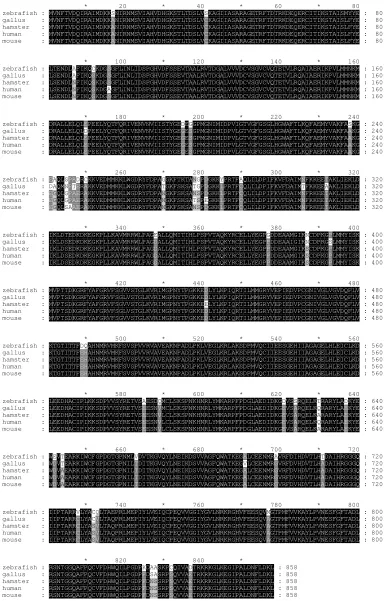
To figure out the structural features of the zebrafish EF-2 protein, its amino acid sequence was compared with those of other EF-2 proteins using clustalw version 1.82 online (http:// www.ebi.ac.uk/clustalw/). The zebrafish EF-2 protein is highly conserved sharing identity with overall 92%, 93%, 93% and 92% of amino acid sequence in human (CAA77750), mouse (NP_031933), Chinese hamster (A25440) and Gallus gallus (NP_990699) EF-2, respectively. Then the alignment was done
with software GeneDoc (shown in Fig. 1). The data shows that the zebrafish EF-2 protein is highly conserved.
Spatiotemporal expression pattern of the zebrafish EF-2 gene
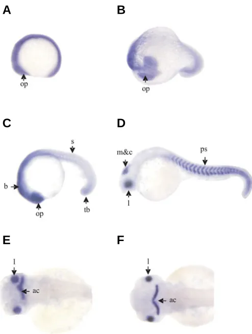
EF-2 transcript was not detected during somitogenesis using sense probe (data not shown). However, the results of whole-mount in situ hybridization using antisense probe showed that a large amount of EF-2 transcripts existed during somitogen-esis. EF-2 transcripts originally appeared strongly throughout envelope at 5-somite stage (Fig. 2A). At 17-somite stage (Fig. 2B) and 22–somite stage (Fig. 2C), EF-2 transcripts were detected still throughout the embryo with especially significant in the optic primordia, the whole brain, the trunk and tail bud. From 22-somite stage the stained cells began to concentrate strongly in the eyes, brain and somites. At prim-15 stage, the stained cells concentrated mainly in the lens, retina, midbrain and cerebellum (Fig. 2D) and posterior somites expression is especially evident. But after prim-25 stage the expression of EF-2 only appeared in the lens and the anterior portion of the cerebellum (including the proliferation zone at the midbrain-hindbrain – boundary) (Fig. 2 E-F).
Overexpression of EF-2
Expressing vector pcDNA3.0-EF2 with various amounts (1.0ng/embryo, 0.67ng/embryo and 0.33ng/embryo) and EF-2 capped mRNA with the amount of 4.5ng/ embryo were injected into each of 1–4-cell-stage embryos, respectively. After inject-ing pcDNA3.0-EF2 expression vector with the amount of 1.0ng/ embryo, most of the zebrafish embryos died (Table 1), showing that the amount of 1.0ng/ embryo was fatal to the zebrafish embryos. However, no abnormal phenotypes appeared when they were injected by other amounts of pcDNA3.0-EF2 (0.67 ng/embryo and 0.53 ng/embryo) or EF-2 capped mRNA (Table 1). So, overexpression of EF-2 had no obvious effect on the zebrafish embryonic development (Fig. 3).
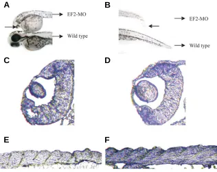
Gene knockdown with morpholino antisense oligo The results of gene knockdown assay with morpholino antisense oligo showed that most embryos injected with anti-MO displayed abnormal phenotypes during embryonic devel-opment while the same dose of sense-MO had no obvious effect on the embryos (Table 1). From 24-hpf EF-2-MO-treated embryos began to show developmental growth arrest. The EF-2-MO treated embryos were shorter, yolk balls were larger, tails were incurvate, lenses were much smaller and the
melano-Fig. 2. Expression patterns of EF-2 in the zebrafish embryo. Side view(A,B,C,D); dorsal view (E,F).(A)5-somite stage; (B)17-somite stage; (C)22 -somite stage; (D) prim-15 stage; (E) prim-25 stage; (F)protruding-mouth stage. Abbreviations: op, optic primordial; b, brain; tb, tail bud; s, somite; m&c, midbrain and cerebellum; l, lens; ps, posterior somites; ac, anterior portion of the cerebellum.
Fig. 3. Overexpression of the EF-2 gene (48 hpf). There is no obvious difference between the pcDNA3.0-EF2 injected zebrafish and the wild type zebrafish.
A
B
C
D
phore spreading on their bodies became lighter than normal level (Fig. 4 A,B). Furthermore cross-sections also revealed that their tails were incurvate and their lenses were much smaller than those of normal embryos (Fig. 4 C–F).
Discussion
We have identified a translation elongation factor 2 (EF-2) in zebrafish with high identity to EF-2 proteins of other animals. From the results of the whole-mount in situ hybridization, it can be presumed that the zebrafish EF-2 gene is developmentally regulated. Because zebrafish EF-2 transcripts appear strongly
Amount of Injected and phenotype
microinjection Whole mount Live embryos for over-expression (ng/embryo) embryos (%) studies (%)
pcDNA3.0-EF2 1.0 263 12(4.6) 2(16.7)
0 88 59(67.0) 0(0)
0.67 248 120(48.4) 7(5.8)
0 60 39(65.0) 0(0)
0.53 169 83(49.1) 3(3.6)
0 64 37(57.8) 0(0)
EF-2 capped mRNA 4.5 211 126(59.7) 7(5.6)
0 73 48(65.8) 0(0)
EF-2 morpholino 4.5 116 87(75) 52(60)
0 96 78(82) 0(0)
control sense-MO 4.5 167 130(77.8) 0(0)
0 155 125(80.6) 0(0)
OVEREXPRESSION AND MORPHOLINO KNOCKDOWN OF THE EF-2 GENE
TABLE 1
at those places when the level of the EF-2 could not maintain normal lens development. Although EF-2 expressed in the anterior portion of the cerebellum, no obvious abnormalities were found here upon the treatments, so we presume that some other important genes participate in the development of the anterior portion of the cerebellum and EF-2 only had a subsid-iary role.
Above all, from the results of the whole-mount in situ hybrid-ization, overexpression and gene “knock-down” experiments, we can believe that the zebrafish EF-2 gene regulates primarily specific aspects of development.
Materials and Methods
Zebrafish and embryo maintenance
Zebrafish were raised and maintained under standard laboratory conditions at 28°C, as described by Westerfield et al. (Westerfield et al., 1995). Embryos used in whole-mount in situ hybridization were
raised in 0.003% PTU (1-phenyl-2-thiourea, Sigma, St.Louis, MO, USA) to prevent pigment formation. The stage of the embryos was determined by morphological features and fixed with 4% PFA (paraform-aldehyde) according to Kimmel et al. (Kimmel et al., 1995).
Cloning and sequence analysis of EF-2
A zebrafish cDNA clone RK115A2C01 containing the EF-2
full-length cDNA was isolated from the zebrafish adult kidney cDNA library (Song et al., unpublished data). The isolated full-length EF-2 cDNA was
subcloned into pBK-CMV vector and the two inserted enzyme sites were EcoRI and XhoI, respectively. Then the full-length EF-2 gene was
sequenced. Its DNA and amino acid sequences were analyzed on National Center for Biotechnology Information (NCBI) blast server. Fig. 4. Morphological and histological analysis of EF2-MO injected embryos at 48 hpf.
(A,B) Morphological comparisons of EF2-MO injected embryos and control embryos. (C,D) Histological analysis of EF2-MO injected embryos through cross-sections. (E,F)The cross-sections of sense-MO injected embryos. It is obvious that in the EF-2-MO injected embryo, the lens is much smaller and the tail is incurvate and incompact.
throughout envelope before prim-15 stage, we consider the EF-2 gene is expressed as a maternal transcript. After prim-15 stage, the expression of zebrafish EF-2 is limited to irre-versibly growth-arrested cells such as lens, cerebellum and somites, so we think zebrafish EF-2 is a terminal differentiation-specific pro-tein just like EF-S propro-tein (Knudsen et al., 1993; Chambers et al., 1998). And we can also ac-cordingly presume that zebrafish EF-2 is poten-tially a multi-gene family and the one which we are reporting is only one isoform of this family and its main function is maintaining the devel-opment of lens, cerebellum and somites but not protein synthsis.
This seems to be the first functional report on the developmental regulation of zebrafish EF-2 gene. Overexpression of EF-2 had no obvious effect on the zebrafish embryonic development. The data of gene knockdown approved that EF-2 surely plays important roles in maintaining the development of zebrafish embryos. As EF-2 proteins are expressed in posterior somites and the tail bud, so in the EF-2-MO treated embryos the low level of EF-2 could not main-tain the production of new somites in the tail buds and finally result in incurvate tails. Simi-larly, EF-2 proteins are also expressed in the lenses and were important to maintain their development, so probably hypoplasia appeared
A
B
C
D
Multiple alignments were performed with clustalw version 1.82 (http:// www.ebi.ac.uk/clustalw/) as well. The graphic presentations were prepared by software GeneDoc.
Whole-mount in situ hybridization
To study the expression pattern of the EF-2 gene during
embryogen-esis, the zebrafish embryos were processed for whole-mount in situ
hybridization after fixation in 4% PFA for 24 h at 4ºC. As described previously, the isolated full-length EF-2 cDNA sequence was subcloned
into pBK-CMV vector, which contains T7 and T3 promoters. Using T3 and T7 RNA polymerases, the sense and antisense RNA probes were synthesized labelled with digoxigenin (Roche) in vitro, respectively. The
whole-mount in situ hybridization procedure was carried out as described
by Westerfield et al. (Westerfield et al., 1995).
Overexpression of EF-2
Vector pBK-CMV-EF2 was cut by Xho I and used as transcription template. Finally the capped EF-2 mRNA was transcribed in vitro using T3
RNA polymerase.
Expressing vector pcDNA3.0-EF2 was constructed as following: EF-2 ORF was cut by Xho I and EcoR I from pBK-CMV-EFEF-2 vector, then
inserted into pcDNA3.0 expression vector (bought from Invitrogen com-pany) that was also digested by Xho I and EcoR I. Capped EF-2 mRNA
and various amounts of expression vector pcDNA3.0-EF2 were injected into each of 1–4 cell stage embryos with a fine glass needle connected with an automatic injector (IM-300, Narishige, Japan), respectively. Phenotypes were observed at 48 hpf (hours post-fertilization). We se-lected three microinjection amounts of expression vector pcDNA3.0-EF2: 1.0ng/embryo, 0.67 ng/embryo and 0.53 ng/embryo. And the amount of capped EF-2 mRNA microinjection is 4.5 ng/embryo.
Gene knockdown with morpholino antisense oligo
Both EF-2 gene-specific antisense and sense morpholino
oligonucle-otides were purchased from Gene Tools LLC (Philomath, OR, USA), as following: anti-MO, 5’-CACCATTTTGACAGATGTTCTTGG-3’; sense-MO, 5’-CCAAGAACATCTGTCAAAATGGTG-3’.
Morpholino oligonucleotides were injected into each of 2-8-cell-stage embryos with a fine glass needle connected with an automatic injector (IM-300, Narishige, Tokyo, Japan). The amount of microinjection was about 4.5ng/embryo (Nasevicius et al., 2000). Some anti-MO and
sense-MO treated embryos at 48 hpf were fixed, dehydrated, embedded in resin and cross-sectioned to 5–10 µm thick with a glass knife. Slides were dyed by 1% toluidine blue.
Acknowledgments
This work was supported by National Natural Science Foundation of China (30370719) and the National Basic Research Program of China (973) (2004CB518803). We acknowledge the help of Professor A.M. Meng in offering the AB strain zebrafish used as experiment material and are grateful to Mr. Yin Sai for his help with sections.
References
CHAMBERS, D.M., PETERS, J. AND ABBOTT, C.M. (1998). The lethal mutation of the mouse wasted (wst) is a deletion that abolishes expression of a
tissue-specific isoform of translation elongation factor 1alpha, encoded by the EEf1a2
gene. Proc. Natl. Acad. Sci. USA. 95: 4463 - 4468.
DJE, M.K., MAZABRAUD, A., VIEL, A., MAIRE, M., H DENIS, H., CRAWFORD, E. AND BROWN, D.D. (1990). Three genes under different developmental control encode elongation factor 1-alpha in Xenopus laevis. Nucleic Acids Res. 18:
3489 - 3493.
HOVEMANN, B., RICHTER, S., WALLDORF, U. AND CZIEPLUCH, C. (1988).
Two genes encode related cytoplasmic elongation factors 1 alpha (EF-1 alpha)
in Drosophila melanogaster with continuous and stage specific expression. Nucleic Acids Res. 16:3175 - 3194.
KIMMEL, C.B., BALLARD, W.W., KIMMEL, S.R., ULLMANN, B. AND SCHILLING, T.F. (1995). Stages of embryonic development of the zebrafish, Dev. Dyn. 203:
253 - 310.
KNUDSEN, S.M., FRYDENBERG, J., CLARK, B.F. AND LEFFERS, H. (1993). Tissue-dependent variation in the expression of elongation factor-1 alpha isoforms: isolation and characterisation of a cDNA encoding a novel variant of human elongation-factor 1 alpha. Eur. J. Biochem. 215:549 - 554.
KOHNO, K., UCHIDA, T., OHKUBO, H., NAKANISHI, S., NAKANISHI, T., FUKUI, T., OHTSUKA, E., IKEHARA, M. AND OKADA, Y. (1986). Amino acid sequence of mammalian elongation factor 2 deduced from the cDNA sequence: Homol-ogy with GTP-binding proteins. Proc. Natl. Acad. Sci. USA. 83:4978 - 4982.
KRIEG, P.A., VARNUM, S.M., WORMINGTON, W.M. AND MELTON, D.A. (1989). The mRNA encoding elongation factor 1-alpha (EF-1 alpha) is a major
tran-script at the midblastula transition in Xenopus. Dev. Biol. 133:93 -100.
LASKO, P. (2000). The Drosophila melanogaster genome: translation factors and
RNA binding proteins. J. Cell Biol. 150: 51 - 56.
MALAVE, T.M. and FORNEY, J.D. (2004). Identification of a developmentally regulated translation elongation factor 2 in Tetrahymena thermophila. Gene.
326: 97 - 105.
MENDOZA, A., SERRAMIA, M.J., CAPA, L. AND GARCIA-BUSTOS, J.F.(1999). Translation elongation factor 2 is encoded by a single essential gene in
Candida albicans. Gene. 229:183 - 191.
NASEVICIUS, A. AND EKKER, S.C. (2000). Effective targeted gene ‘knockdown’ in zebrafish. Nature Genetics. 26: 216 - 220.
RAO, S. AND BODLEY, J.W. (1996). Expression, purification and characterization of the G domain of Saccharomyces cerevisiae elongation factor 2. Protein Exp. Purif. 8:91 - 96.
SONG, H.D., WU, X.Y., SUN,X.J., ZHOU,Y., LIU,T.X., DENG,M., ZHANG,G.W., SHENG,Y., CHEN,Y., RUAN,Z., JIANG,C.L., FAN,H.Y., ZON,L.I., KANKI,J.P., LOOK, A.T. AND CHEN,Z. Gene Expression Profiling in the Zebrafish Kidney Marrow Tissue. Unpublished.
WESTERFIELD, M. (1995). The Zebrafish Book. University of Oregon Press.