CSEIT1833712 | Received : 10 April 2018 | Accepted : 24 April 2018 | March-April-2018 [ (3 ) 3 : 1815-1820 ]
© 2018 IJSRCSEIT | Volume 3 | Issue 3 | ISSN : 2456-3307
1815-
A Study of Different Features and Classification of
Histophathological Image
Prachi Prajapati1, Bijal Talati21PG Student, Sardar Vallabhbhai Patel Institute of Technology, Vasad, Gujarat, India
2Head of Computer Engineering Department, Sardar Vallabhbhai Patel Institute of Technology, Vasad, Gujarat, India
ABSTRACT
Breast cancer is the second largest cause of cancer deaths among women. However, detecting this cancer in its first stages helps in saving lives because it is easier to treat and prevent the tumor from expanded. It has two states, known as benign and malignant. The research focuses on identifying malignancy in histopathological images of breast. In this process, it includes three phases viz: i) Image processing ii) Feature Extraction iii) classification. The proposed work gives complete and automated detection of malignancy using both image processing techniques and classification methods.
Keywords: Breast Cancer, histopathological images, Image Processing, image features, classification techniques
I.
INTRODUCTION
Breast Cancer (BC) is one of the most common type of cancer among female population in the whole world. Death rate for BC is higher than other types of cancer. Diagnosis of Breast Cancer (BC) depends on the visual aspects of tissue or cells removed from a patient and then step by step evaluation are specimen preparation, selection of tumour cell nuclei, image pre-processing, feature extraction according to size, shape, texture and so on [1].
Proper diagnosis can help a patient to get rid of BC risk if the state of cancer type is benign (localized and non - invading) or malignant (invading and life threatening). For diagnosis, different methods like mammograms, CT scan, histopathological image analysis (biopsy image) are commonly used. If there is a positive result of mammograms about BC, then diagnosis with histopathological imaging can be done. A histology image analysis system generally has a combination of hardware and software and it can be divided into two consecutive subsystem tissue preparation and image production and then Image processing analysis. To reduce the death rate among
women two things are very important that is education about BC and proper screening that means diagnosis [3].
Human body is made by number of cells and each cell have its own function when the cell is loss it’s ability and growth then we can clearly say that Something is happen in cell of human body. The extra cell from a mass of tissue is call tumor or storm [7]. Detection of BC in different medical images takes a lot of time because it directly deals with human life. We can improve the methodology and techniques that can be use for finding the Breast Cancer. Reducing the false negative rate we can improve the different techniques and methods for detection of Breast Cancer.we can create the good software for checking the part of the human life.
no widely accepted methods, therefore automatic and reliable methods for tumor detection are of great need and interest.
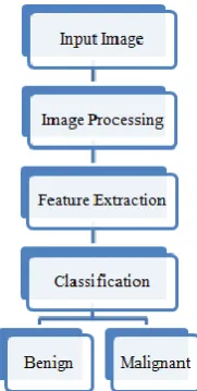
Figure 1. Tumor classification system
1. Image preprocessing - in these process it enhance the picture quality and removes the unwanted noise and background information. 2. Feature Extraction - After Image preprocessing,
positive region will be extraction using the different feature.
3. Classification - After identifying the cancerous nodule classifies the tumor types.
HISTOPATHOLOGICAL IMAGE
Histopathology refers to the examination of tissue in order to study the manifestations of disease. The name Histopathology is derived from the Greek word for tissue = Histos, disease = Pathos and Logos = the study of Histopathology generally involves samples of tissue, in contrast to cytopathology, which studies samples of free cells or tissue fragments. Histopathology can also often yield a more comprehensive view of the disease since the underlying tissue architecture is preserved. histopathological evaluation of tissue samples is critical to many applications, e.g., discovery of biomarkers, treatment planning in a clinical practice,
or cancer research. Characterization and
Figure 2.Histopathological Image
II.
TYPES OF TUMOR
A. BENIGN TUMOR
A benign tumor (benign neoplasm) cannot
metastasize - it cannot spread. "Benign" means it is non-progressive, it remains as it is. Most benign tumors are not harmful to human health. Even though they are not cancerous, some may press against nerves or blood vessels and cause pain or other negative effects. Benign tumors of endocrine tissues may result in the excessive production of some hormones. Tumors that aren’t cancerous are called non-cancerous tumors. Non-cancerous tumors:
stay in one place and don’t spread to other parts of the body
don’t usually come back after they are removed
Tend to have a regular and smooth shape and
have a covering called a capsule.
Figure 3. Benign tumor [9]
B. MALIGNANT TUMOR
tumor is in the middle of the spectrum. Some benign tumors eventually become premalignant, and then malignant. Cancer can start in any part of the body. When cancer cells form a lump or growth, it is called a cancerous tumor. A tumor is cancerous when it:
grows into nearby tissues
has cells that can break away and travel through the blood or lymphatic system and spread to lymph nodes and distant parts of the body
Figure 4. Malignant tumor [9]
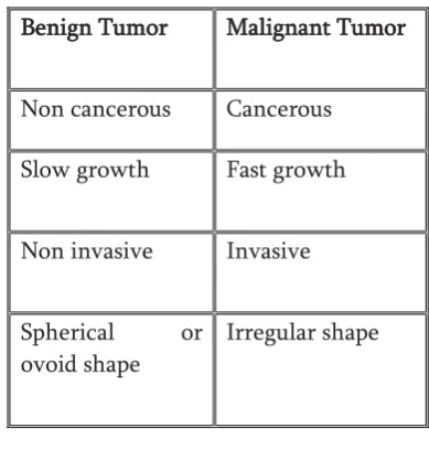
Table 1. Difference between benign and malignant tumor
Benign Tumor Malignant Tumor
Non cancerous Cancerous
Slow growth Fast growth
Non invasive Invasive
Spherical or
ovoid shape
Irregular shape
III.
IMAGE FEATURES
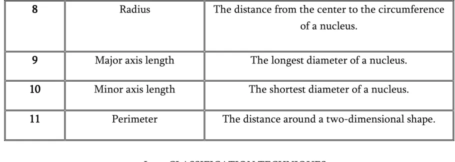
Image feature is the one pieces of information. In the image feature extraction is the process of transfer arbitrary data like image to the relevant numeric data. This numeric data used in the classification process. Following table describes different shape features. Table 2. Features description
No. Feature Description
1 Center The x- coordinates of the point farthest away from
any identified nucleus edge.
2 Mass Displacement The distance between the centers of gravity of the
nucleus in the image. The mass displacement is calculated by taking a weighted average of the
pixels in both X and Y.
3 Mean Intensity Edge Mean of edge pixel intensity values of the nucleus.
4 Mean Intensity Mean of pixel intensity values within the nucleus.
5 Min Intensity Minimum of pixel intensity values within the
nucleus.
6 Standard Intensity The standard deviation of the edge pixel intensities
of a nucleus.
7 Area The size of a surface. The amount of space inside
8 Radius The distance from the center to the circumference of a nucleus.
9 Major axis length The longest diameter of a nucleus.
10 Minor axis length The shortest diameter of a nucleus.
11 Perimeter The distance around a two-dimensional shape.
I. CLASSIFICATION TECHNIQUES
Classification defined as the task categorizes the any of given objects within a given category called class. Following table describes classification techniques with its advantages and
disadvantages, which taken from survey.
Table 3. Image classification method with advantages and disadvantages
No. Technique Advantages Disadvantages
1 Support vector
machine[1,3]
• Maximize the margin between
two classes in the feature space • characterized by a kernel
function
• Difficult to incorporate background knowledge.
• Sensitive to outlier.
2 Neural
network[1,8,9,14]
• Easy to conceptualize. • Provide high speed of
calculation.
• Can solve any machine learning algorithm.
• Neural networks are too much of black box this makes them difficult
to train. • Not probabilistic.
3 Fuzzy logic[1,16] • Allows for modeling and
inclusion of contradiction in a knowledge base
In a high complex system, use of fuzzy logic may become an obstacle
to verification of system reliability.
4 Random
forest[13,16]
• One of the most accurate learning algorithms available for
most data set
• fairly efficiently on large data sets
• Random forest has been observed to over fit for some datasets with noisy
classification task.
• Large number of trees may make the algorithm slow for real time
5 Bayesian Network[16]
• Bayesian methods have support of probability theory • Have well defined semantics for
decision making
• They require significant amount of probability data to construct a
knowledge base.
6 Decision tree[16] • Easy to understand.
• Easy to generate rules.
• May suffer from over fitting. • Classifies by rectangular
partitioning
IV.
CONCLUSION AND FUTURE WORKEarly detection of breast cancer is very important. This paper provides various Image Features and various classification techniques with advantages and disadvantages would give satisfactory results and help patient and pathologist for proper diagnosis of breast cancer and ultimately save a lot of lives.
Furthermore, we intended do survey of optimization algorithms and select suitable one and will be implementing through appropriate software. To increase accuracy of implemented algorithm, it will be optimize with suitable optimization technique.
V.
REFERENCES
[1].Fatema-Tuz Johra, Md. Maruf Hossain Shuvo " Detection of Breast Cancer from Histopathology image and Classifying Benign and Malignant State Using Fuzzy Logic " PP. 101-105 published in IEEE (2016).
[2].Dan C. Cires¸an, Alessandro Giusti, Luca M. Gambardella, Jurgen Schmidhuber "Mitosis Detection in Breast Cancer Histology Images with Deep Neural Networks" PP. 01-08 published in ELSEVIER (2017).
[3].Alexander Brook Ran El-Yaniv Eran Isler Ron Kimmel Ron Meir Dori Peleg "Breast Cancer Diagnosis From Biopsy Images Using Generic Features and SVMs" PP. 01-16 published in Technion (2008).
[4].Murat Karabatak, M. Cevdet Ince "An expert system for detection of breast cancer based on association rules and neural network" PP. 3465-3469 published in ELSEVIER (2017)..
[5].Elif Derya U beyli "Implementing automated diagnostic systems for breast cancer detection" PP. 1054-1064 published in ELSEVIER (2017). [6].J. Dheeba , N. Albert Singh b, S. Tamil Selvi
"Computer-aided detection of breast cancer on mammograms: A swarm intelligence optimized wavelet neural network approach" PP. 1054-1064 published in ELSEVIER (2017).
[7].M.A. Aswathy, M. Jagannath "Detection of breast cancer on digital histopathology images: Present status and future possibilities" " PP. 1054-1064 published in ELSEVIER (2017).
[8].Teresa Arau j0, Guilherme Aresta, Eduardo Castro, Jose Rouco" Classification of breast cancer histology images using Convolutional Neural Networks" PP. 1-14 published in PLOS ONE (June-2017).
[9].Fabio A. Spanhol, Luiz S. Oliveira "Breast Cancer Histopathological Image Classification using Convolutional Neural Networks" PP. 01-08 published in IEEE (2017).
[10]. Fabio A. Spanhol , Paulo R. CavalinY, Luiz S. Oliveira , Caroline Petitjean Z, and Laurent Heutte "Deep Features for Breast Cancer Histopathological Image Classification" PP. 1868-1873 published in IEEE October (2017).
Magnification Important" PP. 17-24 published in IEEE explorer (2017).
[12]. M.A. Aswathy, M. Jagannath "Detection of breast cancer on digital histopathology images: Present statusand future possibilities" PP. 74-79 published in ELSEVIER (2017).
[13]. Moh'd Rasoul A-hadidi,Abdulsalam
Alarabeyyat, Mohannad Alhanahnah "Breast Cancer Detection using K-nearest Neighbor Machine Learning Algorithm" 9th International Conference on Developments in eSystems Engineering, PP. 35-39 published in IEEE (2017).
[14]. Mahendra G. Kanojia, Siby Abraham "Breast Cancer Detection Using RBF Neural Network", PP. 363-369 published in IEEE (2016).
[15]. Dana Bazazeh1 and Raed Shubair
"Comparative Study of Machine Learning Algorithms for Breast Cancer Detection and Diagnosis" PP. 1-4 published in IEEE (2016). [16]. Saurabh Sharma and Dr. Ashish Negi"Breast
Cancer Detection Methods: A Brief Survey"pp.1-7 published in IJCSMS (International Journal of Computer Science & Management Studies) Vol. 25, Issue 01(2016).
[17]. Sangita Bhattacharjee, Jashojit Mukherjee, Sanjay Nag, Indra Kanta Maitra and Samir K. Bandyopadhyay " Review on Histopathological Slide Analysis using Digital Microscopy" pp. 65-96 published in International Journal of Advanced Science and Technology Vol.62 (2014) [18]. "Image Analysis Methods and Tools for
Digital Histopathology Applications Relevant to Breast Cancer Diagnosis"(2014)
[19]. http://web.inf.ufpr.br/vri/breast-cancer-database
[20].