Electronic Thesis and Dissertation Repository
12-10-2012 12:00 AM
Gender Does Not Influence the Relationship Between Posterior
Gender Does Not Influence the Relationship Between Posterior
Cruciate Ligament Design and Patient-Reported Outcomes in
Cruciate Ligament Design and Patient-Reported Outcomes in
Patients Receiving Primary Unilateral Total Knee Arthroplasty
Patients Receiving Primary Unilateral Total Knee Arthroplasty
Ryan Milan
The University of Western Ontario
Supervisor
Femida Gwadry-Sridhar
The University of Western Ontario
Graduate Program in Physiology
A thesis submitted in partial fulfillment of the requirements for the degree in Master of Science © Ryan Milan 2012
Follow this and additional works at: https://ir.lib.uwo.ca/etd
Part of the Health and Medical Administration Commons, Medical Physiology Commons,
Musculoskeletal Diseases Commons, Orthopedics Commons, and the Translational Medical Research Commons
Recommended Citation Recommended Citation
Milan, Ryan, "Gender Does Not Influence the Relationship Between Posterior Cruciate Ligament Design and Patient-Reported Outcomes in Patients Receiving Primary Unilateral Total Knee Arthroplasty" (2012). Electronic Thesis and Dissertation Repository. 1069.
https://ir.lib.uwo.ca/etd/1069
GENDER DOES NOT INFLUENCE THE RELATIONSHIP BETWEEN POSTERIOR CRUCIATE LIGAMENT DESIGN AND PATIENT-REPORTED OUTCOMES IN PATIENTS RECEIVING PRIMARY UNILATERAL TOTAL KNEE ARTHROPLASTY
(SPINE TITLE: IMPACT OF GENDER AND CRUCIATE DESIGN INTERACTION ON KNEE REPLACEMENT)
Thesis format: Monograph
By
Ryan Milan
Graduate Program in Physiology
A thesis submitted in partial fulfillment of the requirements for the degree of Master of Science
The School of Graduate and Postdoctoral Studies The University of Western Ontario
London, Ontario, Canada December 2012
THE UNIVERSITY OF WESTERN ONTARIO School of Graduate and Postdoctoral Studies
CERTIFICATE OF EXAMINATION
Supervisor
_____________________________ Dr. Femida Gwadry-Sridhar
Co-supervisor
_____________________________ Dr. S. Jeffrey Dixon
Supervisory Committee
_____________________________ Dr. Bert Chesworth
_____________________________ Dr. Douglas Naudie
_____________________________ Dr. David Holdsworth
Examination Committee
_____________________________ Dr. Trevor Birmingham
_____________________________ Dr. Rommel Tirona
_____________________________ Dr. Jim Koropatnick
Thesis by:
Ryan Milan
Entitled:
Gender does not influence the relationship between posterior cruciate ligament design and patient-reported outcomes in patients receiving primary unilateral total knee arthroplasty
Is accepted in partial fulfillment of the requirements for the degree of
Master of Science
_____________________________ ________________________________
Date Chair of the Thesis Examination Board
Abstract
The effect of the interaction between gender and posterior cruciate ligament (PCL)
prosthesis design on patient-reported outcomes is an understudied area of research. We
evaluated 1613 patients, from the Ontario Joint Replacement Registry (2001-2006), who
underwent primary total knee replacement. This study investigated the impact of the
gender-PCL design interaction on Western Ontario and McMaster Universities
Osteoarthritis Index (WOMAC) change scores and patient satisfaction, by performing
linear regression analysis, using full-adjusted models that also included the gender-PCL
prosthesis design interaction variable. PCL prosthesis design did not affect WOMAC
change scores or satisfaction (p>0.05). Moreover, gender did not influence either of the
patient-reported outcomes (p>0.05) in the adjusted model. In addition, the interaction
between gender and PCL design did not impact patient-reported outcomes (p>0.05). In
conclusion, the surgical preference to retain or sacrifice the PCL should not be influenced
by the gender of the patient.
Keywords: Total knee replacement, PCL sacrificing, PCL retaining, Gender interactions,
Acknowledgments
I would like to thank my committee members - Drs. Jeff Dixon, David Holdsworth, Bert
Chesworth and Douglas Naudie for their guidance and support and in addition to Dr.
Table of contents
Certification of examination ii
Abstract iii
Acknowledgments iii Table of Contents v List of Figures ix
List of Tables x
List of Appendices xi
List of Abbreviations xii
Chapter 1 – Introduction 1 1.1 Bone and joint anatomy 2 1.1.1 Flexion and extension of the knee 8 1.2.0 The physiology and structure of the joint 9 1.3.0 The physiology and structure of the bone 13
1.3.1 Overview of bone formation 14
1.3.1.1Structure and function of the growth plate 17
1.3.2 Bone repair 21
1.4 Osteoarthritis 22
1.4.1 Treatments 26
1.4.1.1 Non-pharmacological treatments 26
1.4.1.2 Pharmacological treatments 27
1.5 Total joint arthroplasty 28
1.5.1 Posterior cruciate ligament (PCL) prosthesis design 29
1.6 Ontario Joint Replacement Registry 34
1.7 Patient-reported outcomes (PRO) 36
1.8 Variables of interest 37
1.9 Rationale 56
1.10 Objectives 59
1.11 Hypothesis 60
Chapter 2 - Methodology 61
2.1 - Research design 62
2.1.1 - Inclusion criteria 63
2.1.2 - Exclusion criteria 63
2.2 - Preoperative assessment 64
2.3 - Intraoperative assessment 64
2.4 - Postoperative assessment 66
2.5 - Entire experimental group 67
2.6 - Dependent variables of interest 67
2.7 - Self-assessment questionnaires 67
2.8 - Covariates 69
2.9 - Dummy variables 70
Chapter 3 – Results 74
3.1 - Preoperative assessment 75
3.2 - Intraoperative assessment 77
3.3 - Postoperative assessment 78
Chapter 4 - Discussion 86
4.0 - Discussion overview 87
4.1 - Study Conclusions 87
4.1.1 - Gender 87
4.1.2 - PCL prosthesis design 89
4.1.3 - Gender-PCL prosthesis design interaction 90
4.2 - Contribution to current state of knowledge 91
4.3 - Study strengths 94
4.4 - Study limitations 97
4.5 - Future studies 99
4.6 - Study Summary 100
Chapter 5 – Bibliography 101
Appendix 112
Appendix A - REB approval 113
Appendix C – WOMAC and Patient satisfaction self-report 117
Appendix D – OJRR data dictionary 125
Appendix E – Value assignment to variables 150
Appendix F – Permission documents for figures 156
List of Figures
Figure 1.1 – Schematic diagram of a typical synovial joint 3
Figure 1.2 – Comparison of light and heavy load distribution on the knee joint 5
Figure 1.3 – Schematic diagram of the anterior aspect of the knee joint 7
Figure 1.4 – Schematic diagram of a proteoglycan complex 12
Figure 1.5 – Schematic diagram of the epiphyseal plate 20
Figure 1.6 – Anterior view of the right knee with severe degenerative osteoarthritis 23
Figure 1.7 – Progression of degenerative osteoarthritis 25
Figure 1.8 – X-ray of the left knee prosthesis from anterior and medial point of view 30
Figure 1.9 – X ray of PLC retaining prosthesis 32
Figure 1.10 – X ray of PCL sacrificing prosthesis 33
Figure 1.11 – Cement fixated knee 44
Figure 1.12 – Anterior view of the left knee with a valgus deformity 46
Figure 1.13 – Anterior view of the left knee with a varus deformity 48
Figure 1.14 – Schematic diagram of knee flexion range of motion 50
Figure 1.15 – Depiction of knee flexion contracture 52
Figure 1.16 - Path taken by the patient from the decision for surgery to the one-year follow-up point 54
Figure 3.1 - The effect of posterior cruciate ligament (PCL) design on WOMAC change scores stratified based on gender 81
List of tables
Table 2.1 – Summary of data collected as a function of time. 68
Table 3.1 – Clinical characteristics of the arthroplasty
cohort stratified based on gender. 76
Table 3.2 – The impact of gender*PCL prosthesis design interaction on
WOMAC change score 79
Table 3.3 – The impact of gender*PCL prosthesis design interaction on
List of Appendices
Appendix A - REB approval 113
Appendix B - Knee questionnaire 114
Appendix C – WOMAC and Patient satisfaction self-report 117
Appendix D – OJRR data dictionary 125
Appendix E – Value assignment to variables 150
List of Abbreviations
TKA/ TKR – total knee arthroplasty/ Total knee replacement
PCL – Posterior Cruciate Ligament
MCL – Medial collateral ligament
LCL – Lateral collateral ligament
OA – Osteoarthritis
ECM – Extra cellular matrix
GAG – Glycosaminoglycan
WOMAC – Western Ontario and McMaster Universities Osteoarthritis Index
PRO – Patient-reported outcomes
BMI – Body mass index
ADL – Activities of daily living
ASA - American Society of Anesthesiologists
ROM – Range of motion
OJRR – Ontario joint replacement registry
CJRR – Canadian joint replacement registry
CIHI – Canadian institute of health informatics
A good understanding of the underlying anatomy and physiology of the knee is
important to fully realizing the implications of the research conclusions.
1.1. - Bone and joint anatomy
One of the most important components of the human musculoskeletal system is
the joint. The main functions of joints are effective load distribution,
movement/locomotion and provision of stability. A joint is a point of articulation between
two or more bones. This allows for mechanical support and/or movement.
The biggest joint in the human body is the knee joint and it consists of two
articulations. The first articulation is between the femur and tibia and the second is
between the femur and patella. As a pivotal hinge joint, the knee permits extension and
flexion, but very little medial and lateral rotation. Due to its function in supporting the
majority of body weight, the knee joint is susceptible to injuries and degenerative
osteoarthritis.
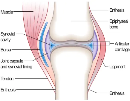
As seen in Figure 1.1, the main components of the joint are cartilage, bone,
synovial capsule (fluid and synovial lining) and meniscus 38
. Moveable joints, such as the
knee joint, are lubricated by synovial fluid, which is kept in the joint by the synovial
capsule 38
. The surfaces of the bones that contact each other are perfectly smooth because
they are lined by articular cartilage composed of hyaline cartilage 38
. In addition,
ligaments limit medial, lateral, posterior and anterior movements of the bones involved in
the joint. The ligaments of the knee joint limit unwanted movements and thereby provide
stability. In addition to the menisci (cushioning cartilage disk in between the tibia and
femur) and bursae, the ligaments of the knee also protect the knee capsule 38
. These
Figure 1.1 - Schematic diagram of a typical synovial joint. A knee joint is an example
of a synovial joint. It consists of two opposing bones. The geometry of bones is
congruent. The tibial component is convex whereas the femur end is concave. This
structure allows for movement and stability. The surface of the bones is covered by
articular cartilage. The elasticity of the articular cartilage allows for load distribution. The
knee joint is encapsulated by a synovial lining (synovium), which is one or two cell layers
thick and is responsible for the production of the joint fluid. This diagram was adapted
The intracapsular ligaments are ligaments that are within the articular capsule of
the synovial joint. The first two ligaments are the anterior cruciate ligaments (ACL) and
the posterior cruciate ligaments (PCL). The ACL connects the lateral condyle of the
femur to the anterior part of the intercondyloid fossa as seen in figure 1.3. The ACL and
the PCL wrap around each other during flexion and unwind in extension. The ACL has
two bundles: the anteromedial and the posterolateral. These bundles are named according
to where they insert into the tibial plateau. The ACL attaches in front of the
intercondyloid eminence of the tibia. The ACL prevents the anterior movement and
medial rotation of the tibia, in relation to the femur.
The PCL connects the medial condyle of the femur to the posterior intercondylar
area 38
. In the average human adult, the PCL is 3.8 cm in length and 1.3 cm in width. This
ligament prevents the posterior movements of the tibia in relation to the femur and also
the anterior movement of the femur on the tibia 38
. It provides 90-95% of the total
restraint required for preventing the posterior displacement of the tibia 38 .
The architecture of the knee joint is very important in effective load distribution.
As seen in Figure 1.2, the structure and flexibility of the articular cartilage and meniscus
of the knee joint, allows for symmetric load distribution. The heavy load demonstration
shows the focus of the weight in the middle of the knee joint. With increased weight
bearing the articular cartilage of the femur and tibia are subject to more pressure and it is
more likely for the patient to experience pain and trauma in their knee. Furthermore,
with increasing load, the articular cartilage of the femur and the tibia make more contact,
but the load is still distributed symmetrically. Some studies suggest that degenerative
Figure 1.2 Comparison of light and heavy load distribution at the knee joint. This
figure shows the distribution of varying loads on the knee joint.The structure of the knee
joint allows for effective load distribution thereby providing stability during gait. In
figure A, only a light load is applied to the knee. In figure B, a heavy load is applied to
the knee. During the heavy loading, the articular cartilage of the femur and tibia make
more contact to distribute the load in an even manner. This figure was adapted from
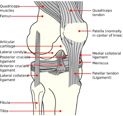
As Figure 1.3 shows, the location of the PCL is near the longitudinal axis of
rotation and it is slightly medial to the center of the knee. Depending on the angle of the
knee, the PCL is more vertical when the knee is extended and it is more horizontal when
the knee is flexed. As the angle of knee flexion increases, there is more tension on the
PCL. That is to say, maximal tension is achieved in full flexion of the knee. Although the
ACL and PCL wrap around one another during flexion (X shape), the PCL takes a more
direct course than the more spiral-shaped ACL fibers 41
. Furthermore, both the ACL and
the PCL stabilize the medial and lateral side of the knee.
Another intracapsular ligament is the transverse ligament, which connects the
anterior portion of the lateral meniscus to the anterior portion of the medial meniscus.
Lastly, the posterior and anterior meniscofemoral ligaments connect the posterior horn of
the lateral meniscus and the lateral surface of the medial femoral condyle.
The extracapsular ligaments are associated with the synovial joint, but are external
to its capsule. The patellar ligament connects the tuberosity of the tibia to the patella. This
ligament is very strong and functions as a cap for the condyles of the knee as well as
supports the patella for mechanical leverage.
The medial collateral ligament (MCL) connects the medial epicondyle of the
femur to the medial tibial condyle 41
. This ligament protects the medial side of the knee
from being bent open when a stress is applied to the lateral side of the knee (valgus
force). The lateral collateral ligament (LCL) connects the lateral epicondyle of the femur
to the head of the fibula. It functions to protect the lateral side of the knee from a medial
Figure 1.3 Schematic diagram of the anterior aspect of the knee joint. Three
components of the knee joint are the femur, the patella and the tibia. The patella can be
seen from the anterior aspect of the knee. When the knee is extended, the PCL becomes
more vertical and when the knee is flexed the PCL becomes more horizontal. The ACL
can also be seen from the posterior aspect, as it wraps around the PCL during flexion and
unwinds during extension. The medial and lateral collateral ligaments prevent medial and
lateral movements that would compromise the stability of the knee joint. This image was
adapted by permission from Wikimedia.com (http://en.wikipedia.org/wiki/
The last two extracapsular ligaments are on the dorsal side of the knee. The
oblique popliteal ligament connects the upper margin of the intercondyloid fossa and the
posterior surface of the femur to the posterior margin of the head of the tibia. The arcuate
popliteal ligament is “Y” shaped and is attached to the fibular head 41. It has two
insertions on the intercondylar area of the tibia and the lateral epicondyle of the femur.
1.1.1 Flexion and extension of the knee
The knee allows not only for flexion and extension of the leg, but also for slight
rotation medially and laterally in the flexed position. In a healthy subject, when the knee
is fully straight we have zero degrees of flexion. As the knee is bent and the heel of the
foot touches the buttocks we have 180 degrees of flexion and zero degrees of extension. It
is important to discuss which ligaments are involved in this range of motion.
In the fully extended position, the medial and lateral collateral ligaments are tense
and stretched (taut). The medial collateral ligament is completely unfolded in the
extended position. Before the knee is fully extended, the knee rotates medially by five
degrees, which is caused by the lateral rotation of the tibia. This is called the obligatory
terminal rotation41
. Stretching of the anterior cruciate ligament causes this rotation. The
anterior and posterior cruciate ligaments are slightly unwound in the extended position,
but the lateral ligaments become tense.
1.2 – Histology of joint tissues
The knee joint is made up of a number of connective tissues. On a macroscopic
and organs. There are two main types of soft connective tissues: loose connective tissues
and dense connective tissues 39
. Loose connective tissues include adipose and material
located between cells throughout much of the body also known as the extracellular matrix
(ECM). The main components of the ECM are proteoglycans, which are large
macropolymers consisting of a protein core that is attached to many carbohydrate chains
called glycosaminoglycans (GAGs). These chains are highly hydrophilic and are
surrounded by water 39
. This gives the tissue its thickness and firmness. Dense connective
tissue or dense fibrous tissue has fibers as its main matrix element. It is mainly composed
of type I collagen. Dense connective tissue makes up the structure of tendons and
ligaments. Tendons attach skeletal muscles to bone, whereas ligaments attach bones to
bones. Consequently, ligaments have more elastic fibers than tendons.
The connective tissue components of the musculoskeletal system come from
mesenchymal cells developmentally. The bone matrix is synthesized by osteoblasts;
cartilage by chondrocytes; and ligaments and tendons by fibroblasts 39
. The fibroblast has
the ability to secrete fibrous material such as collagen and elastin 39
. Collagen is a strong
fibrous material that is found in connective tissues, including tendons, ligaments,
cartilage, bone, intervertebral disks, blood vessels, skin and the cornea. Elastin is a
protein in some connective tissues, and it functions to allow the connective tissue to
stretch and regain its shape (elasticity). Adipocytes are fat cells, which store fat (energy).
Chondrocytes produce and maintain the cartilaginous matrix. Depending on location in
the tissue and the type of cartilage, the organization of chondrocytes can differ. Cartilage
is a strong, but very flexible extracellular tissue. The three main types of cartilage are
hyaline cartilage, elastic cartilage and fibrous cartilage 39
cartilage are that it is avascular, contains no nerves, has few cell-cell contacts, and the
volume of extracellular matrix is much greater than the volume of cells.
[i] Hyaline cartilage
Hyaline cartilage is strong, slightly flexible and is the most abundant type of
cartilage in the human body 39
. For example, the larynx and the trachea are reinforced by
hyaline cartilage. In the embryo, the hyaline cartilage develops from loose connective
tissue when the oxygen supply is low; whereas, bone develops from the same tissue when
oxygen is abundant. In the fetus, the hyaline cartilage forms much of the skeleton and is
replaced by bone during endochondral bone formation. After birth and up to the end of
adolescence, hyaline cartilage makes up the epiphyseal growth plates, which control the
growth and shape of long bones. Hyaline cartilage also lines the articular surfaces of
synovial joints, where it acts as a self-lubricating shock absorber and provides a low
friction surface for movement. Articular cartilage lacks blood vessels and, therefore, can
be irreversibly damaged by overuse or infection. Inflammation of the joint leads to
destruction of the articular cartilage, which causes pain and stiffness. Damaged hyaline
cartilage can only be repaired to a limited extent 39 .
Except where hyaline cartilage serves as articular cartilage and is exposed to
synovial fluid, it is enclosed by the perichondrium, which is essential for the growth of
cartilage. The perichondrium is a layer of dense irregular connective tissue, which
surrounds the cartilage of developing bone. It consists of two separate layers: an outer
fibrous layer and inner chondrogenic layer. The fibrous layer contains fibroblasts, which
form chondroblasts or chondrocytes. Perichondrium can be found around elastic cartilage
and hyaline cartilage. Thus, the perichondrium is thought to have a significant role in the
growth and repair of cartilage. Once vascularized, the perichondrium becomes the
periosteum 39.
Chondrocytes in hyaline cartilage are grouped in pairs of four to six cells known
as an isogenous nest, because they are the progeny of a single chondrocyte during
development. The matrix is composed of type II collagen fibers, water and proteoglycans
(PGs). Chondrocytes of hyaline cartilage synthesize and maintain all components of the
extracellular matrix. The majority of the matrix is water and it is the water and inorganic
salts that give cartilage its resilience and lubricating capabilities. PGs in the matrix are
negatively charged and thus attract large amounts of positively charged ions and water.
The remaining constituents are structural macromolecules: collagens, PGs and
non-collagenous proteins.
Roughly 90-95% of the collagen in hyaline cartilage is type II collagen which
provides a fibrillar meshwork that gives hyaline cartilage its tensile strength. PGs in the
matrix are negatively charged and hold large amounts of positively charged water ions.
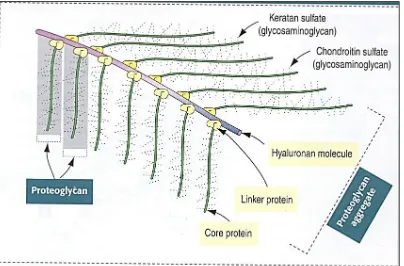
Figure 1.4 shows that PGs are composed of core protein with complex carbohydrates
called glycosaminoglycans (GAGs) that radiate from the core. These GAGs consist of
repeating negatively charged disaccharide units of various lengths, which may or may not
be sulphated. The sulphated PGs such as chondroitin sulphate, dermatan sulphate and
keratin sulphate, attach noncovalently to hyaluronic acid in order to form large PG
aggregates known as aggrecans. The interaction among water, the collagen fibril network
Figure 1.4 - Schematic diagram of a proteoglycan complex. The proteoglycans are
responsible for the integrity of the cartilaginous material in articular joints. The main
components of the extracellular matrix (ECM) are proteoglycans, which are large
polymers consisting of a protein core with many attached carbohydrate chains. The
carbohydrate chains are called glycosaminoglycans (GAGs). There are multiple types of
GAGs, which include keratin sulphate and chondroitin sulphate, and are extremely
important for the integrity of the ECM. Like all carbohydrates, they are very hydrophilic.
Hence, in the body, they are always surrounded by a large amount of water (water of
solvation). This gives tissues their characteristics thickness and firmness. Dehydration
results in decreased hydration of the ECM and thus jeopardizes its integrity. This
diagram was adapted with permission from “Histology and cell biology” by Abraham L.
[ii] – Elastic cartilage
Elastic cartilage possesses a matrix dominated by elastic fibers 39
. The matrix
contains a small number of type II collagen fibers that are covered by proteoglycans,
combined with more abundant elastic fibers. Elastic cartilage is also enclosed by a
perichondrium. Physiologically, elastic cartilage does not calcify with age 39
. Elastic
cartilage and hyaline cartilage are found in structures such as the epiglottis and the outer
ear, where more flexibility is required than hyaline cartilage can provide. In particular,
elastic cartilage contains elastin.
[iii] – Fibrous cartilage
Fibrous cartilage is very rigid and is found in places where structural strength is
important such as the pubic symphysis and the intervertebral disks of the spinal column 39 .
It is a mixture of dense regular connective tissue and hyaline cartilage. It combines the
tensile strength, firmness and durability of tendon with the resistance to compression of
cartilage. In contrast to other types of cartilage, fibrocartilage lacks perichondrium.
Fibrocartilage initially forms from dense connective tissue that is rich in fibroblasts, some
of which differentiate into chondrocytes. Therefore, a mixture of chondrocytes and
fibroblasts is characteristic of this tissue 39 .
1.3. - The structure and development of bone
The osteocyte or bone cell has many functions, which include signal transmission
39
. In particular, osteocytes are thought to be mechanosensory cells that regulate the
which includes type I collagen and proteoglycans. Osteoblasts also express alkaline
phosphatase, which helps to mineralize the osteoid via production of inorganic phosphate
leading to formation of calcium phosphate crystal in the form of hydroxyapatite. Hence
osteoblasts are the bone making cells. Osteoclasts on the other hand, resorb bone and
mineralized cartilage matrix 39 .
Bone is collectively made of spongy/cancellous bone as well as compact/cortical
bone. The human musculoskeletal system is made of long bones (e.g., the femur in the
thigh), short bones (e.g., carpal bones in the wrist), flat bones (e.g., calvaria of the skull)
and irregular bones (e.g., spinal vertebra). The bones of the limbs are long bones and they
are important for support and movement. The main shaft of the long bone is called the
diaphysis. This part is a tube composed of only compact bone. The bone marrow is a
non-bony material found in the shafts of long bones and in the pores of spongy bones. The
flared ends of a long bone are called the epiphyses. Long bones are the main bony
component involved in the knee joint.
1.3.1 - Overview of bone formation
Bone formation is comprised of a series of complex and simultaneous processes,
which include: cell migration, mitosis, differentiation, synthesis and secretion of ECM,
mineralization, and resorption. Bones are formed by either intramembranous or
endochondral ossification. Bone formation can be categorized either by bone forming on
a cartilage (endochondral model) or not (intramembranous model) 39.
Intramembranous ossification occurs in areas of ordinary mesenchyme where
of the cranium, part of the mandible and the clavicles develop in this way. Endochondral
ossification is the method by which long bones and bones in the vertebral column, ribs
and pelvis develop 39. Here, mesenchymal cells differentiate into chondrocytes. A
cartilage template is first formed and then modified to facilitate osteogenesis. After bone
forms, it remains dynamic throughout life to allow for remodelling in response to changes
in functional demands and to serve as a source of mineral ions for homeostasis. Although
this study focused on long bones, it is important to differentiate the two types of bone
formation. Table 1.1 summarizes the two processes side by side. It is primarily
endochondral bone formation that leads to formation of the long bones of the knee joint.
[i] Intramembranous bone formation
Intramembranous bone formation begins during gestation when mesenchymal
cells move to sites of richly perfused/vascularized connective tissue and differentiate into
osteoblasts39
. The osteoblasts secrete osteoid, which consists of Type I collagen fibers,
osteopontin, osteonectin, osteocalcin, fibronectin and a matrix of proteoglycans.
Osteoblasts also secrete alkaline phosphatase, which is an enzyme that facilitates the
mineralization of the osteoid by precipitation of inorganic calcium phosphate salts. That
is to say, hydroxyapatite is the dominant mineral of the bony matrix. During the
ossification process, osteoblasts become trapped and they eventually become osteocytes
in small lacunae. The osteocytes are connected to one another via processes that lie in
small channels called canaliculi. Small islands or trabeculae of newly formed bone are
continue to produce bony matrix. Trabeculae thicken and later merge to produce a
three-dimensional lattice structure of spongy bone39 .
Then the osteoclasts eventually kick in to continue the remodeling process.
Eventually, the deposition of concentric layer or lamellae of matrix around the trapped
blood vessels forms the functional units known as osteons. Lastly, the layer of specialized
connective tissue around the developing bone becomes the periosteum39 .
[ii] Endochondral bone formation
Endochondral bone formation begins with hyaline cartilage replicas of future adult
bone39. Once developed from mesenchyme, the cartilage templates take the shape of the
future bone. The first of the two ossification centers (POC) appears in the diaphysis/shaft
of the cartilage template. Bone is laid down directly by connective tissue perichondrium
of the cartilage. Eventually the perichondrium becomes the periosteum. Once deep into
the new bone, the chondrocytes hypertrophy and die.
From the periosteum blood vessels (periosteal bud) span the diaphysis interior and
bring in mesenchymal and osteoprogenitor cells. At this point, the cartilage in the center
starts to erode and the primitive marrow cavity starts to form. Incoming blood vessels
carry primitive bone marrow cells. The proliferation of remaining chondrocytes causes
the two ends or epiphysis to grow longitudinally. That is to say, the interior diameter of
the diaphysis remains constant. This way, the chondrocytes are arranged in columns and
appear as two fronts on both sides of the central region39.
Eventually the epiphyseal growth plates are formed at the junction between epiphysis
diaphysis growth. After birth and continuing into puberty, ossification centers appear in
the two epiphyses of long bones, until adolescence is reached when the growth plates
eventually close and growth ceases39.
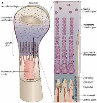
1.3.1.1 - Structure and function of the growth plate
A cartilaginous growth plate in a typical long bone provides temporary
scaffolding on which new bone is laid down, permitting longitudinal bone growth. The
plate stimulates appositional growth of hyaline cartilage at the end facing the epiphysis.
Cartilage destruction in lower regions and replacement with primary spongy bone in the
deepest region follows. Figure 1.5 shows the five distinct transverse zones of the growth
plate reflecting the sequence of events in endochondral ossification.
1. The reserve zone (resting cartilage) consists of small clusters of flattened, rounded
and randomly arranged quiescent chondrocytes 39 .
2. In the proliferation zone, chondrocytes undergo rapid mitosis as a result of stimulation
by growth hormone. No lateral cellular displacement occurs, and the new cells are
stacked into columns. These cells are parallel to the long axis of the growing bone 39 .
3. In the zone of maturation and hypertrophy, mitosis stops. The cells in this region
produce matrix and then enlarge followed by buildup of lipids, glycogen and alkaline
phosphatase 39 .
4. In the zone of provisional calcification, spicules of calcified cartilage matrix are left
vascularized primary marrow extends into the newly opened spaces and osteoblasts
differentiate from mesenchymal cells in the bone marrow 39 .
5. In the ossification zone, the osteoprogenitor cells differentiate into osteoblasts.
Osteoblasts secrete osteoid, which becomes mineralized (calcified), on the surface of
calcified cartilage. Eventually this is followed by the remodeling of the calcified
cartilage/calcified bone complex 39 .
The metaphysis, at the distal end of the growth plate between the epiphysis and
diaphysis is formed by slender calcified cartilage spicules 38
. These project from the
epiphyseal growth plate into the marrow cavity of the diaphysis. The metaphysis is also
filled by many capillary loops that transport cells that become osteoblasts, which deposit
bony matrix on calcified cartilage remnants. The metaphysis is divided into two
functional regions. The upper one or the primary spongiosa contains capillary sprouts
between missed spicules, which consist of a core of calcified cartilage covered by a thin
layer of newly formed bone 38
. The lower end of the metaphysis is the secondary
spongiosa in which the calcified cartilage in the mixed spicules is ultimately resorbed by
osteoclasts followed by secondary remodeling of spongy bone 38
. This results in the
lengths of the spicules remaining almost constant as the marrow cavity volume gradually
increases. Growth in length continues until adulthood under the influence of growth
hormone, thyroid hormone, parathyroid hormone and androgens 38
. At skeletal maturity,
epiphyseal growth stops and the epiphysis and diaphysis join.
Osteoblasts synthesize and secrete osteoid, which consists mainly of type I
collagen and non-collagenous glycoproteins such as osteocalcin, osteopontin, osteonectin
and fibronectin 39
. These cells also synthesize alkaline phosphatase, a cell surface protein
that promotes mineralization. Long branched cytoplasmic processes extend from cell
bodies at the side where bone matrix is formed and penetrate deeply into osteoid to
communicate with osteocyte processes. It is reported that gap junctions between the cells
probably play a role in signal transmission 39
. Osteoblasts respond to parathyroid
hormone, estrogen, progesterone, and a number of other hormones and paracrine factors.
Osteocytes
Osteocytes are essentially mature osteoblasts 39
. They have a high nucleus to
cytoplasm ratio and relatively few cytoplasmic organelles. They reside in lacunae within
the bone matrix. They have many cytoplasmic processes that extend into thin channels or
canaliculi in the mineralized matrix 39
. The processes of one cell are linked to others via
gap junctions. Extracellular fluid in canaliculi permits transfer of molecules, oxygen and
nutrients by diffusion. Some authors have proposed that one of the main jobs of
osteocytes is to actively maintain bone matrix by exchange of calcium ions and other
Figure 1.5 – Schematic diagram of the epiphyseal plate. The first zone is the resting
zone (zone of reserve cartilage) in which the precursor cells are flattened, randomly
arranged and inactive. In the proliferation zone, the chondrocytes become active and
rapidly undergo mitosis when stimulated by growth hormone. The new daughter cells
grow in columns parallel to the long axis (vertical axis) of the future bone. Once in the
hypertrophic zone, mitosis stops and the cells enlarge and accumulate glycogen, lipids
and alkaline phosphatase. This will lead to the mineralized zone where eventually
osteoprogenitor cells develop into osteoblasts and secrete osteoid. This osteoid becomes
mineralized into bone. This figure was adapted with permission, from the nature
Osteoclasts
Actively resorbing osteoclasts reside in or near surface cavities known as
Howship’s lacunae 39
. A characteristic of osteoclasts is their ruffled borders (extensively
folded membrane), which facilitates bone resorption by increasing surface area. The
border membrane contains proton pumps that keep pH low (acidic) in the resorption
cavity. The inorganic minerals of the matrix are dissolved when H+
ions are transported
into the extracellular space. In addition, lysosomes release hydrolytic enzymes (e.g.
collagenase) into the resorption cavity to degrade the organic components of the matrix.
Osteoclasts are capable of resorbing bone and calcified cartilage matrix.
1.3.2 - Bone Healing
Healing of bones can occur by both intramembranous and endochondral
ossification39
. The repair process starts as an inflammatory phase, followed by a
reparative phase. The repair process ends with the bone-remodeling phase 39
. During the
reactive or inflammatory phase, there is blood clot formation and vasoconstriction to stop
the bleeding. This is followed by apoptosis of the surrounding cells, but the mesenchymal
cells survive and replicate 42
. A loose aggregate of new cells are formed, intertwined with
small blood vessels (granulation tissue). During the reparative phase, the periosteal cells
differentiate into chondroblasts to produce hyaline cartilage or osteoblasts to form woven
bone. The hyaline cartilage and woven bone grow in size until the fracture gap is bridged.
The woven bone and hyaline cartilage (collectively known as fracture callus) are
eventually replaced by lamellar bone (higher strength) during the remodeling phase, via
cartilage of the fracture callus are replaced by lamellar bone 42
. This ensures that the new
repaired bone is close to the bone’s original strength. In the last stage, the bone is
remodeled 39
. This is accomplished first via the resorption by osteoclasts and then the
osteoblasts deposit bone within the resorption pits. Eventually the callus is fully
remodeled to the bone’s original shape and strength 42 .
1.4 - Degenerative Osteoarthritis
Osteoarthritis (OA) is a degenerative disease of the joint that can be caused by
both systemic and local factors20
. Figure 1.6 shows the cartilage erosion as well as
asymmetric joint space narrowing. Some of the risk factors for osteoarthritis include
advanced age, female sex, genetics, obesity, and occupations involving overuse or trauma
9, 20
. OA is characterized by pain that typically worsens with weight bearing and activity,
and improves with rest 32
. There is tenderness on palpation as well as bony enlargement of
the involved joint, crepitus on motion, and limitation of joint movement 20 .
OA has a large impact on the Canadian population. It affects three million (or one
in ten) Canadians 4, 7. It is also associated with significant costs to the Canadian economy,
directly through drugs and use of healthcare resources and indirectly through lost
employment time and costs of informal caregiving 8
. On average, OA has an insidious
onset 20
. At a younger age, males are more affected than females; whereas, at an older
age, women are more affected than men 4
. Without treatment, people with OA are left
with significant pain and loss of health-related quality of life 9
. This study focuses on the
Figure 1.6 – The anterior view of the knee with severe degenerative osteoarthritis.
This is a schematic diagram (A) as well as an X-ray (B) of a left knee with OA. On the
left, there is erosion of the cartilage and patches of bone exposed. There is also formation
of bone spurs (osteophytes). In panel B, there is asymmetric joint space narrowing
(presented by the black arrow) as well as sclerosis of the subchondral bone (presented by
the black arrow), which can be seen as parts of the bone that appear more white compared
to the surrounding bone. Subchondral sclerosis associated with OA is localized and can
be caused by injuries that compress the bone. Figure A was adapted with permission
from Nucleus Medical Media F-160
and Figure B was adapted with permission from
Wikimedia (http://en.wikipedia.org/wiki/File:Osteoarthritis_
left_knee.jpg) originally made by Dr. James Heilman F-166 .
B
It is important to note the differences of OA as compared to osteoporosis 30 .
Osteoporosis is a systemic skeletal disease characterized by low bone density and
microarchitectural deterioration of bone tissue with a gradual increase in bone fragility 30 .
OA involves deformation and degradation of joints, including articular cartilage and
subchondral bone. Essentially with OA, there is a degradation of this macromolecular
framework. There is a disruption of collagen through an increase in levels of proteinases
and trauma37 .
In OA, the ECM is initially disrupted through an increase in metalloproteinase
(MMPs), which attracts more water, and it is this abnormality that disorganizes the
meshwork 37
. The increase in edema decreases the elasticity of the cartilage and joint and
causes a loss in lubrication, making simple movements extremely difficult.
The clinical symptoms of degenerative osteoarthritis are joint pain, some swelling,
painful movements, affected small and large joints, both unilateral and bilateral, and
deformity in some joints. This deformity causes a loss of function. These symptoms have
a gradual onset. The main clinical diagnosis of OA is through clinical presentation and
physical examination. Medical imaging may show asymmetric joint space narrowing,
subchondral cysts, sclerosis and osteophyte formation 37 .
Figure 1.7 shows that OA can be subdivided into three stages. In the first stage,
there will be micro cracks in the articular cartilage and there will be areas of chondrocyte
loss, alternating with chondrocyte proliferation. In the second stage, there may be
vascularization of the knee joint, which is not normally evident. There will also be
chondrocyte death and matrix degradation. In the third stage, there will also be synovial
Figure 1.7 – Progression of degenerative osteoarthritis. This is a schematic of the three
stages of OA. Figure A shows a normal healthy synovial joint. In the first stage (B), there
is ECM edema. The cartilage layer is affected and there is a loss of cartilage smoothness.
In the second stage (C), the micro cracks from the first stage deepen with fissure
formation. There are chondrocyte clusters around the fissures. In the third stage (D), the
fissures from the second stage may eventually lead to breaking off of cartilage pieces,
known as loose bodies. More importantly, the subchondral bone is exposed. Eventually
osteophytes surround the area leading to sclerosis. Subchondral ‘cysts’ may form in the
bone. This image was adapted with permission F-161 from thelukinskispineclinic.com
information website.
A
D C
It is important to clarify what is expected with normal aging versus osteoarthritis.
In normal aging, the water content of the cartilage decreases, which decreases the
firmness 43
. There is also a decrease in synthetic activity, which leads to less proteoglycan
content, length and synthesis. Although this is normal, this will lead to decreased strength
and firmness of the cartilage 43
. One important factor is that, with normal aging, the
amount of collagen in the cartilage does not change 43
. On the other hand with OA there is
a substantial reduction in collagen in the cartilage. As well, there is a reduction in
proteoglycan amount. In OA, there is a substantial increase in matrix metalloproteinases
(MMPs), which is not evident in normal aging. Although MMPs have natural roles in the
body, their overproduction leads to destruction of collagen. Eventually deformation of the
joint may result 43 .
1.4.1 – Treatments for osteoarthritis
The main goals for treatment of OA are pain reduction, improved function,
changes in the disease outcome, low cost, and minimal side effects. There are three broad
categories of treatment for OA: non-pharmacological, pharmacological and surgical. The
three treatment categories are described based on the clinical practice guidelines provided
by Osteoarthritis Research Society International (OARSI) recommendations, the
American Academy of Orthopaedic Surgeons (AAOS) guidelines and the National
Institute of Health and Clinical Excellence (NICE) 75-78 .
All three guidelines recommend that a patient diagnosed with osteoarthritis,
should receive education and be taught self-management techniques for treatment75-78 . The
non-pharmacological approaches recommended by the three guidelines are mainly
physical and occupational therapy. Patients are educated on the options available and are
advised to wear proper footwear and use assistive devices when needed. The three
guidelines also recommend strength training as well as weight loss exercises75-78 . In
physiotherapy, patients are given muscle strengthening, aerobic, aquatic and range of
motion exercises. In particular manual therapy includes passive or active movement
techniques that use applied force to improve the mobility of the affected joints. Some of
the techniques that the physiotherapist might use are soft tissue massage, stretching and
passive movements, mobilization and manipulation of the soft tissue and joints75-78 . As
recommended by the OARSI guidelines76
, in occupational therapy, patients are given
orthotics, splints and braces or, more commonly, canes and walkers. The NICE guidelines
also show evidence that walking aids can help can improve stride length and cadence and
thus alleviate walking for knee OA patients78
. For heavier patients (i.e. BMI>25) all three
guidelines75-78 strongly recommend some kind of weight loss program to help with the
functional disability associated with knee OA.
1.4.1.2 - Pharmacological treatments for OA
The pharmacological approach includes oral and topical medications that reduce
pain and inflammation34
. For example, all three guidelines recommend acetaminophen as
an effective oral analgesic for pain relief 75-78
. Intra-articular medications such as
pharmacological treatments that change the course of the disease(i.e. disease modifying
drugs) 28
. The available medications serve mainly to reduce pain and swelling 28
. The most
common pharmacological treatments mentioned by the three guidelines are analgesics
(Tylenol and opioids), Non-Steroidal Anti-Inflammatory Drugs (topical NSAIDs and
oral NSAIDs), glucosamine/chondroitin, articular hyaluronic acid, and
intra-articular steroids to reduce inflammation.
1.4.1.3 - Surgical treatments for OA
Most patients with OA are referred for surgical treatment when other treatments
are ineffective and function is severely impaired. Surgical treatment is usually the last
resort. The most common surgical treatments are arthroscopy and debridement,
osteotomy, ligament repair, joint reconstruction, grafting and total joint replacement. This
study focuses on total joint replacement as a surgical treatment for OA of the knee joint.
In particular, for a primary unilateral total joint replacement, the articular surfaces of a
single knee joint are replaced for the first time 12 .
1.5 – Total Knee Joint Arthroplasty
Total joint arthroplasty remains the top treatment for OA of the knee.5, 15 With
high success rates, total knee arthroplasty (TKA) remains the last resort for patients
suffering from OA. The components used in a total joint replacement are designed to
enable the artificial joint to move like a normal joint. The prosthesis used in TKAs, is
generally composed of two parts: a metal piece that fits closely into a matching sturdy
cobalt and chrome, and titanium. These materials are strong, dense and appear as very
opaque in X-rays, when compared to the surrounding bone. The plastic material
(ultra-high molecular weight polyethylene) is very durable and is resistant to wear, but is not
easily detectable in X-rays.
For the component fixtures, the femur, tibia and patella can be cemented.
Sometimes, the surgeon might choose to cement only the femur and tibia as seen in
Figure 1.8. Depending on the preoperative stage of the patient, it may not be necessary to
cement all three components for fixation of the prosthesis.
1.5.1 - Posterior cruciate ligament (PCL) prosthesis design
Prosthetic design for TKA has evolved into designs that either preserves the
posterior cruciate ligament (PCL retaining) and those in which the ligament is sacrificed
(PCL sacrificing). In patients with a functional PCL, the decision on which design is
chosen depends largely on the preference of the surgeon, but there are many reasons why
the PCL may be sacrificed or retained. Each option has its own advantages and
disadvantages 1. In either case, the implants used have specific geometry to account for
the presence or absence of the ligament.
One of the most important reasons for retaining the PCL is to aid in
proprioception 12
, which is body’s sense of limb position based on stretch receptors or
mechanoreceptors in ligaments and the amount of force employed in movement. This
Figure 1.8– Schematic diagram of the left knee prosthesis from the anterior and
medial point of view. On the left, the anterior view of the knee is shown. The femur and
the tibial component have been cemented in this knee. On the right, the medial view of
the left knee is shown. This image was adapted with permissionF-160
from the Nucleus
The other reason is that the ligament can promote more normal front to back knee
motion. This is important for helping knee flexion. The implant used in these cases
(cruciate retaining) is specifically designed to allow for the presence of the PCL.
For the PCL to perform its function, it must be in a healthy condition. The PCL
must be properly balanced/fitted after the knee prosthesis has been oriented and implanted
by the surgeon. This can sometimes be difficult and, thus, the result is less predictable
from patient to patient. One possible disadvantage of keeping the PCL is that the medial
collateral ligament (MCL) and lateral collateral ligament (LCL) must be properly
balanced as well. This is more difficult when the PCL is retained. Also, tension in the
PCL created by surgery increases the tension on MCL and LCL and other structures 1, 12, 25 .
Figure 1.9 shows an X-ray of a PCL retaining prosthesis.
On the other hand, when the PCL is sacrificed, special geometry in the implant
components substitutes for the function of the ligament 25, 33
. When the surgeon removes
the PCL, the space between the prepared femur and tibia become larger, making access
easier. In the meantime, the MCL and LCL tension created by PCL retention is reduced.
Furthermore, since the ligament is now replaced by very consistent implant geometry,
results tend to be similar among patients. Figure 1.10 is a sample of PCL sacrificing
prosthesis.
For PCL sacrificing prosthesis, the design must provide anteroposterior stability.
This is accomplished by having congruent geometry in flexion in order to avoid sagittal
instability. This geometry allows for uniaxial flexion with less flexion arc and produces
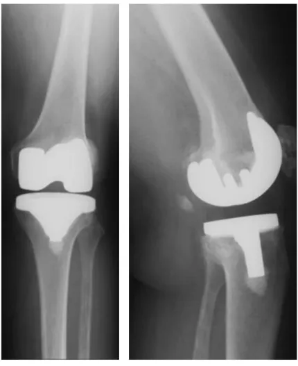
Figure 1.9 – X ray of PCL retaining prosthesis. This is x ray image of the
anteroposterior (A) and lateral (B) view of the knee of a 55-year-old woman who received
a PCL retaining prosthesis. This picture was adapted from an article by Kolisek et al.,
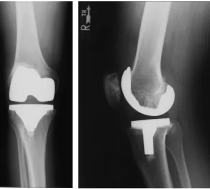
Figure 1.10 – X ray of PCL sacrificing prosthesis. This is an anteroposterior (A) and
lateral (B) radiograph of a knee of a 52-year-old woman who received a PCL sacrificing
prosthesis. This picture was adapted from an article by Kolisek et al., 2009 with
For PCL retaining prosthesis, by keeping the PCL, along with low constraint on
the tibial component, allows for normal roll back of the femur on the tibia with knee
flexion. This roll back allows for a better quadriceps lever arm than the PCL sacrificing
prosthesis. It also allows for more efficient use of the extensor muscles, thereby allowing
the patient to climb stairs easily 63 .
1.6 - Ontario Joint Replacement Registry (OJRR)
There have been numerous registries created that were designed to capture the
preoperative and postoperative states of patients undergoing joint arthroplasty. A few
examples include the American Joint Replacement Registry (AJRR) in the United States,
National Joint Registry (NJR) in United Kingdom and the Swedish knee arthroplasty
register in Sweden. In particular, the Ontario Joint Replacement Registry (OJRR)
captured many variables, which are of prime importance for researchers and health care
practitioners interested in knee arthroplasty outcomes. The OJRR collected data
prospectively from participating orthopaedic surgeons in southwestern Ontario with about
two million people and an ethnically diverse sample. This patient sample is representative
of Canada’s ethnically diverse population. The surgical outcomes reported by the OJRR
are similar to those found in the peer-reviewed literature9
. Furthermore, key demographic
variables in the OJRR database show similar distributions as those found in the Canadian
joint replacement registry9
. Thus the OJRR database has high external validity (whether
our measurement corresponds to the real world). The internal validity of this database is
observational cohort study. Higher internal validity could have been achieved through a
randomized cohort.
The OJRR data are part of the Canadian Joint Replacement Registry (CJRR).
Data from the CJRR can be used for quality improvement, research and statistical
analyses. Conclusions drawn from the analyses can be used as general information to
inform policy and clinical treatment planning and decisions affecting specific patients.
Like the CJRR, the OJRR provides information on distribution and frequency, and
characteristics of total knee replacement surgeries, including the effectiveness of the
surgical methods, long-term outcomes and how to minimize post-operative
complications. The information gathered is intended to help determine which approaches
are effective in specific circumstances and to determine which patients can most benefit
from total knee replacement.
Since 2003, 67% of Ontario orthopaedic surgeons participated in the OJRR. OJRR
is a robust dataset and it is generally applicable to the Canadian healthcare system.
According to CIHI, orthopaedic surgeons in Ontario participated in the CJRR through the
OJRR. Meanwhile, surgeons from other provinces submit data directly to CIHI. Surgical
information in Ontario was collected via hand-held computers in the operating room. The
data was sent electronically to CIHI by the OJRR.
Given the nature of OJRR, the information from the CJRR will help
evidence-based practice and help inform guidelines for total knee replacements in Canada. There
were 496 variables measured in the OJRR database. For the type of variables collected,
please refer to appendix D. Following an extensive literature review (summarized below),
predict surgical outcomes. Two main components measured in the OJRR are
patient-reported outcomes: 1) the Western Ontario and McMaster Universities Osteoarthritis
Index (WOMAC) questionnaire; and 2) the patient satisfaction questionnaire. The
variables included in these questionnaires are extremely important, as they include
measurement of health related quality of life (HRQoL) and functional outcome.
1.7 – Patient-Reported Outcomes
Overall, there are multiple factors responsible for the success of TKA surgery (i.e.
benefit to the patient) 7, 11
. Although these surgeries have different success rates for men
and women, these operations restore function and increase mobility in both men and
women as measured by validated measurements of self-reported functional outcome 29 .
Many studies measure functional outcome/recovery to assess the results of TKA surgeries
8, 11, 29
. Clinically, one method of measuring functional outcome is through patient-reported
outcomes such as the WOMAC. Functional recovery is multifactorial and it correlates
with age, sex, preoperative level of pain and function, number and types of comorbidity,
site of arthroplasty, body mass index (BMI), self-efficacy, patient perceptions concerning
the outcome of surgery and muscle strength (unadjusted models) 11, 12, 14, 16, 17, 21-23, 31, 35, 36 .
Patient-reported outcomes or PROs is a general term given to self-reports by the
patient. Within the last 30 years, enhanced assessments such as health-related quality of
life (HRQoL) and functional outcomes have been used to assess patients’ physical,
mental and social conditions before and after knee arthroplasty surgery21
. Recently,
patient satisfaction forms have also been used, as it has been recognized that surgical
HRQoL evaluation tools to allow health researchers to measure patient health in a
standardized fashion. Clinically, one method of measuring functional outcome is through
patient-reported outcomes such as the WOMAC2, 12
. The WOMAC has three domains
comprising of function, pain and stiffness. In the OJRR, patient-reported outcomes were
collected through WOMAC and postoperative patient satisfaction 8 .
1.8 - Variables of interest for this study
Preoperative predictors of functional recovery are important for patients,
physicians and therapists. Based on a literature review of several studies, we have
provided a list of variables of interest that have been studied for their association with
functional recovery: age, sex, BMI, dependency for activities of daily living (ADL),
living alone status, preoperative WOMAC scores, preoperative comorbidities, site of
arthroplasty, joint deformities and patient expectations regarding the outcome of surgery
11, 12, 14, 16, 17, 21-23, 31, 35, 36
. Below, these variables will be explained in more detail for their
significance in knee arthroplasty.
One important group of variables mentioned above is joint deformities. This term
refers to any misalignment of the knee joint axes. Many studies have looked at varus and
valgus knee deformities. As part of their assessments, these studies have also looked at
increased flexion contracture and reduced flexion range of motion. The valgus deformity,
varus deformity, flexion contracture and flexion range of motion will be explained in
more detail below.
Although there is a hesitance to use total knee arthroplasty on very old patients,
age by itself is not a factor that affects the outcome of surgery and should not be the
limiting factor when considering who should receive this surgery. Jones et al.21
showed
that there are no significant age-related differences in WOMAC pain, function or quality
of life measure pre-operatively or post-operatively (6 months). This study also pointed
out that older patients are more likely to live alone and to be transferred to a rehabilitation
facility. In addition, older patients are more likely to have more pre-operative
comorbidities and depend on others for activities of daily living. Although age alone is
not associated with changes in WOMAC score, some studies 8,9
have chosen to control for
age when there were age-related independent variables in their regression model. It is
even more important to control for age when there is a high variance in the patient age
distribution. Therefore, in our study, we included age as a covariate. The age of the
patient was recorded on the day of the surgery.
Body mass index (BMI)
Body mass index (BMI) is defined as the individual’s weight (kg) divided by the
square of his or her height (m2
). The universal classifications of BMI are as follows
ranging from lowest to highest: underweight (<18.5 kg/m2
), normal weight (18.5 – 25
kg/m2
), overweight (25-30 kg/m2
), obese class 1 (30-35 kg/m2
), obese class 2 (35-40
kg/m2
), obese class 3 (over 40 kg/m2
). Although there is a strong relationship between
obesity and rate of usage of total knee arthroplasty as a measure of OA, many studies
such as Wendelboe et al.35
suggest that there is no evidence of higher risks for knee
receive the same benefit (i.e. change from surgery); but the absolute levels of
self-reported function are lower pre-operatively and lower post-operatively, compared to those
with lower BMI. Wendelboe et al.35
suggest that maintaining ideal weight may prevent
knee replacement due to OA. The BMI of the patient was captured on our study. The
weight and height of the patient was recorded on the day of the surgery, for the
calculation of the BMI.
Gender differences
According to the work of Kennedy et al. 24
, there are substantial pre-operative
differences between men and women. Using functional measures such as the fast
self-paced walk-test, stair climbing, timed up and go, and self-reported measures, Kennedy et
al. 24
, demonstrated that women showed greater disability than men in physical
performance and self-report measures. The works of Ethgen et al. 13
showed that age was
not an obstacle to effective surgery as compared to gender. This team reported that men
benefited more from intervention than did women. Other studies18, 26
show that there are
differences between men and women in physical function and pain, where women
received significant improvement (relative to pre-operative state) in pain scores.
Interestingly, using the Arthritis Impact Measurement Scale (AIMS), Brodie and
Sloman10
showed that men were more mobile than women before and after surgery.
Perhaps this explains the finding that male participants have greater post-operative
WOMAC scores (absolute scores) after surgery with regard to social function, physical
function and pain scores, compared to females 18, 24, 26
. Furthermore, Kennedy et al. 24 have
women. This is related to the finding that men are less likely to delay their knee
replacement surgery. In our study, the gender of the patient was recorded on the decision
day for surgery.
Activities of daily living (ADL)
Muscle atrophy in patients with OA due to long periods of inactivity is an
important factor affecting post-operative functional recovery after TKA. Some studies
suggest that this strength deficit can persist after knee replacement 3, 19
. Muscle atrophy
and worsening muscle strength renders late stage OA patients incapable of accomplishing
activities of daily living on their own. To measure capability of performing ADL, the
OJRR created a dependency variable that was measured pre-operatively. If the patient
checked the dependency box, it meant that the patient was not able to accomplish ADL on
his or her own.
Living alone status
Gandhi et al.17 has shown that patients who live alone are more likely to delay
their knee replacement surgery until an older age. Therefore, they are more likely to have
greater joint pain and dysfunction than those who live with someone else, at the time of
the surgery. Since pre-operative WOMAC is predictive of postoperative outcomes, these
patients are more likely to have poorer one-year outcomes after surgery. This makes
living alone status an important pre-operative predictor of the surgical outcome. The
Patient expectations
Patient expectations are important predictors of post-operative satisfaction9
. In our
study, the expectation variable is measured post-operatively. There are three parts to the
expectation variable. The first part is to assess whether the patient had any expectations
going into the surgery. Then, if the patient did have any expectations, were they met?
Expectations met or not met are known to correlate with patient satisfaction. The patient
also had the opportunity to indicate after the surgery, looking back retrospectively,
whether their expectations going to the surgery were too high, just right or too low.
Patient’s judgment of their pre-operative expectations is important, as patients with very
high expectations were more susceptible to not having their expectations met after the
surgery. Patients should be advised to have realistic expectations going into surgery,
based on their pre-operative diagnostics 27 .
Pre-operative comorbidities
One method of quantifying preoperative comorbidities is the American Society of
Anesthesiologists (ASA) physical status classification system. In Ontario,
anesthesiologists routinely record this value in the patient’s surgical records because the
value recorded impacts their remuneration. This classification system is designed for
assessing the well-being or pre-operative health of the patient. In our study, this was
captured as a four-category variable. Category one corresponds to a patient with normal
health. Category two is a patient with mild systemic disease. Category three is a patient
with severe systemic disease. The fourth category is a patient with severe systemic