RAFAA ISMAEL YAHYA
TISSUE-LIKE P SYSTEM FOR REGION-BASED AND EDGE-BASED IMAGE SEGMENTATIONS
RAFAA ISMAEL YAHYA
A thesis submitted in fulfilment of the requirements for the award of the degree of
Doctor of Philosophy (Computer Science)
Faculty of Computing Universiti Teknologi Malaysia
This thesis is dedicated to:
✓ The living memories of my father, my mother
✓ My Husband and two beautiful daughters (Nadeen and Zahraa)
ACKNOWLEDGEMENT
I thank Almighty Allah SWT for guiding me and giving me the strength to finish
my PhD. There are no words to describe my whole hearted gratitude and appreciation to
my supervisor Prof. Dr. Siti Mariyam Shamsuddin, for her guidance, encouragement,
immense knowledge, and sheer patience.
Also, I would like thank to my co-supervisor Dr. Shafaatunnur Hasan for her
guidnes and supporting, and I would like to expand my thanks to my peers in the Soft
Computing Research Group and UTM Big Data Centre for their invaluable assistance
and advice throughout the course of the research. I would like to express a great deal of
thanks to the UTM and Faculty of Computing for providing a good environment to carry
out this research, and to our professors and staff for their friendly support and help
during this study. I would like to express my deepest gratitude to the ministry of higher
education of Iraq and Al-Mustansiriyah University for their generous financial support
during this study.
Special thanks to Prof. Dr. Salah Ismael Yahya for his supporting and
encouragement. This journey would never have been completed without the incredible
encouragement, unwavering faith and unending support of my father, my mother,
brothers and sisters. I would like to express my extremely profound gratitude to my
family (my husband and my two lovely daughters, (Nadeen and Zahraa)) for their
understanding, prayers, unconditional love, and enormous support throughout.
I would like to express my gratitude to my friends, who have always been there
for me. I would like to take this opportunity to thank those who have contributed therein
ABSTRACT
Membrane Computing (MC), a relatively recent branch of natural computing is an
emerging field in molecular computing. MC aims at abstracting models, called membrane
systems or P systems, which mimic the function and structure of a biological cell. Many
studies have utilized MC in various applications such as image segmentation. Due to the
high computational cost of conventional segmentation techniques, bio-inspired models
including MC may be applicable to tackle this limitation. In this study, tissue-like P systems,
a variant of MC,with sophisticated communication rules were developed to improve
region-based and edge-region-based segmentation algorithms for manual and automatic segmenting of 2D
artificial and real images. Manual segmentation was applied for artificial images, whereas,
the automatic segmentation was applied for artificial and real medical images. The manual
segmentation of 2D artificial images was achieved using four, six and eight adjacency pixel
connectivity relationships, whereas, the automatic segmentation of 2D artificial and real
medical images were achieved using four and eight adjacency pixel connectivity
relationships. Two methods were used to realize the automatic edge-based and region-based
segmentations. The first method is for 2D artificial images using P-lingua linked to Java
Netbeans using the P-linguaCore4 Java Library. The second method is for the 2D real and
real medical images using C# linked to P-linguaCore4 Java library. The results of the second
method demonstrated the ability of the system to automatically segment 2D real and real
medical images with arbitrary sizes and different image formats. The experimental results
statistically proved that the methods markedly outpaced the state-of-the-art methods of 2D
real image segmentation using the same data set. Furthermore, the methods showed better
segmentation accuracy and ability to deal with images of different sizes and types. Extra
efficient results such as reducing the number of rules and computational steps were achieved
for 2D hexagonal artificial images based on Tissue-like P systems. The main contributions of
this study are automatic loading and codifying of the input image as well as automatic
visualization of output images after segmentation. Furthermore, six and eight adjacency pixel
connectivity relationships should be considered for reducing computational steps, number of
ABSTRAK
Pengkomputeran Membran (MC), secara relatifnya adalah cabang terkini pengkomputeran asli dalam bidang baharu pengkomputeran molekul. MC bertujuan untuk
model pengabstrakan, dipanggil sebagai sistem membran atau sistem P, yang meniru fungsi
dan struktur sel biologi. Banyak kajian telah menggunakan MC dalam pelbagai aplikasi seperti pengsegmenan imej. Oleh kerana kos pengkomputeran yang tinggi bagi teknik
pengsegmenan konvensional, model bio-inspirasi termasuk MC mungkin boleh diterima
pakai untuk menangani kekangan ini. Dalam kajian ini, sistem P seperti tisu, satu varian MC, dengan kaedah-kaedah komunikasi canggih telah dibangunkan untuk meningkatkan
algoritma pengsegmenan berasaskan kawasan dan pinggir untuk pengsegmenan manual dan
automatik imej tiruan 2D dan imej sebenar. Pengsegmenan manual telah digunakan untuk imej tiruan, manakala, pengsegmenan automatik telah digunakan untuk imej perubatan tiruan
dan sebenar. Pensegmenan manual imej tiruan 2D dicapai menggunakan empat, enam dan
lapan hubungan sambungan pixel bersebelahan, manakala pengsegmenan automatik imej perubatan 2D tiruan dan sebenar dicapai dengan menggunakan empat dan lapan hubungan
sambungan piksel bersebelahan. Dua kaedah telah digunakan untuk merealisasikan
pengsegmenan automatik berasaskan pinggir dan berasaskan kawasan. Kaedah pertama adalah bagi imej tiruan 2D menggunakan P-lingua dipautkan kepada Java Netbeans
menggunakan Perpustakaan P-linguaCore4 Java. Kaedah kedua adalah bagi imej sebenar 2D
dan imej perubatan sebenar mengunakan C# yang dipautkan kepada perpustakaan P-linguaCore4. Keputusan kaedah kedua menunjukkan keupayaan sistem untuk secara
automatik mengsegmen imej perubatan 2D sebenar dan imej sebenar dengan saiz arbitrari
dan format imej yang berbeza. Keputusan eksperimen secara statistik membuktikan bahawa kaedah yang ketara mengatasi kaedah canggih pengsegmenan imej 2D sebenar
menggunakan set data yang sama. Tambahan pula, kaedah menunjukkan ketepatan dan
keupayaan pengsegmenan yang lebih baik untuk berurusan dengan imej pelbagai saiz dan jenis. Hasil kecekapan tambahan seperti mengurangkan bilangan peraturan dan
langkah-langkah pengkomputeran telah dicapai untuk imej heksagon 2D tiruan berdasarkan sistem P
seperti tisu. Sumbangan utama kajian ini adalah pemuatan dan pengekodan automatik imej input serta visualisasi automatik imej output selepas pengsegmenan. Tambahan pula, enam
dan lapan hubungan sambungan bersebelahan pixel patut dipertimbangkan bagi
TABLE OF CONTENTS
CHAPTER TITLE PAGE
DECLARATION ii
DEDICATION iii
ACKNOWLEDGEMENT iv
ABSTRACT v
ABSTRAK vi
TABLE OF CONTENTS vii
LIST OF TABLES xii
LIST OF FIGURES xv
LIST OF ABBREVIATIONS xx
LIST OF SYMBOLS xxii
LIST OF ALGORITHMS xxiii
LIST OF APPENDICES xxiv
1 INTRODUCTION 1
1.1 Introduction 1
1.2 Problem Background 5
1.3 Problem Statement 10
1.4 The Aim of the Study 12
1.5 Research Objectives 12
1.6 Scope of the Study 13
2 LITERATURE REVIEW 16
2.1 Introduction 16
2.2 From Cell Biology to Computing 18
2.3 Membrane Computing 20
2.3.1 Description of Membrane Computing 20 2.3.2 Membrane Computing Elements 22
2.4 Types of Membrane Computing 24
2.5 Traditional Image Segmentation Techniques 25 2.6 Membrane Computing in Image Processing 27 2.6.1 Membrane Computing Image Segmentation 29 2.7 Analytical View in the Literature Review 45
2.8 Summary 48
3 METHODOLOGY 49
3.1 Introduction 49
3.2 Research Framework 51
3.3 Evaluation using the Benchmark of the Real
Segmented Image 56
3.3.1 Edge-based Segmentation 56
3.3.2 Region-based Segmentation 57
3.4 Research Methodology: Overview 57
3.5 Data Sets 60
3.5.1 Artificial Image 60
3.5.2 Real Image 62
3.6 P-Lingua for Segmentation of Images 63
3.7 Summary 65
4 TISSUE-LIKE P SYSTEM BASED ON
REGION-BASED SEGMENTATION 66
4.1 Introduction 66
4.2 Tissue-Like P system 67
4.3 Tissue Simulator to Simulate Region-Based
Segmentation 74
4.3.1 Create an Input Image and Rules 74 4.3.2 Simulate the Rules by Tissue Simulator and
the Output of Segmentation 77
4.4 Experimental Results 97
4.5 Summary 99
5 TISSUE-LIKE P SYSTEM FOR REGION-BASED
AND EDGE-BASED IMAGE SGMENTATION 101
5.1 Introduction 101
5.2 Types of Images 102
5.2.1 Image with Square Pixels 103
5.2.2 Image withHexagonal Pixels 104 5.3 Tissue-like P System for Artificial Image
Segmentation 105
5.3.1 Generating P-Lingua File in P-Lingua
Format 106
5.3.2 Input Image based on P–Lingua Format 107 5.3.3 Segmentation Rules using P-Lingua 110
5.3.4 Segmentation Process using P-Lingua 123 5.3.5 Output of Segmentation in file.txt 124
5.3.6 Image Visualization 124
5.4 Experimental results 125
5.5 Summary 130
6 TISSUE-LIKE P SYSTEM FOR AUTOMATIC
IMAGE SEGMENTATION USING REGION-BASED
AND EDGE-BASED 133
6.1 Introduction 133
6.2 Tissue-Like P System based on Automatic Image
Segmentation 134
6.3 Segmentation with Membrane Computing using
6.3.1 P-Lingua Library (PLinguaCore4) to the
Java Platform 137
6.3.2 Generating P-Lingua File (outrulesRGB.pli) 137
6.3.3 Input Image Type (png) 137
6.3.4 Rules Segmentation using P-Lingua 140 6.3.5 P-Lingua Rules Simulation using
PLinguaCore4 (outrulesRGB.pli) 140 6.3.6 Output of Segmentation in file.txt
(outrulesRGB.txt) 140
6.3.7 Reading Output and Visualization from
Cell One 143
6.3.8 Reading Output and Visualization from
Cell Two 146
6.4 Performance Comparison between 4-Adjacency And
8-Adjacency 150
6.5 Summary 151
7 AUTOMATIC SEGMENTATION OF REAL
IMAGES BASED ON REGION-BASED
SEGMENTATION AND EDGE-BASED
SEGMENTATION 154
7.1 Introduction 154
7.2 Tissue-Like P System for Real Medical Image
Segmentation 155
7.3 Segmentation with Tissue-Like P System 157
7.3.1 Image Pre-processing 159
7.3.2 Converting an Image to P-Lingua Syntax
Format 161
7.3.3 Writing Rules of Segmentation in the
P-Lingua Format 162
7.3.4 Compile and Simulate Process using
P-Lingua 162
7.3.5 The Output in Text File 162
7.3.7.a Reading Output and Visualization from
Cell One (Region-based) 163
7.4 Experiment Results and Analysis 169 7.5 Performance of the Proposed Methods Compared to
Ground Truth Image 179
7.5.1 Edge-Based Segmentation Benchmarking
with The Ground Truth 181
7.5.2 Region-Based Segmentation Benchmarking
with The Ground Truth 182
7.6 Summary 184
8 CONCLUSION AND FUTURE WORK 185
8.1 Research Overview 185
8.2 Research Contributions 186
8.3 Future Work 187
REFERENCES 189
LIST OF TABLES
TABLE NO TITLE PAGE
2.1 Summarised studies on MC based segmentation 42
4.1 Types of rules and their description 76
5.1 Output of segmentation with time and memory space
of image segmentation (4-adjacency) 126 5.2 Output of segmentation with time and memory space
of image segmentation (8-adjacency) 127
5.3 Comparison between 4-adjacency and 8-adjacency 128 5.4 Output of segmentation with time and memory space
of image segmentation for region-based segmentation and edge-based segmentation
(6-adjacency) 129
5.5 Comparison between 4-adjacency, 6-adjacency, and 8-adjacency in terms of time, memory, and
computational steps 129
5.6 Shows the visualization evaluation between
Christinal’s study and this study 130
6.1 Time and memory space of 4-adjacency image
segmentation 150
6.2 Time and memory space of 8-adjacency image
segmentation 150
6.3 Jaccard Index Accuracy Comparisons between 4-adjacency and 8-adjacency of region-based
6.4 Jaccard Index Accuracy Comparisons between 4-adjacency and 8-adjacency of edge-based
segmentation. 151
7.1 Image resizing based on region-based segmentation
and 4-adjacency 170
7.2 Image resizing based a region-based segmentation
using 8-adjacency 171
7.3 Image resizing based on edge-based segmentation
and 4-adjacency 172
7.4 Image resizing based on edge-based segmentation
and 8-adjacency 173
7.5 The impact of image format type on region-based
segmentation using 4-adjacency 174
7.6 The impact of image format type on region-based
segmentation using 8-adjacency 174
7.7 Image with different format types based on
edge-based segmentation and 4-adjacency 175
7.8 Images with different format type based on
edge-based segmentation using 8-adjacency 175 7.9 Comparing the performance of 4-adjacency and
8-adjacecy using two different platforms based on region-based segmentation in terms of time and
memory space 177
7.10 Comparing the performance 4-adjacency and 8-adjacency using two different platforms based on edge-based segmentation in terms of time and
memory space 178
7.11 Jaccard Index accuracy for 4-adjacency and
8-adjacency (region- based segmentation) 179 7.12 Jaccard Index accuracy for 4-adjacency and
8-adjacency of edge-based segmentation 179 7.13 Jaccard index value of the ground truth image for 4
7.14 Evaluating the ground truth image, 4 and
LIST OF FIGURES
FIGURE NO TITLE PAGE
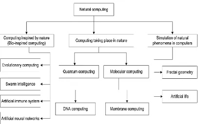
1.1 Taxonomy of natural computing branches (Alsalibi et
al., 2014) 2
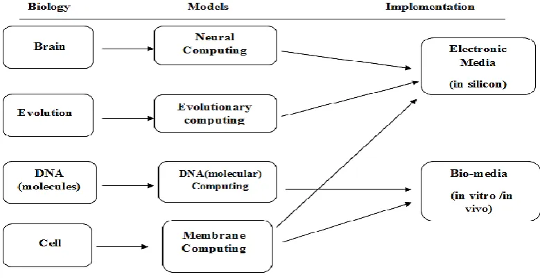
1.2 Natural Computing inspiration in various studies
(Woodworth, 2007) 3
2.1 Membrane structure with its associated tree (Păun,
2002) 21
2.2 Taxonomy of traditional image segmentation methods 26 2.3 Taxonomy of MC based on published papers related
to MC-based segmentation 30
3.1 Research framework 50
3.2 Edge-based segmentation dataset benchmark
(Arbelaez et al., 2011) 56
3.3 Region-based segmentation dataset benchmark (Rahtu
et al., 2010) 57
3.4 General research design 58
3.5 Artificial manual images 61
3.6 Three artificial images 62
3.7 Natural real images 62
3.8 Real medical images 63
3.9 Workflow of PLinguaCore4 Java simulator
(Garcıa-Quismondo et al., 2009) 65
4.2 Type 2 of 4-adjacency rules with Red and Green
(G<R) 72
4.3 Type 2 of 4-adjacency rules with Green and Blue
(G<B) 72
4.4 Type 3 of 4-adjacency rules with Blue and Red (B <
R) 73
4.5 Type 3 of 4-adjacency rules with green and blue (G <
R) 73
4.6 Type 3 of 4-adjacency rules with Green and Blue (G <
B) 74
4.7 Input image 75
4.8 Initial configuration of the system (configuration 0) 78 4.9 Drawing image manually to present initial step 0 79
4.10 First step of the computation (step1) 81
4.11 Manual drawing of image to visualize the first step of
the computation (step1) 81
4.12 Second step of the computation (step2) 83
4.13 Manual drawing of image to present second step of
the computation (step2) 84
4.14 Third step of the computation (step 3) 85
4.15 Manual drawing of image manually to present the
third step of the computation (step 3) 86
4.16 Fourth step of the computation (step 4) 87
4.17 Manual drawing of the image to present fourth step of
the computation (step 4) 87
4.18 Fifth step of the computation (step 5) 89
4.19 Manual drawing of image to present fifth step of the
computation (step 5) 89
4.20 Sixth step of computation (step 6) 91
4.21 Manual drawing of image to present sixth step of the
computation (step 6) 91
4.22 Seventh step of the computation (step 7) 93 4.23 Manual drawing of the image to present seventh step
4.24 Eighth step (step 8) 94 4.25 Manual drawing of image to present eighth step (step
8) 95
4.26 Final step of the computation (step 9) 96
4.27 Manual drawing of image to present final step of the
computation (step 9) 96
4.28 The results of segmentation image when (R>G>B) 98 4.29 The results of segmentation image when (R<G<B) 98 5.1 4-adjacency (square) neighborhoods (Efford, 2000) 103 5.2 8-adjacency (square) neighborhoods Efford, 2000) 104
5.3 6-adjacent (hexagonal) neighborhoods (Snyder et al.,
1999) 105
5.4 Framework of manual segmentation of artificial
images using P-Lingua 106
5.5 Typical manual artificial images 107
5.6 Sample of P-Lingua form for artificial apple image 108 5.7 Sample of P-Lingua syntax for artificial apple
hexagonal image 109
5.8 Example of input artificial apple image based on
P-Lingua 109
5.9 Type Two of 4-adjacency rules with Green and Blue
(G<B) 112
5.10 Type Two of 4-adjacency rules with Red and Blue
(B<R) 113
5.11 Type Two of 4-adjacency rules with Red and Green
(G<R) 113
5.12 Type Three of 4-adjacency rules with green and blue
(G < B) 114
5.13 Type Three of 4-adjacency rules with Blue and Red
(B < R). 115
5.14 Type Three of 4-adjacency rules with with green and
red (G< R) 115
5.15 Type Two of 8-adjacency rules with Green and Blue
5.16 Type Two of 8-adjacency rules with Blue and Red
(B<R) 118
5.17 Type Two of 8-adjacency rules with Green and Red
(G<R) 119
5.18 Type Two of 6-adjacency rules with Blue and Red
(B<R) 121
5.19 Type Two of 6-adjacency rules with Green and Blue
(G<B) 122
5.20 Type Two of 6-adjacency rules with Green and Red
(G<R) 123
5.21 Snapshot of segmentation for hexagonal image 124 5.22 Hexagonal segmentation of region-based and
edge-based 125
6.1 Flowchart of automatic segmentation of artificial
image 135
6.2 Input artificial images (.png) 138
6.3 Converting the pixels of the artificial image to
hexadecimal 139
6.4 Conversion process of pixel for P-Lingua syntax 139 6.5 Output of segmented images using 4-adjacency 141
6.6 Output of segmented face images using 8-adjacency 142 6.7 Automatic region-based visualization of artificial
image 145
6.8 Results before and after region-based segmentation 146 6.9 Automatic edge-based visualization of artificial image 149 6.10 Results before and after edge-based segmentation 149 7.1 Methodology of automatic segmentation of real image 159
7.2 Sample Images 160
7.3 Binary image conversion in P-Lingua 161
7.4 Snapshot of a generated output file 164
7.5 Effect of the value of threshold parameter on region and edge-based segmentation performance; (a) and (b) skin cancer image, (c) and (d) lungs image, and (e)
7.6 Edge-based segmentation results between orginal
image, ground truth and proposed method 181
7.7 Threshold comparesion between the ground truth benchmark with proposed 4 and 8-adjacency
edge-based segmentation 182
7.8 Region-based segmentation results between orginal
image, ground truth and proposed method 183 7.9 Threshold comparison between the ground truth
benchmark with proposed 4 and 8-adjacency
LIST OF ABBREVIATIONS
2D - Two dimensional
2D - Two dimensional
2D-ES - Edge-based segmentation of 2D
3D - Three dimensional
3D - Three dimensional
ACO - Ant Colony Optimization
AGP - A Graphical P segmentator
API - Application Program Interface
BBC - Black Connected Component
BMP - Bitmap Picture
BSDS - Berkeley Segmentation Dataset
CT - Computed Tomography
CT - Computed Tomography
CUDA TM - Compute Unified Device Architecture CUDA TM - Compute Unified Device Architecture FPGA - Field-Programmable Gate Array unit FPGA - Field-Programmable Gate Array unit
FPGAs Field Programmable Gate Arrays
FPGAs Field Programmable Gate Arrays
GA - Genetic Algorithm
GPU - Graphics Processing Unit
GPU - Graphics Processing Unit
HSV - Hue-Saturation-Value
JPEG - Joint Photographic Experts Group
Subalgorithms
MAQIS - Membrane Algorithm with Quantum-Inspired Subalgorithms
MATLAB - Matrix Laboratory
MC - Membrane Computing
MC - Membrane Computing
NMS - Nested Membrane Structure
PC Personal Computer
PNG - Portable network graphics
PSNR - Peak Signal to Noise Ratio PSO - Particle Swarm Optimization
RGB - Red, Green and Blue
RGB - Red, Green and Blue
TIFF - Tagged Image File Format
TSP - Travelling Salesman Problem
VHDL - Very High-Level Design Language
WBCs - White Blood Cells
WBCs - White Blood Cells
LIST OF SYMBOLS
- Whole system
- A finite alphabet whose symbols
- Input alphabet
- Objects in the environment
- Multisets of objects
- Rules
- Input of the system
- Output of the system
- Initial mmbrane
- Represent counter
- Blue object
- Green object
- Red objects
X - Marked pixel
RX - Marked Red pixel
GX - Marked Green pixel
BX - Marked Blue Pixel
S - Segmented results
LIST OF ALGORITHMS
ALGORITHM TITLE PAGE
6.1 Algorithm of artificial image segmentation 136 6.2 Algorithm of drawing region-based artificial
image segmentation 144
6.3 Algorithm for drawing edge-based artificial
image segmentation 147
7.1 Algorithm of real image segmentation 158
7.2 Algorithm of drawing region-based real image
segmentation 165
7.3 Algorithm for drawing edge-based real image
LIST OF APPENDICES
FIGURE NO TITLE PAGE
A Rules of segmentation using 6-adjacency in
P-Lingua syntax (Three colors Red, Blue and Green) 200
B Programming and tools details 202
C Example of drawing region-based image
segmentation for real image 205
D Example of drawing edge-based image
CHAPTER 1
INTRODUCTION
1.1 Introduction
In recent years, computing systems inspired by biological systems, or bio-inspired computing, has been considered as a promising area of theoretical computer science for the next generation computing devices (Ishdorj, 2007).
computing represents a paradigm for the substitution or supplementation of current silicon-based computers, the fields of which are composed of fractal geometry and
artificial life (De castro and Von zuben, 2005).
Figure 1.1 Taxonomy of natural computing branches (Alsalibi et al., 2014)
Figure 1.2 Natural Computing inspiration in various studies (Woodworth, 2007)
The above-mentioned natural computing areas do not take into account the internal structure of the cell. This is the start point of membrane computing. In 1998, Păun studied the behaviour of living cells and introduced membrane
computing, or P systems, in honor of their initiator G. Păun, with the latest version related to his initial research studies published in 2000 (Păun, 2000). The primary
models for MC began with a single cell and its hierarchical structure of organized compartments, or membranes, where localized 'biochemistry' took place. The resulting computing device comprised a distributed parallel model with multisets of objects or “chemicals" placed in regions (tree-like nodes) processed as “reactions"
similar to those of natural biochemistry. The model was extended according to
different biological suggestions to include the processing of objects by means of operations patterned after bio-symport/antiport functions, or as computational motivations extended from single cells to cell populations, or from tree-like membrane arrangements to arbitrary graph techniques, as well as other biological processes such as neuro-pathways (Păun, 2002).
programmability; (iii) scalability and extendibility (major challenges when using differential equations in biological applications); (iv) transparency (multi-set
rewriting rules are little more than equations that mimic chemical reactions); (v) parallelism (a computer science dream, but commonly observed in biology); (vi) non-determinism; and (vii) communication with the marvellous and yet not perfectly understood life phenomenon that coordinates numerous processes within a cell. These approaches all stand in stark contrast to the costly, parallel, coordinating, and synchronizing computations of electronic computing architectures (Păun, 2005).
MC is an emerging field of research due to its inherent parallelism and it has attracted widespread attention from all over the world since its introduction. Its purview includes computer science, biology, biomedicine, bioinformatics, and multiple disciplines such as mathematics, artificial intelligence, automation, and economics. Extensive models of computation involving mathematics, computer science, and biology are derived from cell-type membrane systems, group weave-type membrane systems and neuronal membrane systems. They have applications in computer graphics, approximate optimization, cryptography, parallel computing, and many other areas (Ciobanu and Păun, 2006).
Researchers of MC have applied useful techniques from cell biology in computer science, such as using cell organization in tissues as populations of cells (bacteria) as well as using organizational schemes similar to the pattern of neurons in the brain. P systems can be broadly classified into three fundamental models (Gelenbe, 2009). The first is modelled after a membrane structure or tree-like group and is called cell-like P systems (Păun, 2000). The main component of cell-like P systems is that membranes are structured in a hierarchical arrangement of three dimensional vesicles. The second model is tissue-like P systems (Martın-Vide et al.,
The motivation of using tissue-like P systems is that tissue-like P systems have two biological inspirations: intercellular communication and neuron
cooperation. These two mechanisms have a common mathematical model that utilizes a network of processors that work with symbols through specific channels. The basic feature of the tissue-like P system is that the cell is not polarized and the structure of the graph is general (Christinal et al., 2009).
1.2 Problem Background
P systems have a number of interesting features that open new lines of research that were recently launched to solve several problems related to digital imagery such as the encapsulation of data, the simple representation of information and parallelism, all of which are appropriate for digital images ( az-Pernil et al., 2010). According to these features, MC is used in image segmentation by an extensive number of researches as shown in the related works.
Segmentation in Digital images has features that are parallel and/or local and can be solved regardless of image size. Parallel implementation becomes more
practical at different local areas. Another feature is that the basic information can be easily encoded according to bio-inspired representations. These features make digital imaging flexible and amenable for nature-inspired techniques (Díaz-Pernil et al., 2013).
Limited work on segmentation methods based on P systems has been published and two types of MC methods have been used in segmentation, which are MC rules and MC algorithm.
In 2009, Christinal et al. (Christinal et al., 2009) proposed a new and promising line of research for a family of tissue-like P systems using communication rules to perform edge-based segmentation for 2D images by employing 4-adjacency and 26-adjacency relationships as has been used for 3D digital images. Their results show a constant number of steps (9 steps) of computation to segment a image.
Later (Christinal et al., 2010a) calculated some algebraic-topological information for two-dimensional (2D) and three-dimensional (3D) images in a general and parallel manner with P systems by presenting two areas implemented by tissue-like P systems using communication rules. First, the edge-based segmentation segmentation of 2D images by employing 4-adjacency and 26-adjacency has been used for 3D digital images. Their results show a constant number of steps (9 steps) of computation to segment an image. Second, Homology, this work paved the way for another areas of study in which efficiency and power are used in topological processes for the first time by presenting new rules to achieve the homologous groups of 2D digital images in logarithmic time with respect to input data.
designed an MC approach to solve the threshold problem by using cell-like P system rules where in the massive parallelism of MC allowed the solution to be reached in
linear time. In the second (Christinal et al., 2012), tissue-like P systems have been proposed for the parallel colour segmentation of simple artificial images. The images were segmented such that a threshold was employed to search for edge pixels.
For region-based segmentation (Christinal et al., 2011) proposed new tissue-like P system rules for region-based segmentation, in which a 4-adjacency relationship between pixels was adapted to segment 2D digital images, and a 6-adjacency relationship was used for 3D digital images.
Many limitations can be found in the works of Christinal et al., like the need to manually codify the input image and the need to manually visualize output images. They did not consider that the time of segmentation is not feasible when dealing with big real images, that only one type of segmentation was obtained, that only 4-adjacent relations were used, and that no evaluation procedure has been considered to validate the quality of segmentation.
tissue-like P systems with new rules for segmenting images by the use of gradient-based edge-detection to enhance the classical methods of segmentation.
In (Isawasan et al., 2014), tissue-like P system rules were used to perform the region-based segmentation of 2D hexagonal images using a minimum number of steps (7 steps). Whether or not P-Lingua was used to perform the segmentation is unclear and the details that backup the usage were not illustrated, nor did they not consider the time of segmentation. In addition, only region-based segmentation has been considered and no evaluation procedure has been performed to validate the segmentation results.
In the work of (Sheeba et al., 2011), the authors constructed a family of tissue-like P systems based on Christinal’s work (Christinal et al., 2009) using edge-based segmentation to segment medical images (nuclei of the white blood cells, or WBCs). This technique was implemented through Matlab, only edge-based segmentation was obtained, 4 and 8-adjacency are used, the differences of both types of adjacency has not been illustrated, and the evaluation procedure and the method to compute the success rates have not been mentioned.
Reina-Molina et al. (Reina-Molina et al., 2011) proposed new tissue-like P system rules by replacing single cells with multiple auxiliary cells to deal with
segmentation problems and to exploit potential parallelization. No evaluation procedure has been performed to validate the segmentation results.
(Zhang and Peng, 2012) proposed MC algorithms using cell-like P systems for a novel infrared object segmentation technique based on the thresholding method to quickly get the best set of parameters.
(Peña-Cantillana et al., 2011) presented two types of MC methods to solve the threshold problem. First, MC rules suggested by the authors in (Peña-Cantillana et al., 2011) using tissue-like P systems with an innovative device architecture called CU A™. Second, MC algorithm in which the authors in (Peng et al., 2012) and (Peng et al., 2015) proposed MC algorithms based on cell like-P systems to improve the performance of threshold segmentation. However, (Peng et al., 2015) proposed a multi-level thresholding with enhanced computation efficiency.
In region-growing, another work of (Peng et al., 2014) proposed new tissue-like P system rules based on region-growing based colour image segmentation. The proposed image segmentation has the advantage of fast segmentation. The experimental results also show improved segmentation performance. They have not explained the programming language that has been used in their work.
From the literature review, there is a lack of region-based segmentation for real images. In other words, researchers have not obtained two types of
segmentation using the same rules at the same time. Despite extensively cited efforts and achievements, there remains room for numerous improvements for MC in image segmentation. Difficulties for newcomers to understand the concept means that its complex methodology has yet been clarified.
1.3 Problem Statement
Traditional segmentation methods have several drawbacks including the high computational time. Hence, a recent research trend has been shifted towards using MC for image segmentation (Christinal et al., 2009). Based on observations of previous studies that are mentioned in the previous section, MC has reduced iteration time and computational cost for region-based segmentation and edge-based segmentation.
The majority of the literature lacks a comprehensive explanation of the methodological entities of the proposed approaches, which makes understanding the literature a tedious task. Thus, a more comprehensive illustration of MC models pertaining to image segmentation is of great importance.
The main limitation of the previous studies pertaining to MC based image segmentation is the manual processing of images which makes its practical applications not straightforward.
Furthermore, the majority of the works use only the tissue simulator tool to perform segmentation. However, P-lingua programming language has only been used in few
studies and without much explanation. Another limitation of the literature is that there is no standard evaluation procedure to validate the efficiency of segmentation.
This thesis proposes a bio-inspired MC technique for the automatic segmentation of artificial and real images. This will be accomplished through the development of a new technique based on MC rules. This study will address the following issues:
a. Large number of rules is required to segment large images (Christinal et al., 2011).
b. Lack of region-based segmentation for real images (Christinal et al., 2011).
c. The lack of edge-based segmentation for hexagonal images (Isawasan, et al., 2014).
d. Difficulties in manually codifying input images (Christinal et al., 2011).
e. Difficulties in manually visualizing output images (Christinal et al., 2011).
f. The lack of attention paid to real images because of tissue simulator limitations and drawbacks (Christinal et al., 2011).
This proposed study will answer the following research questions:
1. How can tissue-like P systems be effectively adapted for hexagonal-artificial image segmentation using edge-based segmentation and region-based segmentation?
3. How can the proposed tissue-like P system be improved to automatically segment real images using edge-based segmentation and region-based
segmentation?
4. How can the intrinsic parallelism inherent in the structure of MC models be fully exploited to enhance segmentation results (improve the segmentation accuracy) and reduce computation time?
5. How can the proposed tissue-like P system be evaluated using image segmentation datasets and benchmarks?
1.4 The Aim of the Study
The goal of this thesis is to propose a parallel tissue-like P system framework for image segmentation that draws inspiration from cell biology and membranes. The proposed approach will enhance segmentation quality and obtain better results than previous methods.
1.5 Research Objectives
The goal of this study is to use tissue-like P system concepts and models to improve image segmentation performance using less effort, time and computational steps. The main objectives of this thesis are:
2. To propose a novel tissue-like P system for the segmentation of square and hexagonal images using three types of adjacency relationships
between pixels, 4-adjacency, 6-adjacency, and 8-adjacency.
3. To design automatic region-based and edge-based image segmentation using both 4-adjacency and 8-adjacency relationships using tissue-like P systems.
4. To improve the proposed tissue-like P system to handle real medical images using region-based and edge-based segmentation. A fair comparison between different adjacency relationships will be conducted to assess the efficiency of the tissue-like P system.
1.6 Scope of the Study
This thesis centers on the basic structure or framework of a tissue-like P system. All of the communication rules used in this thesis are based on the work of Christinal et al. (2011). The image datasets that have been used in this thesis are as
follows:
a. The first dataset is associated with skin cancer images. The Dataset was used with permission from the Central Laboratories of the Ministry of Health in Iraq.
b. A bone image available at CTisus was used (Computed Tomography, 2013). [avilable online; http://www.ctisus.com]
c. A tree image available at the Friedrich-Alexander-Universität was used (Technische Facultat, 2016). [avilable online;
https://www5.cs.fau.de/research/data/msseg]
d. A lung image (CT scans) available at CTisus was used (Computed Tomography, 2013). [avilable online; http://www.ctisus.com]
Note that the artificial images used in this thesis and their characteristics are as follows: RGB apple image with size 13 × 13, face with size 14 × 14 and house
with size 30 × 30). These simple apple images have three colors (Red, Blue, and Green) which have been drawn manually.
The Jaccard index method was used for the evaluation of the proposed segmentation approach. Furthermore, two different machine platforms (i7 and i5) have been used to implement the system. In the first part of the study, the tissue simulator tool has been used to implement and execute the proposed P system. Then, P-lingua, the official programming language of MC has been used instead. P-Lingua programming language has been linked to Java (PLinguaCore4 Java library) and C# environments.
1.7 Organization of the Thesis
REFERENCES
Albert, B., Shamoo, A. E., Johnson, A., Khin-Maung-Gyi, F. A., Lewis, J., Raff, M., and Roberts, K. (2002). Molecular Biology of the Cell. (2002). ISBN-10,815332181.
Alberts, B., Bray, D., Hopkin, K., Johnson, A., Lewis, J., Raff, M., and Walter, P. (2013). Essential cell biology. Garland Science.
Alsalibi, B., Venkat, I., Subramanian, K. G., and Christinal, H. A. (2014). A Bio-Inspired Software for Homology Groups of 2D Digital Images. In Membrane Computing (ACMC), 2014 Asian Conference on (pp. 1-4): IEEE.
Aneja, K. R. (2003). Experiments in microbiology, plant pathology and biotechnology. New Age International.
Arbelaez, P., Maire, M., Fowlkes, C. and Malik, J. (2011) Contour Detection and Hierarchical Image Segmentation. IEEE TPAMI, Vol. 33, No. 5, pp. 898-916. Ardelean, I., Díaz Pernil, D., Gutiérrez Naranjo, M. Á., Peña Cantillana, F., Reina
Molina, R., and Sarchizian, I. (2012). Counting Cells with Tissue-like P Systems. Proceedings of the Tenth Brainstorming Week on Membrane Computing, 69-78. Sevilla, ETS de Ingeniería Informática, 30 de Enero-3 de Febrero, 2012.
Ausubel, F. M., R. Brent, R. E. Kingston, D. D. Moore, J. Seidman, J. A. Smith and K. Struhl (2002). Short Protocols in Molecular Biology: A Compendium of Methods from Current Protocols in Molecular Biology, Wiley New York. Berciano, A., ıaz-Pernil, D., Christinal, H. A., Venkat, I., and Subramanian, K. G.
Blankenship, R. E. (2013). Molecular mechanisms of photosynthesis. John Wiley and Sons.
Blaschke, T., Burnett, C., and Pekkarinen, A. (2004). Image segmentation methods for object-based analysis and classification. In Remote sensing image analysis: Including the spatial domain (pp. 211-236). Springer Netherlands. Borrego Ropero, R.,Diaz Pernil,D.,Perez Jimenez,M., (2007).Esuela tecnica de
ingeniria informatica de sevilla.
forja.rediris.es/frs/download.php/374/index_ing.html.
Cardelli, L. (2005, May). Brane calculi. In International Conference on Computational Methods in Systems Biology (pp. 257-278): Springer Berlin Heidelberg.
Carnero, J., Christinal, H. A., Daniel, D., Reina-Molina, R., and Subathra, M. S. P. (2014). Improved Parallelization of an Image Segmentation Bio-Inspired Algorithm. In Proceedings of the Second International Conference on Soft Computing for Problem Solving (SocProS 2012), December 28-30, 2012 (pp. 75-82): Springer India.
Carnero, J., Díaz-Pernil, D., Abril, H. M., and Jurado, P. R. (2010). Image Segmentation Inspired by Cellular Models using Hardware Programming. Paper presented at the 3rd International Workshop on Computational Topology in Image Context Image-A, 1(3), 143-150.
Carnero, J., ıaz-Pernil, D., and Gutiérrez-Naranjo, M. A. (2011). Designing
Tissue-like P Systems for Image Segmentation on Parallel Architectures. Ninth Brainstorming Week on Membrane Computing, 43-62.
Ceterchi, R., Gramatovici, R., Jonoska, N., and Subramanian, K. G. (2003). Tissue-like P Systems with Active Membranes for Picture Generation. Fundamenta Informaticae, 56(4), 311-328.
Ceterchi, R., Mutyam, M., Păun, G., and Subramanian, K. G. (2003). Array-rewriting P systems. Natural Computing, 2(3), 229-249.
Chao, J., and Nakayama, J. (1996, August). Cubical singular simplex model for 3D objects and fast computation of homology groups. In Pattern Recognition, 1996., Proceedings of the 13th International Conference on(Vol. 4, pp. 190-194): IEEE.
Christinal, H. A., Berciano, A., Díaz-Pernil, D., and Gutiérrez-Naranjo, M. A. (2014). Searching Partially Bounded Regions with P Systems. In Proceedings
of the Third International Conference on Soft Computing for Problem Solving (pp. 45-54): Springer India.
Christinal, H. A., Díaz-Pernil, D., and Jurado, P. R. (2009, November). Segmentation in 2D and 3D Image Using Tissue-like P System. In Iberoamerican Congress on Pattern Recognition (pp. 169-176): Springer Berlin Heidelberg.
Christinal, H. A., Díaz-Pernil, D., and Jurado, P. R. (2010a, November). Using Membrane Computing for Obtaining Homology Groups of Binary 2D Digital Images. In International Workshop on Combinatorial Image Analysis (pp. 383-396). Springer Berlin Heidelberg.
Christinal, H. A., DíAz-Pernil, D., and Real, P. (2010b). P Systems and Computational Algebraic Topology. Mathematical and Computer Modelling, 52(11), 1982-1996.
Christinal, H. A., Díaz-Pernil, D., and Real, P. (2011). Region-based segmentation of 2D and 3D Images with Tissue-like P Systems. Pattern Recognition Letters, 32(16), 2206-2212.
Christinal, H. A., ıaz-Pernil, D., Gutierrez-Naranjo, M. A., and Perez-Jimenez, M.
J. (2010). Thresholding of 2D Images with Cell-like P Systems. Romanian Journal of Information Science and Technology (ROMJIST), 13(2), 131-140. Christinal, H. A., Díaz-Pernil, D., Jurado, P. R., and Selvan, S. E. (2012). Color
Segmentation of 2D Images with Thresholding. In Eco-friendly Computing and Communication Systems (pp. 162-169). Springer Berlin Heidelberg. Ciobanu, G., and Păun, G. (2006). Applications of Membrane Computing (Vol. 17).
M. J. Pérez-Jiménez (Ed.). Berlin: Springer.
Ciobanu, D., and Meer, P. (2012). Robust analysis of Feature Spaces: Color Image Segmentation.
Computed Tomography, 2013. CTisus Everything You Need to Know About Computed Tomography (CT). [online] Available at: <http:// http://www.ctisus.com/> [Accessed 10 October 2015].
Dass, R., and Priyanka, S. (2012). Image Segmentation Techniques. IJECT. Volume 3 (issue 1), ISSN: 2230-7109 (Online) | ISSN: 2230-9543 (Print).
De Castro, L. N., and Von Zuben, F. J. (Eds.). (2005). Recent Developments in Biologically Inspired Computing. Igi Global.
De Silva, A. P., and Uchiyama, S. (2007). Molecular logic and computing. Nature Nanotechnology, 2(7), 399-410.
Díaz-Pernil, D., Berciano, A., PeñA-Cantillana, F., and GutiéRrez-Naranjo, M. A. (2013). Segmenting Images with Gradient-Based Edge Detection using Membrane Computing. Pattern Recognition Letters, 34(8), 846-855.
Díaz-Pernil, D., Christinal, H. A., Gutiérrez-Naranjo, M. A., & Real, P. (2012a). Using Membrane Computing for Effective Homology. Applicable Algebra in Engineering, Communication and Computing, 23(5-6), 233-249.
Díaz-Pernil, D., Gutiérrez-Naranjo, M. A., Molina-Abril, H., and Real, P. (2012b). Designing a New Software Tool for Digital Imagery based on P Systems. Natural Computing, 11(3), 381-386.
Diaz-Pernil, D., Molina-Abril, H., Real, P., and Gutiérrez-Naranjo, M. A. (2010, September). A bio-inspired software for segmenting digital images. In Bio-Inspired Computing: Theories and Applications (BIC-TA), 2010 IEEE Fifth International Conference on (pp. 1377-1381): IEEE.
Díaz-Pernil, D., Peña-Cantillana, F., and Gutiérrez-Naranjo, M. A. (2013). A Parallel Algorithm for Skeletonizing Images by Using Spiking Neural P Systems. Neurocomputing, 115, 81-91.
Díaz-Pernil, D., Pérez-Hurtado, I., Pérez-Jiménez, M. J., and Riscos-Núnez, A. (2008, July). A P-Lingua Programming Environment for Membrane Computing. In International Workshop on Membrane Computing (pp. 187-203): Springer Berlin Heidelberg.
Efford, N. (2000). Digital image processing: a practical introduction using java (with CD-ROM). Addison-Wesley Longman Publishing Co., Inc.
Ehrenfeucht, A., Harju, T., Petre, I., Prescott, D. M., and Rozenberg, G. (2003). Computation in living cells: gene assembly in ciliates. Springer Science and Business Media.
El-Rewini, H., and Abd-El-Barr, M. (2005). Advanced Computer Architecture and Parallel Processing (Vol. 42). John Wiley and Sons.
Freixenet, J., Muñoz, X., Raba, D., Martí, J., and Cufí, X. (2002, May). Yet Another Survey on Image Segmentation: Region and Boundary Information
Integration. In European Conference on Computer Vision (pp. 408-422): Springer Berlin Heidelberg.
Freund, R., and Paun, A. (2002, August). Membrane systems with symport/antiport rules: universality results. In Workshop on Membrane Computing (pp. 270-287). Springer Berlin Heidelberg.
Frisco, P., Gheorghe, M., and Pérez-Jiménez, M. J. (Eds.). (2014). Applications of Membrane Computing in Systems and Synthetic Biology: Springer.
García Quismondo, M., Gutiérrez Escudero, R., Pérez Hurtado de Mendoza, I., and Pérez Jiménez, M. D. J. (2009). P-Lingua 2.0: New Features and First Applications. Proceedings of the Seventh Brainstorming Week on Membrane Computing, Sevilla, Spain, 1, 141-167.
García-Quismondo, M., Gutiérrez-Escudero, R., Martínez-del-Amor, M. A., Orejuela-Pinedo, E., and Pérez-Hurtado, I. (2009). P–Lingua 2.0: A Software Framework for Cell–like P Systems. International Journal of Computers Communications and Control, 4(3), 234-243.
García-Quismondo, M., Gutiérrez-Escudero, R., Pérez-Hurtado, I., Pérez-Jiménez, M. J., and Riscos-Núñez, A. (2010, August). An Overview of P-Lingua 2.0. In International Workshop on Membrane Computing (pp. 264-288). Springer Berlin Heidelberg.
Gelenbe, E. (2009). Fundamental Concepts in Computer Science (Vol. 3). Imperial College Press.
Gheorghe, M. (Ed.). (2005). Molecular Computational Models: Unconventional Approaches. IGI Global.
Gupta, S., Bhardwaj, S., and Bhatia, P. K. (2011). A reminiscent study of nature inspired computation. International Journal of Advances in Engineering & Technology, 1(2), 117-125.
Hamadani, N. A. (1981). Automatic Target Cueing in IR Imagery (No. AFIT/GEO/EE/81D-3). Air Force Inst of Tech Wright-Patterson Afb Oh School of Engineering.
Hoek, C., Mann, D., and Jahns, H. M. (1995). H: an introduction to phycology. Cambridge university press.
Hooke, R. (2006). Micrographia (pp. pp-216). IndyPublish. com.
Ibarra, O. H., and Păun, G. (2006). Membrane Computing: A General View. Ann Eur
Acad Sci. EAS Publishing House, Liege, 83-101.
Ionescu, M., Păun, G., and Yokomori, T. (2006). Spiking Neural P Systems.
Fundamenta Informaticae, 71(2, 3), 279-308.
Isawasan, P., Venkat, I., Subramanian, K. G., Khader, A. T., Osman, O., and Christinal, H. A. (2014, September). Region-based Segmentation of Hexagonal Digital Images using Membrane Computing. In Membrane Computing (ACMC), 2014 Asian Conference on (pp. 1-4): IEEE.
Ishdorj, T. O. (2007). Membrane Computing, Neural Inspirations, Gene Assembly in Ciliates (Doctoral dissertation, Ph. D. Thesis, Sevilla University).
Khalid, N., Ahmad, S., Noor, N., Fadzil, A., and Taib, M. (2011). Parallel Approach of Sobel Edge Detector on Multicore Platform. International Journal of Computers and Communications Issue, 4, 236-244.
Kirk, D. B., and Wen-mei, W. H. (2012). Programming massively parallel processors: a hands-on approach. Newnes.
Lodish, H., Baltimore, D., Berk, A., Zipursky, S. L., Matsudaira, P., and Darnell, J. (1995). Molecular cell biology (Vol. 3). New York: Scientific American
Books.
Macías–Ramos, L. F., Pérez–Hurtado, I., García–Quismondo, M., Valencia–Cabrera, L., Pérez–Jiménez, M. J., and Riscos–Núñez, A. (2011, August). AP–Lingua based Simulator for Spiking Neural P Systems. In International Conference on Membrane Computing (pp. 257-281) Springer Berlin Heidelberg.
Martín-Vide, C., Păun, G., Pazos, J., and Rodr guez-Patón, A. (2003). Tissue P Systems. Theoretical Computer Science, 296(2), 295-326.
Mazzarello, P. (1999). A unifying concept: the history of cell theory. Nature Cell Biology, 1(1), E13-E15.
Milton Jr, H., and Stiles, C. D. (2012). Molecular dynamics in biological membranes. Springer Science and Business Media.
Nexusmods, 2016. Nexusmods SKYRIM. [online] Available at: <http://www.nexusmods.com/skyrim/mods/74627/? [Accessed 10 October
2015].
Nguyen, V., Kearney, D., and Gioiosa, G. (2012). An Implementation of Membrane Computing using Reconfigurable Hardware. Computing and Informatics, 27(3+), 551-569.
Nicolescu, R. (2011, August). Parallel and distributed algorithms in P systems. In International Conference on Membrane Computing (pp. 35-50). Springer Berlin Heidelberg.
Nishida, T. Y. (2004). An Application of P System: A New Algorithm for NP-complete Optimization Problems. In Proceedings of the 8th World Multi-Conference on Systems, Cybernetics and Informatics (Vol. 5, pp. 109-112). Păun, G. (2000). Computing with Membranes. Journal of Computer and System
Sciences, 61(1), 108-143.
Păun, G. (2001). From cells to computers: computing with membranes (P systems).
Biosystems, 59(3), 139-158.
Păun, G. (2002). Introduction: Membrane Computing—What It Is and What It Is
Not. In Membrane Computing (pp. 1-6): Springer Berlin Heidelberg.
Păun, G. (2005, June). Membrane Computing: Power, efficiency, applications. In
Conference on Computability in Europe (pp. 396-407): Springer Berlin Heidelberg.
Păun, G. (2006). Introduction to Membrane Computing. In Applications of Membrane Computing (pp. 1-42): Springer Berlin Heidelberg.
Paun, G. (2007). Tracing Some Open Problems in Membrane Computing. Romanian Journal of Information Science and Technology, 10(4), 303-314.
Păun, G. (2012). Membrane Computing: An Introduction. Springer Science and Business Media.
Păun, G. (2014). Looking for Computers in the Biological Cell. After Twenty Years⋆. 13th Brainstorming Week on Membrane Computing, 251.
Păun, G., and Păun, R. (2006). Membrane computing and economics: Numerical P
systems. Fundamenta Informaticae, 73(1, 2), 213-227.
Păun, G., and Pérez-Jiménez, M. J. (2006). Membrane Computing: Brief
Păun, G., and Rozenberg, G. (2002). A Guide to Membrane Computing. Theoretical Computer Science, 287(1), 73-100.
Păun, G., Rozenberg, G., and Salomaa, A. (2010). Handbook of Membrane Computing. Oxford University Press.
Peña Cantillana, F., Díaz Pernil, D., Christinal, H. A., and Gutiérrez Naranjo, M. Á. (2011). Smoothing Problem in 2D Images with Tissue-like P Systems and Parallel Implementation.In Proceedings of the Ninth Brainstorming on membrane computing.Seville, Spin.317-328.
Peña-Cantillana, F., Berciano, A., Díaz-Pernil, D., and Gutiérrez-Naranjo, M. A. (2012). Parallel Skeletonizing of Digital Images by Using Cellular Automata. In Computational Topology in Image Context (pp. 39-48): Springer Berlin Heidelberg.
Peña-Cantillana, F., Díaz-Pernil, D., Berciano, A., and Gutiérrez-Naranjo, M. A. (2011, August). A Parallel Implementation of the Thresholding Problem by using Tissue-Like P Systems. In International Conference on Computer Analysis of Images and Patterns (pp. 277-284): Springer Berlin Heidelberg. Peng, B., Zhang, L., and Zhang, D., (2013). A survey of graph theoretical approaches
to image segmentation, Pattern Recognition, Volume 46, Issue 3. Pages 1020-1038, ISSN 0031-3203
Peng, H., Shao, J., Li, B., Wang, J., Pérez Jiménez, M. D. J., Jiang, Y., and Yang, Y.
(2012). Image Thresholding with Cell-Like P Systems. Proceedings of the Tenth Brainstorming Week on Membrane Computing, 2, 03.
Peng, H., Wang, J., and Pérez-Jiménez, M. J. (2015). Optimal Multi-Level Thresholding with Membrane Computing. Digital Signal Processing, 37, 53-64.
Peng, H., Wang, J., Pérez-Jiménez, M. J., and Shi, P. (2013). A Novel Image Thresholding Method Based on Membrane Computing and Fuzzy Entropy. Journal of Intelligent and Fuzzy Systems, 24(2), 229-237.
Peng, H., Yang, Y., Zhang, J., Huang, X., and Wang, J. (2014). A Region-based Color Image Segmentation Method Based on P Systems. Sceince and Technology, 17(1), 63-75.
Pham, D. L., Xu, C., and Prince, J. L. (2000). Current methods in medical image segmentation 1. Annual review of biomedical engineering, 2(1), 315-337.
Pohani, M., Pathania, M., and Varma, A. (2009). The Cell. A Textbook of Molecular Biotechnology, 47.
Popa, B. D. (2006). Membrane Systems with Limited Parallelism (Doctoral Dissertation, Louisiana Tech University).
Rahtu, E., Kannala , J ., Salo , M., and Heikkila, J., “Segmenting salient objects
from images and videos,” ECCV, pp. 366–379, 2010.
Reina Molina, R., Carnero Iglesias, J., and Díaz Pernil, D. (2011). Image Segmentation Using Tissue-Like P Systems with Multiple Auxiliary Cells. Image-A. 1(3), 143-150.
Reina Molina, R., Díaz Pernil, D., and Gutiérrez Naranjo, M. Á. (2012). Cell Complexes and Membrane Computing for Thinning 2D and 3D Images. Proceedings of the Tenth Brainstorming Week on Membrane Computing,(2) 167-186. Sevilla, ETS de Ingeniería Informática, 30 de Enero-3 de Febrero. 2012.
Richmond, M. L. (2000). TH Huxley's criticism of German cell theory: an epigenetic and physiological interpretation of cell structure. Journal of the History of Biology, 33(2), 247-289.
Ruusuvuori, P., Lehmussola, A., Selinummi, J., Rajala, T., Huttunen, H., &
Yli-Harja, O. (2008, August). Benchmark set of synthetic images for validating cell image analysis algorithms. InSignal Processing Conference, 2008 16th European(pp. 1-5). IEEE.
Šafa ı k, I., and Šafa ı kov , M. (1999). Use of magnetic techniques for the isolation of cells. Journal of Chromatography B: Biomedical Sciences and
Applications, 722(1), 33-53.
Shapiro, L., and Stockman, G. C. (2001). Computer Vision. 2001. ed: Prentice Hall. Sharma, N., Mishra, M., and Shrivastava M.. (2012). Color Image Segmentation
Techniques and Issues: An Approach. International Journal of Science &
Technology Research. Volume 1 (issue 41), ISSN 2277-8616.
Senthilkumaran, N. and Rajesh, R. (2009). Edge Detection Techniques for Image
Sheeba, F., Thamburaj, R., Nagar, A. K., and Mammen, J. J. (2011, September). Segmentation of Peripheral Blood Smear Images Using Tissue-Like P
Systems. In Bio-Inspired Computing: Theories and Applications (BIC-TA), 2011 Sixth International Conference on (pp. 257-261) IEEE.
Shi, R., Ngan, K. N., and Li, S. (2014, October). Jaccard Index Compensation for Object Segmentation Evaluation. In 2014 IEEE International Conference on Image Processing (ICIP) (pp. 4457-4461) IEEE.
Snyder, W. E., Qi, H., and Sander, W. A. (1999, May). Coordinate system for hexagonal pixels. In Medical Imaging'99 (pp. 716-727). International Society for Optics and Photonics.
Somasundaram, S. K., and Alli, P. (2012). A Review on Recent Research and Implementation Methodologies on. In Medical Image Segmentation, Journal of Computer Science, 8(1), 170-174.
Sonka, M., Hlavac, V., and Boyle, R. (2014). Image processing, analysis, and machine vision. Cengage Learning.
Stein, W. D., and Litman, T. (2014). Channels, carriers, and pumps: an introduction to membrane transport. Elsevier.
Technische Facultat, 2016. Friedrich-Alexander-Universität. [online] Available at: <https://www5.cs.fau.de/research/data/msseg/> [Accessed 10 October 2015]. Tirunelveli, G., Gordon, R., and Pistorius, S. (2002). Comparison of Square-Pixel
and Hexagonal-Pixel Resolution in Image Processing. In Electrical and Computer Engineering, 2002. IEEE CCECE 2002. Canadian Conference on (Vol. 2, pp. 867-872): IEEE.
Woodworth, S. (2007). Computability Limits in Membrane Computing. ProQuest. Xue, J., and Liu, X. (2012, May). Applied Membrane Computation in Creative
Design. In Computer Supported Cooperative Work in Design (CSCWD), 2012 IEEE 16th International Conference on (pp. 265-267): IEEE.
Yahya, R. I., Hasan, S., George, L. E., & Alsalibi, B. (2015). Membrane Computing for 2D Image Segmentation. Int. J. Advance Soft Compu. Appl, 7(1), 35-50. Yahya, R. I., Shamsuddin, S. M., Hasan, S., & Yahya, S. I. (2016). Tissue-like P
Yang, S., and Wang, N. (2012). A novel P systems based optimization algorithm for parameter estimation of proton exchange membrane fuel cell model.
International Journal of Hydrogen Energy, 37(10), 8465-8476.
Yang, Y., Peng, H., Jiang, Y., Huang, X., and Zhang, J. (2013). A Region-Based Image Segmentation Method under P Systems. J. Inf. Comput. Sci, 10(10), 2943-2950.
Zhang, G. X., Cheng, J. X., & Gheorghe, M. (2011). A membrane-inspired approximate algorithm for traveling salesman problems.Romanian Journal of Information Science and Technology, 14(1), 3-19.
Zhang, G. X., Liu, C. X., and Rong, H. N. (2010). Analyzing Radar Emitter Signals with Membrane Algorithms. Mathematical and Computer Modelling, 52(11), 1997-2010.
Zhang, G., Gheorghe, M., and Li, Y. (2012). A Membrane Algorithm with Quantum-Inspired Subalgorithms and Its Application to Image Processing. Natural Computing, 11(4), 701-717.
Zhang, G., Liu, C., Gheorghe, M., and Ipate, F. (2009, October). Solving Satisfiability Problems with Membrane Algorithms. In Bio-Inspired Computing, 2009. BIC-TA'09. Fourth International Conference on (pp. 1-8): IEEE.
Zhang, Z., and Peng, H. (2012). Object Segmentation with Membrane Computing.