A Review to Biomedical Image Analysis
Aditi
1, Sheetal Chhabra
2M-Tech Student, Dept. of CSE, Maharishi Ved Vyas Engineering College, Kurukshetra University, Haryana, India1 Associate Professor, Dept. of CSE, Maharishi Ved Vyas Engineering College, Kurukshetra University, Haryana, India2
ABSTRACT:In this research paper a review of biomedical image processing is proposed. Medical images like ultrasound images, CT scan image, MRI images and X-ray images are discussed in this article. How to analyze and process biomedical images and its procedural study is proposed.
KEYWORDS:CT scan, MRI Images, Biomedical Imaging, Ultrasound Images
I. INTRODUCTION
In medical field for the diagnosing of diseases, the ultrasound B-Scan pictures play a vital role. These pictures are obtained with an easy linear or sector scan ultrasound probe, that show a granular look referred to as speckle. The medical specialty pictures obtained by ultrasound (US) systems are considerably poorer compared to different medical imaging systems [1]. But, North American country pictures are thought-about to be non -invasive, portable, accurate, much harmless to the citizenry, and comparatively low-priced imaging modality. These options create the ultrasound B-Scan imaging be the foremost common medical diagnostic tool in hospitals round the world. The medical imaging devices specifically X-ray, CT/MRI and ultrasound are manufacturing long pictures that are employed by medical practitioners within the method of diagnosing. The most drawback featured by them is that the noise introduced owing to the consequence of the coherent nature of the wave transmitted. (i.e., totally different phases of mirrored signals). These noises corrupt the image and sometimes result in incorrect diagnosing. Every of those medical imaging devices is stricken by differing types of noise. As an example, the x-ray pictures are typically corrupted by Poisson noise, whereas the ultrasound pictures are stricken by speckle noise. Speckle could also be a fancy development that degrades image quality with a backscattered wave look that originates from several microscopic subtle reflections that meet up with internal organs and makes it tougher for the observer to discriminate fine detail of the photographs in diagnostic examinations. Thus, denoising or reducing these speckle noise from a loud image has become the predominant step in medical image process. In recent years there has been a truthful amount of study on riffle thresholding and threshold selection for signal and image denoising [2, 3, 7] as a results of riffle provides academic degree applicable basis for separating noisy signal from image signal. Two threshold operators used throughout denoising are soft thresholding and exhausting thresholding. Soft thresholding is a lot of oft used as a result of it reduces the abrupt sharp changes and provides a picture whose quality isn't affected. Applied mathematics filters like Frost filter, Kuan filter, Lee Filter and riffle filters are chosen for this study owing to their economical speckle reduction property [4-6]. Additionally, a mixture of riffle filter with soft thresholding techniques are tried that exhibits higher results than the quality filters.
II. IMAGE PROCESSING
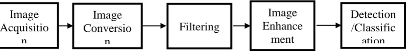
Image processing includes capturing video and images from camera using command „videoinput‟ and getsnapshot. Image processing procedures are shown as in fig. 1.
Fig. 1: Basic Steps in Image Processing
Image
Acquisitio
n
Image
Conversio
n
Filtering
Image
Enhance
ment
Image Acquisition
Digital image process deals with manipulation of digital pictures through a computing machine. It‟s a subfield of signals and systems however focus significantly on pictures. DIP focuses on developing a computer system that's ready to perform processing on a picture. The input of that system may be a digital image and therefore the system method that image mistreatment economical algorithms, and provides a picture as associate output. The foremost common example is Adobe Photoshop. It‟s one amongst the wide used applications for process digital pictures. Image acquisition commands are
(a) imaqhwinfo: Extracting installed adaptor name (b) videoinput: To open video object using adaptor name (c) getsnapshot: To capture single frame
(d) closepreview: To close video object
Types of Images:
There are many types of images, and we will look in detail about different types of images, and the color distribution in them.
The Binary Image: The binary image as it name states, contain only two pixel values. 0 and 1. Gray Scale image: This is two dimensional matrix in behind with values ranging between 0 and 255.
Colored Image:Color image is also known as RGB image. This is three dimensional image, it has three values, l,m,n. l for red ranges 0-255, m for green ranges from 0-255 and n for blue ranges from 0-255.
Color to Gray Scale Conversion: Command is „rgb2gray‟. Gray Scale to binary Conversion: Command is „gray2bw‟. Intensity Image to an Indexed Image: Command is „gray2ind‟.
III.BIOMEDICAL IMAGING
Biomedical images are CT scan image, X-ray image, MRI image, Ultrasound image.
CT stands for computed axial tomography. It‟s a computerized setup that examines the body. The scanner is comprised of a table and a framing. The framing is that the fried cake form half that homes the ray supply. The X-ray supply rotates within the framing because the patient moves through. Information is obtained and processed by a pc to supply a 2 dimensional image.
Magnetic Resonance Imaging (MRI) may be a painless non-invasive procedure that permits physicians to ascertain elaborated pictures of the interior structures of your body while not victimization X-rays. This technology uses an outsized magnet, radio waves and a pc to scan your body.
MRI scans illustrate additional clearly than was ever before doable, the distinction between healthy and unhealthy tissue, and might offer necessary info concerning the brain, spine, joints and internal organs. It will cause early detection and treatment of sickness and has no renowned facet effects. Consequently, your doc are going to be higher able to confirm the foremost acceptable treatment for you.
St. Michael's Hospital‟s tomography department is provided with 2 progressive 2.5 tesla tomography scanners, one in all that may be a vessel magnet. Each scanners are high speed and are used for neurovascular imaging, medical science imaging, abdominal imaging and analysis. The vessel magnet is additionally used for heart and tube distinction increased studies.
Ultrasound technology is that the application of high-frequency sound waves to supply diagnostic pictures. These pictures are often employed in each a diagnostic and therapeutic manner. Sound waves emitted from high-frequency probes or transducers into the patient's body are either mirrored into the probe or attenuated by the body. The acoustic wave that's remitted into is then reborn into electrical signal. The strength of that signal corresponds to the brightne ss on the monitor.
Doppler studies use a similar ultrasound application to show motion in Associate in Nursing audio (continuous wave physicist), visual (colour Doppler) or graphical (pulsed wave Doppler) kind. This can be helpful within the study of arteries and veins to notice blood flow (hemodynamics).
this same spectrum includes X-rays, gamma rays and radiation (alpha and beta waves that are employed in nuclear medicine).
Angiography is that the study of blood vessels and organs by injecting distinction media (X-ray dye) into arteries or veins and taking footage because the distinction media flows through these blood vessels. Sure sickness processes, we have a tendency to able to provide treatment within the angio suite instead of causing our patients to the operative rooms. This cluster of procedures is termed "interventional procedures".
IV.MRIIMAGE PROCESSING
Normally, the anatomy of tumour is examined by imaging scan or CT scan. The most advantage of imaging over CT scan is, it's not contain any radiation. Imaging give correct visualize of bodily structure of tissues. Owing to that‟s imaging not have an effect on bod. Therefore basically imaging is healthier compared to CT scan. Imaging could be a one variety of scanning device that use field of force and radio waves. It‟s additionally use pc to form pictures of the brain on film [8]. Owing to the complicated structure of brain tissues like nervous tissue (WM), gray substance (GM) and humor (CSF) within the brain pictures, extracting of helpful feature could be an elementary task [8].
Tumor is outlined because the abnormal growth of the tissues. Tumor is associate degree abnormal mass of tissue during which cells grow and multiply uncontrollably, ostensibly unbridled by the mechanisms that management traditional cells. Brain tumors is primary or pathologic process, and either malignant or benign. A pathologic process tumour could be a cancer that has unfold from elsewhere within the body to the brain [9].
Usually Image process system includes treating pictures as 2 dimensional signals whereas applying already set signal process strategies to them. Tumor is caused by associate degree abnormal growth of cell in brain. Commonly tumour emerges from brain cells, blood vessels or nerves that are gift within the brain. Early detection of tumour is important as death rate is higher among humans having tumour. Today there are many methodologies for classifying adult male pictures that are fuzzy strategies, neural networks, atlas strategies, data primarily based techniques, form strategies, variation segmentation. Imaging consists of T1 weighted, T2 weighted and Pd (proton density) weighted pictures and are processed by a system that integrates fuzzy primarily based technique with multispectral analysis [9]. The subsequent figure embraces the elemental steps in image process system.
V. CTBRAIN IMAGING
Attractive CAT (CT) image is associate examining contrivance that uses enticing fields and machines to catch photos of the mind on film. It does not utilize x-beams. It provides photos from completely different planes, which permit specialists to create a 3 dimensional image of the tumour. X-ray locates signs transmitted from typical and strange tissue, giving clear photos of most tumors [10]. It‟s become a usually used system for excellent therapeutic imaging, notably in mind imaging wherever delicate tissue distinction and non-obtrusiveness square measure clear points of interest. X-ray is analyzed by radiologists targeted around visual translation of the films to acknowledge the neighborhood of bizarre tissue. Mind photos are chosen for the image reference for this examination on the grounds that the injuries to the neural structure have a bent to influence substantial ranges of the human brain half.
VI. IMAGE DENOISING
Median Filter
The median filter may be a well-liked nonlinear digital filtering technique, usually wont to take away noise. Such noise reduction may be a typical pre-processing step to boost the results of later process (for example, edge detection on AN image). Median filtering is incredibly wide utilized in digital image process as a result of underneath bound conditions, it preserves edges whereas removing noise [13]. Typically called a rank filter, this spatial filter suppresses isolated noise by commutation every pixel‟s intensity by the median of the intensities of the pixels in its neighbourhood. It‟s wide utilized in de-noising and image smoothing applications. Median filters exhibit edge-preserving characteristics (cf. linear strategies like average filtering tends to blur edges), that is incredibly fascinating for several image process applications as edges contain necessary data for segmenting, labelling and protective detail in pictures. This filter may be represented by eq (1).
𝐺 𝑢, 𝑣 = 𝑚𝑒𝑑𝑖𝑎𝑛 𝐼 𝑥, 𝑦 , 𝑥, 𝑦 𝜖𝑤𝐹 eq (1)
Where 𝑤𝐹 = 𝑤𝑥𝑤 Filter window with pixel (𝑢,) as its middle
Gaussian filter
Gaussian filter could be a filter whose impulse response is mathematician perform [14]. Mathematician filters are designed to provide no overshoot to a step perform input whereas minimizing the increase and fall time. This behavior is closely connected to the actual fact that the mathematician filter has the minimum attainable cluster delay. Mathematically, a mathematician filter modifies the signal by convolution with a mathematician function; this reformation is additionally referred to as the Weierstrass transform. Smoothing is often undertaken mistreatment linear filters like the mathematician perform (the kernel relies on the traditional distribution curve), that tends to provide smart leads to reducing the influence of noise with relevancy the image. The 1D and 2D Gaussian distributions with standard deviation for a data point (x) and pixel (x,y), are given by eq (2) and eq (3), respectively [15].
𝐺 𝑥 = 1
2𝜋𝜎2𝑒 𝑥 2
2𝜎 2eq (2)
𝐺 𝑥, 𝑦 = 1
2𝜋𝜎2𝑒 𝑥 2+𝑦 2
2𝜎 2 eq (3)
The kernel could be extended to further dimensions as well. For an image, the 2D Gaussian distribution is used to provide a point-spread; i.e. blurring over neighboring pixels. This is implemented on every pixel in the image using the convolution operation. The degree of blurring is controlled by the sigma or blurring coefficient, as well as the size of the kernel used (squares with an odd number of pixels; e.g. 3×3, 5×5 pixels, so that the pixel being acted upon is in the middle). The processing can be speeded up by implementing the filtering in the frequency rather than spatial domain, especially for the slower convolution operation (which is implemented as the faster multiplication operation in the frequency domain).
Wiener filter
Wiener filters are a category of optimum linear filters that involve linear estimation of a desired signal sequence from another connected sequence. It‟s not associated to adaptive filter. The wiener filter‟s main purpose is to cut back the quantity of noise gift in a picture by comparison with associate estimation of the specified quiet image. The Wiener filter may additionally be used for smoothing. This filter is that the mean squares error-optimal stationary linear filter for pictures degraded by additive noise and blurring. It‟s sometimes applied within the frequency domain (by taking the Fourier transform) [17], because of linear motion or unfocussed optics Wiener filter is that the most vital technique for removal of blur in pictures. From an indication process stance. Every component in a very digital illustration of the photograph ought to represent the intensity of one stationary purpose before of the camera. Sadly, if the shutter speed is simply too slow and therefore the camera is in motion, a given component are associate amalgram of intensities from points on the road of the camera's motion.
1. Assumption: signal and (additive) noise are stationary linear random processes with known spectral characteristics. 2. Requirement: the filter must be physically realizable, i.e. causal (this requirement can be dropped, resulting in a non-causal solution).
3. Performance criteria: minimum mean-square error. Wiener Filter in the Fourier Domain as in eq (4).
𝐺 𝑢, 𝑣 = 𝐻∗(𝑢,𝑣)𝑃𝑠(𝑢,𝑣)
𝐻 𝑢 ,𝑣 2𝑃𝑠 𝑢,𝑣 +𝑃𝑛 𝑢 ,𝑣 eq (4)
Where
(𝑢,𝑣) = Fourier transform of the point spread function
(𝑢,𝑣) = Power spectrum of the signal process, obtained by taking the Fourier transform of the signal autocorrelation (𝑢,𝑣) = Power spectrum of the noise process, obtained by taking the Fourier transform of the noise autocorrelation
VII. IMAGE SEGMENTATION
Unsupervised image segmentation algorithms have matured to the aim that they provide segmentations that adjust to associate degree outsize extent with human intuition. The time has arrived for these segmentations to play an even bigger role in seeing. It‟s clear that unattended segmentation is also accustomed facilitate cue and refine various recognition algorithms. However, one in all the obstacles that keep is that it's unknown specifically but well these segmentation algorithms perform from degree objective posture. Most shows of segmentation algorithms contain superficial analysis that simply show footage of the segmentation results and charm to the reader‟s intuition for analysis. There‟s an even lack of numerical results, thus it's powerful to know that segmentation algorithms gift useful results and at intervals that things they're doing so. Appealing to human intuition is convenient, however if the formula goes to be used in associate degree automatic system then objective results on huge datasets are to be desired. Throughout this paper we tend to tend to gift the results of degree objective analysis of two commonplace segmentation techniques: mean shift segmentation [18], and additionally the economical graph-based segmentation formula bestowed in [19]. As well, we tend to look at a hybrid variant that mixes these algorithms. For each that algorithms, we tend to tend to look at three characteristics:
(a) Correctness: the pliability to supply segmentations that settle for as true with human intuition. That is, segmentations that properly establish structure at intervals the image at neither too fine nor too coarse grade of detail.
(b) Stability with respect to parameter choice: the pliability to supply segmentations of consistent correctness for a diffusion of parameter selections.
(c) Stability with respect to image alternative: the pliability to supply segmentations of consistent correctness exploitation an analogous parameter selection on a decent varies of varied footage. If a segmentation theme satisfies these three characteristics, then it's going to give useful and inevitable results which can be reliably incorporated into an even bigger system. The live we tend to tend to use to evaluate these algorithms is that the recently planned Normalized Probabilistic Rand (NPR) index [21]. we tend to tend to choose to use this live as a result of it permits a principled comparison between segmentation results on fully totally different footage, with differing numbers of regions, and generated by completely different algorithms with different parameters. Also, the NPR index of one, segmentation is critical as degree absolute score, not merely compared therewith of segmentation. These characteristics are all necessary for the comparison we tend to tend to would really like to perform. Our dataset for this analysis is that the Berkeley Segmentation information [20], that contains 300 natural footage with multiple ground truth hand segmentations of each image. To verify a sound comparison between algorithms, we tend to tend to cypher an analogous choices (pixel location and colour) for every image and every segmentation formula. This paper is organized as follows. We tend to begin by presenting some previous work on examination segmentations and clump. Then, we tend to tend to gift each of the segmentation algorithms and additionally the hybrid variant we tend to tend to thought-about. Next, we tend to tend to explain the NPR index and gift the reasons for exploitation this live, followed by a top level view of our comparison methodology.
VIII.BIOMEDICAL IMAGE PROCESSING
In classification here the act of forming the images will classify into which stage the tumor is present, either in normal, begin or malignant.
DWT is mostly used in the jpg files. Mostly the segmentation image will contain high pass filter, here the high pass filter image will refined by changing the brightness, then it is easy to extract the image features. Always the DWT will define in Fourier transform, where it captures the frequency and location information. The feature extraction of CT images is acquiring using DWT sub images. The DWT is implemented by using bilateral filter banks in which loss pass and high pass filter satisfy a particular constraints for feature extraction only the sub images LL is used for DWT decomposition of next scale. The LL sub merge at the last level is used as output feature.
K-Nearest neighbor
The K-Nearest Neighbor is one of the most simple pattern recognition algorithms [17]. Existing technique used KNN classifier for classification of brain images as normal or abnormal. The KNN algorithm is mainly based on wavelet transform, which utilize the information hiding in the transform to reduce the computerized tomography (CT) image is determined then no more processing is required. The KNN algorithm is a slow running technique.
SVM training and testing
SVM is one of the best known routines in example characterization and picture order. The concentrated peculiarities are taken as information to the preparation stage. Help vector machine prepares a picture utilizing concentrated DWT characteristic. Utilizing a fore-mentioned courses of action, prepare the picture for different tumor levels like low, direct and high. In the wake of finishing the preparation procedure, test the forecast exactness of our proposed characterization instrument. In every CT image, we apply a classifier to figure out if it is ordinary or strange. The utilization of SVM includes preparing and testing the SVM, with a specific portion capacity, which thusly has particular piece parameters. Preparing a SVM is the most vital piece of the machine learning procedure. The standard preparing and testing sets are made. Second request surmised wavelet coefficients of typical and anomalous pictures utilized for preparing and testing the SVM. The RBF and polynomial capacities have been utilized for non-straight preparing and testing with degrees 2, 3 and 4.the direct part was likewise utilized for SVM preparing and testing, however it shows lower grouping rate than the polynomial and RBF portions. The precision of arrangement is high in RBF portion contrasted and polynomial and straight parts.
IX. CONCLUSION
In this review paper basics of image processing and image acquisition in matlab functions are reviewed. Types of image in MATLAB and types of image in medical field are discussed in this article. Image denoising techniques and image segmentation is discussed briefly and finally image classification techniques are specified.
REFERENCES
[1] O. V. Michailovich and A. Tannenbaum, “Despeckling of Medical Ultrasound Images”, IEEE Transactions on Ultrasonics, Ferroelectrics and Frequency Control Vol. 53, No. 1, pp. 64-78, 2006.
[2] R. Sivakumar and D. Nedumaran, “Comparative study of Speckle Noise Reduction of Ultrasound B-scan Images in Matrix Laboratory Environment”, International Journal of Computer Applications, Vol. 10, No. 9, pp. 46-50, 2010.
[3] K. Karthikeyan and C. Chandrasekar, “Speckle Noise Reduction of Medical Ultrasound Images using Bayesshrink Wavelet Threshold”,
International Journal of Computer Applications, Vol. 22, No. 9, pp. 8-14, 2011.
[4] S.C. Kang and S.H. Hong, “Experimental and Theoretical Analysis of Wavelet based denoising filter for Echocardiographic images”,
Studies in Health Technology and Informatics, Vol. 84, No. Part 2, pp. 906-909, 2001.
[5] S. Sudha, G.R. Suresh and R. Sukanesh, “Speckle noise reduction in ultrasound images using context – based adaptive wavelet Thresholding”, IETE Journal of Research, Vol. 55, No. 3, pp. 135, 2009.
[6] Manish Goyal and Gianetan Singh Sekhon, “Hybrid Threshold Technique for Speckle Noise Reduction using wavelets for Grey scale images”, International Journal of Computer Science and Technology, Vol. 2, No. 2, pp. 620-625, 2011.
[7] David L. Donoho, “De-Noising by Soft-Thresholding”, IEEE Transactions on Information Theory, Vol. 41, No. 3, pp. 613- 627, 1995. [8] Jay Patel and Kaushal Doshi,“ A Study of Segmentation Methods for Detection of Tumor in Brain MRI” Advance in Electronic and Electric
Engineering, ISSN 2231-1297, Volume 4, Number 3 (2014), pp. 279-284© Research India Publications.
[9] Rajesh C. Patil and Dr. A. S. Bhalchandra, “Brain Tumour Extraction from MRI Images Using MATLAB”, International Journal of Electronics, Communication & Soft Computing Science and Engineering, ISSN: 2277-9477, Volume 2, Issue 1.
[10] http://www.braintumor.org/TumorTypes.
[11] D. Selvathi, R.S. Ram Prak ash, Dr. S.ThamaraiSelvi. 2007. Performance Evaluation of Kernel Based Techniques for Brain MRI Da ta Classification, International Conference on Computational Intelligence and Multimedia Applications.
[12] Mohd FauziBin Othman, NoramalinaBt Abdullah, NurulFazrenaBt Kamal. 2010. MRI BrainClassification Using Support vector machine , Cairo University, Malaysia.
[13] H. Raymond Chan, Chung-Wa Ho, and M. Nikolova, “Salt-and-Pepper noise removal by median-type noise detectors and detail-preserving regularization”. IEEE Trans Image Process 2005; 14: 1479-1485.
[15] R. Fisher, S. Perkins, A. Walker, E. Wolfart. Gaussian smoothing, Hypermedia image processing reference (HIPR2), 2003. Availa ble from:URL:http:// homepages .inf. ed.ac.uk / rbf/ HIPR2/gsmooth.htm.
[16] Kazubek, “Wavelet domain image de-noising by thresholding and Wiener filtering”. M. Signal Processing Letters IEEE, Volume: 10, Issue: 11, Nov. 2003 265 Vol.3.
[17] S. Kumar, P. Kumar, M. Gupta and A. K. Nagawat, “Performance Comparison of Median and Wiener Filter in Image De-noising,” International Journal of Computer Applications 12(4):27–31, December 2010.
[18] D. Comaniciu, P. Meer, “Mean shift: A robust approach toward feature space analysis”, IEEE Trans. on Pattern Analysis and Machine Intelligence, 2002, 24, pp. 603-619.
[19] P. Felzenszwalb, D. Huttenlocher,“Efficient Graph-Based Image Segmentation”, Intl Journal of Computer Vision, 2004, 59 (2).
[20] D. Martin, C. Fowlkes, D. Tal, J. Malik, “A Database of Human Segmented Natural Images and its Application to Evaluating Segmentation Algorithms and Measuring Ecological Statistics”, Intl Conf on Computer Vision, 2001.