Comparative Study on Implementation of
Segmentation Algorithm to Detect Brain
Tumor
Snehal B Rase1, Prof. Komal B Bijwe2
M.E Student, Dept. of CSE, PRPCEM, Amravati University, Amravati, India1
Assistant Professor, Dept. of CSE, PRPCEM, Amravati University Amravati, India 2
ABSTRACT: Brain tumor detection and segmentation is one in every of the foremost difficult and time overwhelming task in medical image process. magnetic resonance imaging (Magnetic Resonance Imaging) may be a medical technique, in the main utilized by the radiotherapist for visualization of internal structure of the body with none surgery. Magnetic resonance imaging provides plentiful info regarding the human soft tissue that helps within the designation of neoplasm. In this paper median filter will be used to remove noise. Segmentation will be done by using proposed algorithm and extract the tumor pattern. Taking in to account the said challenges, this analysis is concentrated towards highlight the strength and limitations of earlier projected classification techniques mentioned within the up to date literature.
KEYWORDS: median filter, segmentation, MRI, tumor, gabor filter, DICOM image, image processing.
I. INTRODUCTION
The human body is consisting of various types of cells and every cell has its own specific function. On losing the ability to grow properly, cells start growing in an improper fashion and in an unordered manner. Extra cells are grouped together to form a mass of tissue which is called tumor .A tumor is an abnormal growth caused by cells reproducing themselves in an uncontrolled manner. These cells can be categorized in two ways: Benign and malignant. Benign tumors do not contain cancer cells and malignant tumors contain tumor cells [1]. Brain tumor is one of the prime causes behind the increase in mortality among people. A tumor also known as neoplasm is a growth in the abnormal tissue which can be differentiated from the surrounding tissue by its structure [2].Brain tumor can occur in different parts of the brain. Brain tumors either include tumors in the central spinal canal or inside the cranium. The most common brain tumors are Gliomas, Oligodendroglioma, Meningiomas, and Glioblastoma.
At present with the involvement of high resolution diagnostic techniques e.g. Magnetic Resonance Imaging (MRI), Computed Tomography (CT), functional MRI (fMRI), Positron Emission Tomography (PET), and Single Photon Emission Computed Tomography (SPECT) in medical imaging we have gained more knowledge about brain tumor than last hundred years. Now imaging facilitates secondary prevention by offering the detection of tumors in developing stages before symptoms start appearing. Early detection usually results in less extensive treatment and better outcomes. Detection of tumors by image processing is known to reduce the mortality subsequently [3]. MRI is a medical imaging technique, and radiologists use it for visualization of the internal structure of the body. Magnetic resonance images are used to produce images of soft tissue of human body. Automatic defects detection in MRI is quite useful in several diagnostic and therapeutic applications computed tomography and MRI are two imaging modalities that help researchers and medical practitioners to study the brain by looking at it noninvasively [2].
they grow, they will become more conspicuous and increase the probability of showing their characters. A person with tumor usually has certain symptoms and this will bring that person to a physician. [4].
II. RELATED WORK
The images were acquired in MINC format, which are then converted to JPEG format before the processing in [5]. Image enhancement techniques were used to improve the visual appearance of the MRI, by eliminating high frequency components from the images. The proposed system describes the image enhancement done using Histogram Equalization (HE) and Center Weighted Median (CWM) filter for removing high frequency components such as impulsive noise, salt and pepper noise, etc. In this, the modified artificial bee colony optimization and modified fuzzy possibility c-means (MFPCM) algorithm was used to detect brain tumor. This work is mainly focused to reduce the computational complexities, which are found in existing methods and aimed at getting higher accuracy [5].
In [6] proposed two main clustering based segmentation methods named Fuzzy C-Means and K-Means to detect brain tumor. By changing the image mode to gray-scale, remove the color components for FCM and K-MEANS. The Enhancement of the image was done by using median filter. Author has been Plotted the histogram to study and analyze the intensity distribution of the pixel. In [7], a novel approach was applied to MRI Brain Image segmentation based on the Hybrid Parallel Ant Colony Optimization (HPACO) with Fuzzy Algorithm. In this the block based non rigid registration has been applied, Author has been applied clustering methods PSO with FCM, HPACO with FCM to detect the tumor region. HPACO was given the best accuracy comparing with other optimization techniques. Overall accuracy of tumor Gray Level Similarity Measure using HPACO is 95.16%. Author achieved the lowest error Rate was about 20%.
The previous methods for brain tumor segmentation are thresholding, region growing & clustering. Thresholding is the simplest method of image segmentation. From a grayscale image, thresholding can be used to create binary images. During the thresholding process, individual pixels in an image are marked as "object" pixels if their value is greater than some threshold value and as "background" pixels otherwise. This convention is known as threshold above. The major drawback to threshold-based approaches is that they often lack the sensitivity and specificity needed for accurate classification. The first step in region growing is to select a set of seed points. Seed point selection is based on some user criterion. [8].
In [9], the image slicing exploitation bit plane technique and reconstructed exploitation Principal was used to extract the brain tumor. The dimensions of tumor and also the quantitative relation of tumor and non tumor space have been calculated using 2by2 neighborhood technique, which can also be used to monitor the efficacy of radiotherapy and chemotherapy sessions. Quadrant method has been developed to find the direction in which the tumor is spreading. This approach will group the kind of mind tumor and consequently help radiologists to arrange the sessions. The results were checked on all 16 images manually and found the efficiency as 93.75%.
S. Mahalakshmi et.al in [10] implemented the PSO algorithm with image segmentation techniques. In this the Digital Imaging and Communication in Medicine (DICOM) file format converted into image file format. PSO algorithm was implemented to detect the tumor. The automation of MRI brain images to detect the brain tumor is based on the criteria of elapsed time and the segmentation level. The process of segmenting the MRI brain image by PSO algorithm is better for the coronal plane than the axial plane.
In [11], the brain tumors have been segmented with the help of three methods Fuzzy C Means, K Means and Hierarchical clustering algorithm. The execution time for Hierarchical clustering was less compared to the other clustering methods. In this the three clustering algorithms were tested with a database of MRI brain images for non-medical format images. The K-means and Fuzzy C means clustering achieved about 70% result. By using Hierarchical clustering achieved about 66.66% and for DICOM images overall efficiency for all algorithms proved to be improved up to 86%.Tumour location detection algorithm increases overall efficiency of the system as it reduces FP(False Positive) output form segmentation algorithm.
and MSE value was minimum in rough entropy. This work illustrates that the rough entropy has higher accuracy compared with Renyi Entropy, Shannon Entropy, Min Entropy and Log Energy
III. PROPOSED METHODOLGY
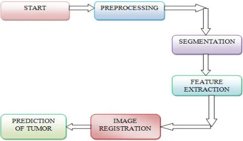
In this research work there are mainly three phase preprocessing, segmentation and image registration. The already converted DICOM image into bmp file format will take as input image. The background of the image will be removed to get ROI region. Then convert the RGB image into gray image for better segmentation process. This study will be done on gray scale images so the RGB image is converted into gray image. Then contrast the image by using matlab function imadjust to manage the brightness of an image. The image will be smoothening by using median filter to remove to noise so that tumor can be easily extracted. The user will have a noise free image for processing.
The segmentation will be done by using clustering techniques proposed method, K-means, Fuzzy C means and PSO method. In clustering the pixel having similar characteristics are grouped into one unit. After clustering the user will get an ROI which is nothing but tumor region. This extracted region will be stored in database as per the pattern of the tumor. Then the image registration process will be done by registering the image. In this stage the location of the tumor in the original image will be display for recognizing the tumor location. Finally, it displays the type of tumor which was stored earlier. The architecture of detection of brain tumor by using proposed algorithm is as shown in figure 1.
Figure 1: Architecture of detection of brain tumor
IV. RESULT ANALYSIS
In this research work, the proposed techniques have been carefully implemented to produce robust, accurate and increased accuracy. There are three different segmentation approaches have been used for detection of tumor in brain MRI images for the comparative analysis. The techniques were tested over real brain MRI images and results were obtained and compared with existing popular algorithms. All experiments were done on a PC with 2.20 GHz Intel(R) Core(TM) 2 Duo CPU, 3 GB of RAM with Matlab 2013a .
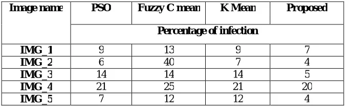
Image name PSO Fuzzy C mean K Mean Proposed
Percentage of infection
IMG_1 9 13 9 7
IMG_2 6 40 7 4
IMG_3 14 14 14 5
IMG_4 21 25 21 20
IMG_5 7 12 12 4
Table 1: Comparison between PSO, Fuzzy C mean and k Mean algorithm in terms of percentage of infection found
The table 2 shows the comparison between the PSO, Fuzzy C means and K Means with the proposed technique in terms of CPU time needed in seconds.
Image name PSO Fuzzy C mean K Mean Proposed
CPU time needed(seconds)
IMG_1 2.58 3.5 3.60 1.62
IMG_2 2.58 2.99 2.93 2.41
IMG_3 3.02 2.15 3.02 2.34
IMG_4 1.85 1.62 2.99 1.35
IMG_5 1.82 1.85 1.82 1.45
Table 2: Comparison between PSO, Fuzzy C mean and k Mean algorithm in terms of CPU time needed
V. CONCLUSION AND FUTURE WORK
In this research work, equal emphasis is given for image segmentation in the context of abnormal MR brain image analysis. Different segmentation techniques and fuzzy techniques are studied in this work. A comparative analysis between three techniques is given in this paper to bring out the merits and demerits of each technique. Different set of feature values may be used to enhance the performance of the proposed systems. The proposed segmentation technique gives the better result as well as better accuracy than previously implemented technique. The proposed methodology requires less CPU time to extract the tumor region. In future Ground truth can be collected for real-time images in order to perform multi-level segmentation with real-time images. The user can process the DICOM images in matlab to improve the accuracy of the detection process also can use various data mining technique to obtained the better result.
REFERENCES
[1] Komal Sharma1, Akwinder Kaur2, Shruti Gujral3,” A review on various brain tumor detection techniques in brain MRI images” IOSR Journal of Engineering (IOSRJEN) www.iosrjen.org, Vol. 04, Issue 05, PP 06-12, May. 2014.
[2] K.R.Yasodha M.Sc1, Dr. V.Thiagarasu M.Sc., PGDCA, M.Phil, B.Ed, Ph.D ,” Automatic Segmentation of Brain Tumor from MRI Images- A Review”, International Journal of Computer Trends and Technology (IJCTT) ,volume 4 Issue 9, PP 3110-3116, Sep 2013.
[3] Rana Bainik, Md. Rabiul Hasan, Md. Saif Iftekhar,” Automatic Detection, Extraction and Mapping of Brain Tumor from MRI Scanned Images using Frequency Emphasis Homomorphic and Cascaded Hybrid Filtering Techniques”, 2nd Int'l Conf. on Electrical Engineering and Infonnation & Communication Technology (ICEEICT) 2015.
[6] Jinal A. Shah , S. R. Suralkar,” Brain Tumor Detection from MRI Images using Fuzzy C-Means Segmentation”, International Journal of Advanced Research in Computer and Communication Engineering Vol. 5, Issue 6,PP 178-183, June 2016.
[7] Dr.N. NandhaGopal,” Automatic Detection of Brain Tumor through Magnetic Resonance Image”, International Journal of Advanced Research in Computer and Communication Engineering Vol. 2, Issue 4,PP 1647-1651, April 2013.
[8] A.S.Bhide1 Priyanka Patil2, Shraddha Dhande,” Brain Segmentation using Fuzzy C means clustering to detect tumour Region”, International Journal of Advanced Research in Computer Science and Electronics EngineeringVolume 1, Issue 2, PP 85-90, April 2012.
[9] Madhav Kurupl, Abhijith Bailur, Pavithra Rajeswaran, Madhuneka Sundararajan, Abhinav,” An innovative approach to monitor brain tumor propagation and track the efficacy of treatment by processing MR Images,” International Conference on Industrial Instrumentation and Control (ICIC), PP 897-901,2015.
[10] S. Mahalakshmi* and T. Velmurugan, “ Detection of Brain Tumor by Particle Swarm Optimization using Image Segmentation”, Indian Journal of Science and Technology, Vol 8(22), PP 13-19,September 2015.
[11] Kshitij Bhagwat, Dhanshri More, Sayali Shinde, Akshay Daga, Assistant Prof Rupali Tornekar, “Comparative Study of Brain Tumour Detection Using K means, Fuzzy C Means and Hierarchical Clustering Algorithms”, International Journal of Scientific & Engineering Research, Volume 4, Issue 6,PP-626-632 June-2013.