University of Windsor University of Windsor
Scholarship at UWindsor
Scholarship at UWindsor
Electronic Theses and Dissertations Theses, Dissertations, and Major Papers
9-10-2019
Structure-function relationship in S-nitrosoglutathione reductase
Structure-function relationship in S-nitrosoglutathione reductase
and the development of fluorogenic pseudo-substrates
and the development of fluorogenic pseudo-substrates
Nneamaka Chinwendu Onukwue University of Windsor
Follow this and additional works at: https://scholar.uwindsor.ca/etd
Recommended Citation Recommended Citation
Onukwue, Nneamaka Chinwendu, "Structure-function relationship in S-nitrosoglutathione reductase and the development of fluorogenic pseudo-substrates" (2019). Electronic Theses and Dissertations. 7829. https://scholar.uwindsor.ca/etd/7829
This online database contains the full-text of PhD dissertations and Masters’ theses of University of Windsor students from 1954 forward. These documents are made available for personal study and research purposes only, in accordance with the Canadian Copyright Act and the Creative Commons license—CC BY-NC-ND (Attribution, Non-Commercial, No Derivative Works). Under this license, works must always be attributed to the copyright holder (original author), cannot be used for any commercial purposes, and may not be altered. Any other use would require the permission of the copyright holder. Students may inquire about withdrawing their dissertation and/or thesis from this database. For additional inquiries, please contact the repository administrator via email
Structure-function relationship in S-nitrosoglutathione reductase and the development of fluorogenic pseudo-substrates
By
Nneamaka Chinwendu Onukwue
A Thesis
Submitted to the Faculty of Graduate Studies through the Department of Chemistry and Biochemistry
in Partial Fulfillment of the Requirements for the Degree of Master of Science
at the University of Windsor
Windsor, Ontario, Canada
2019
Structure-function relationship in S-nitrosoglutathione reductase and the development of fluorogenic pseudo-substrates
by
Nneamaka Chinwendu Onukwue
APPROVED BY:
__________________________________________________ J. Hudson
Department of Biomedical Sciences
__________________________________________________ D. Marquardt
Department of Chemistry & Biochemistry
__________________________________________________ B. Mutus, Advisor
Department of Chemistry & Biochemistry
iii
DECLARATION OF ORIGINALITY
I hereby certify that I am the sole author of this thesis and that no part of this
thesis has been published or submitted for publication.
I certify that, to the best of my knowledge, my thesis does not infringe upon
anyone’s copyright nor violate any proprietary rights and that any ideas, techniques,
quotations, or any other material from the work of other people included in my thesis,
published or otherwise, are fully acknowledged in accordance with the standard
referencing practices. Furthermore, to the extent that I have included copyrighted
material that surpasses the bounds of fair dealing within the meaning of the Canada
Copyright Act, I certify that I have obtained a written permission from the copyright
owner(s) to include such material(s) in my thesis and have included copies of such
copyright clearances to my appendix.
I declare that this is a true copy of my thesis, including any final revisions, as
approved by my thesis committee and the Graduate Studies office, and that this thesis has
iv
ABSTRACT
S-nitrosation is the attachment of a nitric oxide moiety to the thiol side chain of
cysteine. S-nitrosoglutathione (GSNO) acts as a bioactive reservoir for NO to maintain an
equilibrium in the concentration of NO in the body. Due to this, the study of the enzyme
S-nitrosoglutathione reductase has of great interest because of its ability to metabolize
GSNO. S-nitrosoglutathione reductase’s activity has been linked to a number of human
diseases. Chapter 1 of this thesis presents a proposed allosteric binding domain on
GSNOR. Positive cooperativity (sigmoidal deviation) was observed from steady state
analysis of GSNOR which indicated an affinity for the binding of GSNO at this site. The
presence of such a site was further supported by Molecular docking simulations and
HDX-MS which showed that the amino acids Gly321, Lys323, Asn185 and Lys188
interact with molecules bound at this site.
Chapter two introduces four reagents that can function as probes or
pseudo-substrates for the monitoring of enzymatic activity as well as measuring concentrations of
free thiols in vitro and live cells. These reagents are N,N
-di(thioamido-fluoresceinyl)-cystine (DTFCys2), N,N-di(thioamido-fluoresceinyl)-homocystine (DTFHCys2),
N-amido-O-aminobenzoyl-S-nitrosoglutathione (AOASNOG), and N
-thioamido-fluoresceinyl-S-nitroso-glutathione (TFSNOG). They are easy to prepare and purity and
v
DEDICATION
vi
ACKNOWLEDGEMENTS
I would like to thank my supervisor, Dr. Bulent Mutus for his guidance and support
throughout my graduate studies. He provided the opportunity for me to grow as a
researcher and as an individual.
I would also like to extend my gratitude to my committee members, Dr. Hudson
and Dr. Marquardt. This project would not have been completed without their insight and
critical evaluation of my thesis.
A special thanks to the Mutus Lab members, both past and present for their
assistance and support throughout my graduate studies. Thank you, Cody Caba, Katie
Fontana, Scott Smith, Leslie Ventimiglia, Dave Ure, Mark Potter, Sara Aljoudi, Mitchell
Dipasquale, and Angela Awada. I would also like to thank Justin Roberto and Ashley
DaDalt for being excellent troubleshooters. To our graduate secretary,
Mrs. Marlene Bezaire, thank you for looking out for me during my graduate years.
Finally, I would like to thank my Family for the unwavering support throughout
vii
TABLE OF CONTENTS
DECLARATION OF ORIGINALITY ... iii
ABSTRACT ... iv
DEDICATION ...v
ACKNOWLEDGEMENTS ... vi
LIST OF TABLES ...x
LIST OF FIGURES ... xi
LIST OF APPENDICES ... xii
LIST OF ABBREVIATIONS/SYMBOLS ... xiii
CHAPTER 1 ...1
Proposed Allosteric site on S-nitrosoglutathione Reductase ...1
Chapter summary ... 2
1.1 Nitric Oxide ... 3
1.1.1 NO Synthases ... 3
1.1.2 NO signaling Mechanism ... 4
1.2 Protein S-nitrosation ... 5
1.3 S-nitrosoglutathione Reductase (GSNOR) ... 6
1.3.1 GSNOR as a product of ADH 5 ... 6
1.3.2. GSNOR protein structure ... 9
1.3.3. Functions of GSNOR ... 12
1.3.4. Physiology and Inhibitors of GSNOR ... 15
1.4 Computational study of the proposed allosteric site ... 16
viii
1.5.1 (HDX) MS Results ... 18
1.6 Method and Materials ... 21
1.6.1 GSNOR WT cloning and Protein isolation ... 21
1.6.2 GSNO synthesis ... 22
1.6.3 GSNOR Kinetics ... 23
1.7 Results ... 24
1.7.1 GSNOR Kinetics ... 24
1.8 Discussion ... 28
1.9 Conclusion ... 30
1.10 Future direction ... 31
CHAPTER 2 ...32
Development of fluorogenic pseudo-substrates ...32
Chapter Summary ... 33
2.1 Fluorescence: A brief introduction ... 34
2.2 Fluorescein ... 36
2.3 The redox probes ... 37
2.4 Materials ... 39
2.5 Methods ... 39
2.5.1 N,N-di(thioamido-fluoresceinyl)-cystine (DTFCys2, figure 2.3.1A) ... 39
2.5.2 N,N-di(thioamido-fluoresceinyl)-homocystine (DTFHCys2, figure 2.3.2B) ... 40
2.5.3 N-amido-O-aminobenzoyl-S-nitrosoglutathione (AOASNOG, figure 2.3.2D) ... 40
2.5.4 N-thioamido-fluoresceinyl-S-nitroso-glutathione (TFSNOG, figure 2.3.2C) ... 41
2.6 Results ... 43
2.6.1 Free thiol determination ... 44
2.6.2 Kinetic Characterization of disulfide reductases in vitro and in live cells ... 45
2.6.3 Kinetic characterization of S-nitrosoglutathione reductase in vitro and live cells ... 47
2.7 Discussion ... 48
ix
REFERENCES/BIBLIOGRAPHY ...50
APPENDICES ...66
APPENDIX A – Recombinant GSNOR ... 67
APPENDIX B – Mass Spectrometry to identify peptides of GSNOR 79 ... 69
APPENDIX C – Supplementary Data for the development of pseudo-substrate ... 78
x
LIST OF TABLES
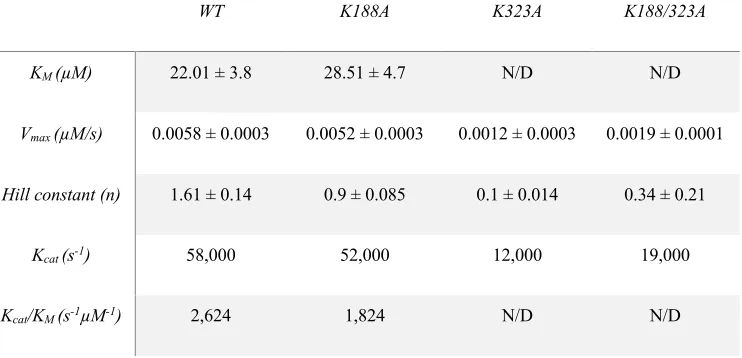
1.5.1. Summary of experimental data obtained for the KM, Vmax, Hill constant, Kcat and the catalytic efficiency of GSNOR WT and three mutants
xi
LIST OF FIGURES
1.3.1 GSNORs role in the detoxification of formaldehyde 8
1.3.2 GSNOR Crystal structure 11
1.3.3 Reaction scheme showing how GNSOR works 14
1.4 Molecular docking simulations 17
1.7.1 (i-ii) Plots of kinetic data obtained for GSNOR 26-27 2.1 Jablonski diagram of absorbance, non-radiative decay, and fluorescence 34
2.2 Fluorescein molecule 36
2.3 MM2 energy 38
2.6 UV/Vis spectrum of reagents 43
2.6.1 Free thiol measurements 44
2.6.2a Kinetic characterization of PDI + ARPE cells (DTFCys2) 45
2.6.2b Imaging of ARPE cells 46
xii
LIST OF APPENDICES
APPENDIX A – Recombinant GSNOR 69
Figure A.1: Recombinant wild type GSNOR protein sequence. Figure A.2: Recombinant GSNOR Plasmid Map.
APPENDIX B – Mass Spectrometry to identify peptides of GSNOR 71 Figure B.1: GSNOR peptide Map
Table B.1: Full peptide list resulting from MS-MS identification.
Table B.2: Representative peptide to visualize deuterium uptake
Table B.3: Deuterium uptake results of two seconds reaction time
Table B.4: Deuterium uptake results of four seconds reaction time
Figure B1: HDX-MS heat map after two seconds of deuterium exchange
Figure B2: HDX-MS heat map after four seconds of deuterium exchange
Figure B3 (i-iv): HDX-MS heat maps with dimerized GSNOR
APPENDIX C – Supplementary Data for the development of fluorogenic pseudo-substrates. 80
Table C1: 1H-NMR chemical shifts for the outlined reagents
Table C2: 1H-NMR chemical shift for GSNO and AOASNOG (OAbz-GSNO)
xiii
LIST OF ABBREVIATIONS/SYMBOLS
NO Nitric Oxide
NOS Nitric Oxide Synthases
nNOS Neuronal Nitric Oxide Synthases
iNOS Inducible Nitric Oxide Synthases
eNOS Endothelial Nitric Oxide Synthases
NADH nicotinamide adenine dinucleotide phosphate
FAD Flavin adenine dinucleotide
GSNO S-nitrosoglutathione
GSNOR S-nitrosoglutathione reductase
GSH-FDH Glutathione-dependent formaldehyde dehydrogenase
GSH Glutathione
GSSG Glutathione persulfide
NH2OH Hydroxylamine
Glu Glutamic acid
Arg Arginine
Cys Cysteine
His Histidine
Asn Asparagine
Lys Lysine
2x YT Yeast extract Tryptone
MS Mass Spectroscopy
1
CHAPTER 1
2
Chapter summary
S-nitrosoglutathione reductase, an enzyme in the alcohol dehydrogenase family, is
responsible for the metabolism of GSNO as well as the detoxification of formaldehyde in
the body. Steady state assay of the WT of this enzyme revealed a deviation from the
classical Michaelis Menten kinetics, fitting more into a sigmoidal curve. This led to the
hypothesis that the enzyme has an allosteric site that binds the substrate GSNO and
increases the activity of the enzyme. Molecular docking was used to visualize the
possible location of such a site, and it was found adjacent to the structural zinc. Four
amino acid residues were implicated to have an interaction with the substrate bound at the
site. They are Lys188, Lys323, Asn185 and Gly321. Mutations to the Lysine residues
were performed to monitor how the changes to the environment around this site would
affect the activity of the enzyme and its affinity for binding the substrate. To further
confirm the presence of the proposed allosteric site Hydrogen Deuterium exchange mass
spectroscopy was performed. This exchange showed decreased uptake of deuterium by
three of the four residues at the proposed site identified on the peptide list. Asn185 was
not identified and will be part of the further investigations with respect to this
experiment. All results obtained from the steady state analysis and HDX-MS support the
hypothesis that there is an allosteric site on the enzyme GSNOR located adjacent to the
3
1.1Nitric Oxide
Nitric Oxide (NO) is a diatomic free radical gasotransmitter. It is the first
identified gasotransmitter discovered to be the endothelium-derived relaxing factor
(EDRF) that is responsible for vascular smooth muscle relaxation. 1-4 NO is a
well-established signaling molecule involved in many physiological processes. These include
the control of vascular tone and blood pressure, promoting angiogenesis, mediating
neurotransmission, immune response, and wound healing.5 NO is also involved in the
regulation of growth, immunity, environmental and root development of Plants.6
1.1.1 NO Synthases
NO regulation involves the control of nitric oxide synthases (NOS), a group of
enzymes responsible for the biosynthesis of NO. Endogenous NO is enzymatically
synthesized in mammalian tissues by three isoforms of NOS. These isoforms are
neuronal NOS (nNOS, NOS 1), inducible NOS (iNOS, NOS 2) and endothelial NOS
(eNOS, NOS 3) according to basal level of activity and constitutive expression in tissues.
NOSs produce NO in eukaryotic cells which are found in animals, and some algae. Other
organisms utilize alternative methods such as nitrite reduction in the production of NO.7-8
The isoforms of NOS function as homodimers, catalyzing the oxidation of
L-arginine to L-citrulline and NO in the presence of molecular oxygen with reduced
nicotinamide adenine dinucleotide phosphate (NADPH), as a co-substrate.9-10 NO
productions catalyzed by NOS require a number of cofactors/coenzymes such as flavin
4
and calmodulin. 11 nNOS is natively expressed in the neurons of the peripheral and
central nervous system, in addition to the epithelial cells of various organs.12 nNOS
activity is Ca2+ dependent and NO derived by nNOS mediates synaptic plasticity
affecting complex physiological functions 13-14 and is a part of the central regulation of
blood pressure.15-16 iNOS is expressed in the presence of inducing stimulants like
cytokines. Once induced, it produces substantial amounts of NO and is completely Ca2+
independent. iNOS was discovered in macrophages and is involved in the immune
system and inflammatory bowel disease (IBSs).17-18 eNOS from the name is expressed in
the endothelial cells and just like nNOS is Ca2+ dependent. NO produced by this isoform
results in vasorelaxation and protects blood vessels from thrombosis by the inhibition of
platelet adhesion and aggregation.19-21
1.1.2NO signaling Mechanism
There are several NO signaling mechanisms that have been identified. The most common
are the classical, nonclassical and less classical mechanisms.22 Classical signaling
involves the activation of soluble guanylate cyclase which then converts guanosine
triphosphate (GTP) to cyclic guanosine monophosphate (cGMP). This leads to the
activation of cGMP-dependent protein kinases subsequently facilitating down-stream
effects. Nonclassical signaling suggests the formation of NO-induced posttranslational
modifications (PTMs) such as S-glutathionylation, S-nitrosation and tyrosine nitration.
5
metabolism due to the interaction between NO and the mitochondrial cytochrome c
oxidase.23-26
1.2 Protein S-nitrosation
S-nitrosation is the covalent attachment of a nitric oxide (NO) group to the thiol
side chain of the amino acid cysteine resulting in the formation of S-nitrosothiols
(SNOs).27 The post-translational modification of Cysteine residues in proteins has
been regarded as a primary mechanism which NO uses to regulate cell signaling.28-30
S-nitrosation of proteins is not directly catalyzed by enzymes, however protein
catalyzed S-nitrosation and denitrosation pathways have been discovered and are yet
to be understood in the context of global proteomic analysis.29 There has been
increasing evidence that suggests the participation of S-nitrosation in both normal
physiology and pathogenesis of several human diseases.31 For instance, in GSNOR
knockout mice increased levels of SNO-proteins were observed which demonstrates
the role of GSNO/GSNOR in SNO-protein homeostasis. First step in S-nitrosation
process is NO oxidized to higher oxides of nitrogen such as dinitrogen trioxide
(N2O3) when reacted with molecular oxygen (O2). N2O3 can react directly with the
thiols as shown in equation 1.2.1.
6
A proposed alternative mechanism for the formation of SNO is the reacting of NO
with reduced thiol forming a radical intermediate.31 Recent evidence shows that the
reaction of NO with molecular oxygen (O2) is not only a primary pathway for
S-nitrosation of thiols but might also yield products of thiol oxidation sometimes greater
than RSNOs.30,32
S-nitrosation of proteins occurs through transnitrosation. Transnitrosation
reactions are all fully reversible and often include SNOs of low molecular weight
such as GSNO.29,33
S-nitrosylation is involved in the physiology and dysfunction of cardiac, airway and
skeletal muscle as well as the immune system showing wide-ranging functions in cell
and tissues.34-37
1.3 S-nitrosoglutathione Reductase (GSNOR)
1.3.1 GSNOR as a product of ADH 5
S-nitrosoglutathione Reductase (GSNOR) is an enzyme in the alcohol
dehydrogenase (ADH) family. The alcohol dehydrogenase family has been evolutionarily
conserved from bacteria to man with five distinct classes containing seven known
isoforms.38 ADH is involved in several important roles in the body but the most studied is
the metabolism of short chain alcohols. GSNOR is a member of the class III alcohol
7
dehydrogenase family. GSNOR is encoded by the ADH5 gene located on the reverse
strand of chromosome 4 (4q23-Chr4: 99,993,567-10,000,985).38
GSNOR also known as ADH5, FALDH, GSH-FDH and Formaldehyde
dehydrogenase is a ubiquitously expressed NADH dependent enzyme with the ability to
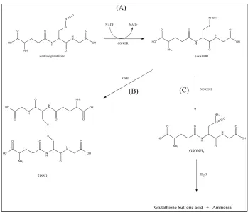
oxidize medium-chain alcohols and the GSH adduct S-hydroxymethylglutathione
(HMGSH). It also reduces GSNO using NADH as a cofactor producing GSNHOH as an
intermediate which can further react with GSH to produce GSSG and NH2OH. GSNOR
when acting as a glutathione-dependent formaldehyde dehydrogenase is critical in the
metabolic elimination of formaldehyde (Figure 1.3.1). This formaldehyde is eliminated
by reacting with glutathione to produce the adduct S-hydroxymethylglutathione which
can then be oxidized to S-formylglutathione. This reaction requires NAD+ as a cofactor
which leads to the production of NADH, a cofactor necessary for the metabolism of
GSNO.39-41 GSNOR in plants is important for growth and root development. Most of the
studies on GSNOR done on plant are mainly focused on the Arabidopsis thaliana plant
however, there have been papers published on sunflower (Helianthus annuus L.), pepper
(Capsicum annuum L.), maize and rice.42 GSNOR in humans can be found in the
endometrium, ovary, fat, esophagus, prostate, liver and kidney.43
8
9
1.3.2. GSNOR protein structure
GSNOR functions as a homodimer with 40kDa subunits containing 347 amino
acids per subunit (Figure 1.3.2, page 11). In each subunit there is a catalytic domain and a
coenzyme binding domain. GSNOR is a metalloprotein with two zinc atoms per
monomer for a total of four zinc atoms per functional enzyme. The zinc atoms are both in
the catalytic domain however the catalytic zinc acts as a lewis acid during catalysis while
the structural zinc is critical for the maintenance of proper protein structure. GSNOR
requires a coenzyme that may vary depending on the substrate. These include NADH,
NADPH+H+ and, NAD(P)+.44
The amino acid residues involved in the binding of substrates are highly
conserved, particularly Glu68 and Arg379 as they are integral to the catalysis
mechanism.45,46 The catalytic zinc is coordinated by the residues Cys45, His67, Cys174
and Glu68 or a water molecule. Similarly, the structural zinc is coordinated by four
closely spaced cysteine residues Cys97, Cys100, Cys103 and Cys111.47,48 Although the
structural zinc is not involved in the catalytic mechanism mutations to any of the cysteine
residues results in the enzymes inactivity. The catalytic domain moves towards the
coenzyme binding domain during the formation of the complex HMGSH with NADH
present.
We hypothesize the presence of an allosteric site on GSNOR that binds the
substrate GSNO and enhances the activity of the enzyme. This site is postulated to be
10
local with molecules bound at this site. The amino acid residues involved are Lys323,
Gly321, Asn185 and Lys188 with both Lysine residues proposed to interact indirectly
with molecules while the Glycine and Asparagine residues interact directly with bound
molecules. This was Supported by molecular docking (MD) simulations performed on the
11
12
1.3.3. Functions of GSNOR
GSNOR as previously mentioned is highly conserved in eukaryotic and
prokaryotic organisms and expressed in all tissues studied.49,50 Multiple studies have
provided documentation of the involvement of GSNOR in a number of pathways from
metabolism of GSNO,51-55 to w-hydroxy fatty acid oxidation,56,57 and formaldehyde
detoxification.58-61 The most characterized and studied functions of GSNOR are the
glutathione-dependent formaldehyde oxidation and the NADH-dependent GSNO
reduction (fig 1.3.1 and fig 1.3.3). The glutathione-dependent formaldehyde
dehydrogenase activity is important for the elimination of formaldehyde, a classified
carcinogen because of its high reactivity with DNA and proteins.62 Studies have shown
that GSNOR is localized in the cytoplasm and as a condensed chromatin in the nucleus,63
this supports its function in the elimination of formaldehyde and protecting DNA from its
toxicity.
The substrates that are preferred by GSNOR are HMGSH and GSNO. However,
GSNO is considered the best substrate due to its catalytic efficiency of metabolism being
twice that of HMGSH.64 GSNO is metabolized by the reductase activity of GSNOR with
the first step being the reduction of GSNO in the presence of the NADH to the unstable
intermediate glutathione N-hydroxysulfenamide (GSNHOH). In the presence of excess
GSH in the system, GSNHOH will react with the GSH to produce a glutathione dimer
(GSSG) and a side product of Hydroxylamine (NH2OH) (Figure 1.3.3). When no GSH is
13
glutathione sulfonamide (GSNONH2) which can then be hydrolyzed under acidic
conditions to glutathione sulfonic acid (GSOOH) and ammonia (NH4+). The above
reaction schemes are all irreversible.
Under physiological conditions, the ratio of NADH/NAD+ present is typically
low.65 This is not favorable for reductive pathways that require NADH implying that the
reductase activity of GSNOR may depend on the availability of the cofactor. An increase
in cellular NADH levels can be triggered by various factors like the inhibition of NADH
dehydrogenase.66 GSNOR deficient mice exhibit substantial increases in protein
s-nitrosation.67 In the cellular environment GSNO is observed to be in equilibrium with
S-nitrosated proteins via reversible transnitrosation, while the observation of increased
levels of S-nitrosation places GSNOR in a crucial role for the maintenance of SNO
14
15
1.3.4. Physiology and Inhibitors of GSNOR
GSNOR plays a crucial role in the variation of NO in the cells. NO is highly
reactive and will form stable RSNO equilibrium in the presence of GSH in the form of
GSNO, a reservoir of bioavailable NO. NO and by extension GSNO plays a critical role
in the relaxation of smooth muscle,68-70 cardiopulmonary regulation71-73, and several other
intra/extracellular functions.74 GSNOR dysregulation has been implicated in numerous
diseases such as asthma, cystic fibrosis and intestinal lung disease. GSNOR knockout
mice have been used to obtain valuable data related to GSNOR function. Canonical
NO-mediated pathways and RSNO levels are severely modified when GSNOR activity is
modulated. GSNOR plays an important protective role in the immune system’s
development of lymphocytes. GSNOR knockout mice show a decrease in CD4
thymocyte development and an increase in lymphocytic apoptosis.75 GSNOR’s regulation
activity in the brain affects a broad swath of cellular functions ranging from neural
development to other neurodegenerative diseases seen in adults.
GSNO has been identified as a long-lived and potent relaxant of human airway
in pulmonary physiology, due to its role as a reservoir for NO. This is characterized by
inflammation and hyper-responsiveness in the airway.76 GSNO has been linked to other
diseases like IBS, autoimmune encephalomyelitis and more. An increase in SNO levels in
knockout mice with asthma lead to the study of therapeutic approaches for the inhibition
of GSNOR activity to restore SNO levels. Investigations into inhibitors of GSNOR are
16
in the regulation of GSNOR is the development of N9115, an inhibitor marketed as
Cavosonstat, that assists cystic fibrosis (CF) patients with the DF508-CFTR mutation.
This mutation occurs within the gene for the cystic fibrosis transmembrane conductance
regulator (CFTR) and accounts for most of cases of CF.77,78 N91115 ensures the
availability of GSNO to promote CFTR development and plasma membrane stability via
its inhibition of GSNOR.
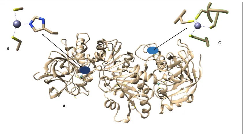
1.4 Computational study of the proposed allosteric site
A computational study of the enzyme to identify the location of the proposed
allosteric site via the measurement of minimum energy between the substrate and the
enzyme was conducted using molecular docking simulations. This study was done as a
collaboration between Sahar Nikoo, Dr. James Gauld, Dr. Bei Sun, and Dr. Bulent
Mutus. The crystal structure for GSNOR utilized was obtained from the PDB library
(ID:3QJ5) and was loaded onto the Molecular Environment Software (MOE). This study
revealed that at the speculated site, the bound ligand interacts with four amino acid
residues. These are lysine188, lysine323, glycine321 and asparagine185 (Figure 1.4). The
docking results showed that when GSNO is bound at the proposed site, it interacts
directly with the residues Gly321 and Asn185. The results also show that Lys188 and
Lys323 interact with GSNO via a solvent network of hydrogen bonds.5 Docking scores
obtained from this study show a score of -8.60 when GSNO is bound to the known active
site of the enzyme and a score of -10.4 when GSNO binds to the postulated allosteric site.
17
showing the system is stable and shows the interaction is favorable. This study further
confirms the presence of the proposed allosteric binding site for the substrate GSNO.
18
1.5 Hydrogen deuterium exchange (HDX) MS
A Hydrogen deuterium exchange experiment was carried out using a Synapt G1
that was properly fitted with custom TRESI apparatus as per Wilson et al.53 The reagents
used for this are reported in appendix B from the work done by Kathleen Fontana. The
experiment was performed using 5- and 10-mm reaction spaces corresponding to 2 and 4
seconds, respectively. Data collection was done in IMS mode within the range of
400-1500 m/z. Experimental uptake of deuterium for each peptide was calculated using a
software that was custom-built for this purpose. Data was collected on the same day for
all trials. These include 5-minute spectrums of GSNOR without deuterium exchange,
with two- and four-seconds exchanges. This was followed by 5 min spectrums of
GSNOR + 20x GSNO (excess) with the same conditions as mentioned above.
1.5.1 (HDX) MS Results
The results from the HDX experiments seen in Appendix B supports the presence
of the proposed allosteric binding site. The incorporation of deuterium shows a shift in
the distribution of peaks due to the addition of the heavier isotope. This shift
corresponded to the amount of deuterium being acquired by the peptide. 20x GSNO was
used to ensure the availability of the substrate in the reaction to enable easy observation
of the interaction with the active site and the proposed allosteric site. Three of the four
amino acid residues that were implicated to be involved in the interaction at the proposed
site by computational studies are represented by the identified peptide list (appendix B).
19
experiment showed that most of the peptides had an increase in the uptake of deuterium
and is summarized in Table 1.5.1. All other relevant results are in appendix B.
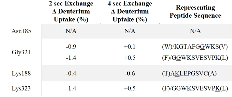
Table 1.5.1 Change in Deuterium uptake by the residues implicated in allosteric binding.
The Gly321, Lys188 and Lys323 residues that are associated with the allosteric site
were identified by peptide MS-MS. These residues as well as those that surround the
GSNO molecule from the computational study all showed a decrease in the uptake of
deuterium. Lys323 showed the highest rate of decrease at 1.4%, Lys188 a decrease of
0.4% and Gly321 which was represented twice on the peptide list showed decreases of
0.9% and 1.4%. Peptides leading to the active site pocket as well as those involved in the
binding of NADH and the catalytic zinc also showed a decrease in the uptake of
-20
1.8% to + 3.4% and -5.2% to +5.1% for deuterium uptake, respectively. These results
21
1.6 Method and Materials
1.6.1 GSNOR WT cloning and Protein isolation
The ADH5 cDNA used for this project was initially cloned by Dr. Bei Sun, who also
performed mutagenesis on said gene that resulted in the recombinant GSNOR with
6x-histidine tags at each terminus as outline in appendix A. The pET28b_ADH5 was then
transformed into BL21(DE3) E.coli cells to facilitate purification of the enzyme.
Protein purification begins with a single colony from the transformed BL21 cells
grown on LB Kanamycin agar plates. The colony is grown in a sterile polypropylene
culture tube containing 4ml of 2x YT media with 50 µg/ml of Kanamycin. The culture
tubes are left to grow overnight at 37ºC while shaking for further use in growing a starter
culture at a rate of 1ml of colony per 100ml of media under the same conditions. The
starter culture is used to inoculate 1.5L of 2x YT media containing 50 µg/ml of
Kanamycin and grown at 37 ºC until an optical density of 0.5-0.6 is reached. At this
point, GSNOR expression is induced by the addition of IPTG to a final concentration of
0.4mM. The induced culture was left for 24 hours at room temperature with shaking to
incubate. The cells were then collected by centrifugation at 6000rpm, 4ºC for 30 minutes.
The supernatant was discarded, and the pellet resuspended with lysis buffer, composed of
50mM Tris-HCl, 150mM NaCl, 15mM imidazole, 1mM DTT, 1mM PMSF, 1% Triton
X-100, 75µg/ml DNase I and 100µg/ml Lysozyme at pH8. The lysate was incubated on
ice for 30mins and further lysed by pulse sonification (20 seconds on and 20 seconds off
22
performed and the supernatant further purified using a HIS-select Nickel Affinity Gel
from Sigma-Aldrich (P6611).
The Nickel column was equilibrated with a wash buffer (with no imidazole) and
Affinity purification was performed by following the manufacturer’s protocol published
by Sigma with modifications to buffer compositions. The wash buffer was composed of
50mM Tris-HCl, 150mM NaCl and 40mM imidazole, while the elution buffer was
composed of 50mM Tris-HCl, 150mM NaCl and 300mM imidazole both at pH 8. The
eluted protein was then buffer exchanged into a storage solution at pH 7.4, composed of
58mM Na2HPO4, 17mM of na2H2PO4, 68mM NaCl and 15% glycerol, using Amicon
centrifugal 30,000 NMWL filter (Millipore sigma UFC 903008) and stored at -80ºC.
1.6.2 GSNO synthesis
Reduced glutathione was dissolved in a solution of cold water and 2 M HCl. An
equal amount of sodium nitrite was added, and the reaction mixture was stirred at 4ºC in
the dark for 40 minutes. GSNO was then precipitated using 10 mL of cold acetone and
the final pink product was washed with cold water and cold acetone before it was
lyophilized for storage at -20ºC. All steps followed as per Hart’s method.80 The final
23
1.6.3 GSNOR Kinetics
Steady state analysis was performed on GSNOR WT and three mutants by varying
GSNO as the substrate while keeping enzyme and NADH constant. A stock solution of
20mM NADH was prepared using milli-Q water. GSNO stock solutions of 1mM and
10mM were freshly prepared for each experiment using PBS buffer. GSNO of increasing
concentration was added to a cuvette containing 80µM of NADH and PBS to a final
reaction volume of 500µl. The reaction was initiated by the addition of 2µg of enzyme.
The consumption of NADH during the reaction was monitored using the decrease in
absorbance at 340nm over a period of 60 seconds using the Agilent 8453 UV/Vis
spectrophotometer. The rate of the initial linear decrease corresponding to NADH uptake
was plotted in correlation to the concentration of GSNO. From this, KM, Vmax and
24
1.7 Results
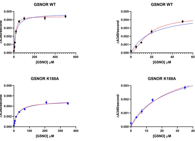
Steady state analysis of GSNOR WT revealed a sigmoidal deviation [Figure 1.7.1
(i)] resulting in positive cooperativity. This led to the proposal of a possible allosteric site
on the enzyme. Molecular docking simulations were performed to further investigate the
presence of the allosteric binding site for the substrate GSNO. Hydrogen-Deuterium
exchange experiments were also performed by Kathleen Fontana to investigate direct
ligand interactions as per appendix B.
1.7.1 GSNOR Kinetics
Steady state kinetic assays of the WT were performed with the concentrations of
NADH and enzyme kept constant while the concentration of GSNO was varied from
0uM to 700uM. Upon analysis of experimental data obtained, the apparent KM, and Vmax
were calculated and are summarized in Table 1.5.1. The Hill coefficient was used to
quantitatively determine the cooperativity of ligand binding. The Hill constant observed
was 1.61 ± 0.14. Mutations at the proposed allosteric binding site were used to observe
changes in the rate of ligand binding and how important the Lysine residues are for
binding. Lysine (K) residues at position 188 and 323 were mutated to alanine (A). Three
mutants were studied; a double mutant and single mutants at each position. For each
mutant KM, Vmax were determined. Hill constant (n) obtained from the data set are
25
enzyme was displayed positive cooperativity for the WT while the mutant K188A loses
the sigmoidal activity. Negative cooperativity can be seen for the mutants K323A and
K188/323A. This shows that both WT and K188A have an affinity for the ligand to bind
at this site however once the mutation at the other position as well as the double mutation
occurs all ligand binding affinity was lost.
Table 1.7.1: Summary of experimental data obtained for the KM, Vmax, Hill constant, Kcat and the catalytic efficiency of GSNOR WT and three mutants
WT K188A K323A K188/323A
KM (µM) 22.01 ± 3.8 28.51 ± 4.7 N/D N/D
Vmax (µM/s) 0.0058 ± 0.0003 0.0052 ± 0.0003 0.0012 ± 0.0003 0.0019 ± 0.0001
Hill constant (n) 1.61 ± 0.14 0.9 ± 0.085 0.1 ± 0.014 0.34 ± 0.21
Kcat (s-1) 58,000 52,000 12,000 19,000
26
27
28
1.8 Discussion
The data obtained from the computational, steady state kinetics and HDX-MS studies
of GSNOR support the hypothesis that there is indeed an allosteric site for the binding of
the substrate GSNO present on the enzyme GSNOR.
The steady state assay first performed on GSNOR WT showed a slight deviation
from the Michalis Menten plot when a Hill constant was applied to fit a sigmoidal curve
which led to the hypothesis of the presence of the proposed allosteric site and the desire
to study it. In an attempt to understand how the presence of an allosteric site affects
enzyme function, mutational study was done.
The steady state assay performed gave results that show difference in KM, Vmax, and
Hill constant. As table 1.5.1 shows the WT and mutant K188A show KM values that are
close to the literature value of 27µM81 while the K
M values of K323A and K188/323A
decreases drastically. This shows that the mutations occurring at these sites lead to an
almost complete loss in affinity for substrate binding that could be because of
conformational changes in the enzyme structure. This was further confirmed by the Hill
constant which showed positive cooperativity for the WT, non-cooperativity for the
mutant K188A and negative cooperativity for the other mutants. The graphs show a slow
rate moving towards the KM but a much faster rate on its way to the Vmax. The steady
state data shows that although the computational study suggests that Lys323 does not
interact directly with the substrate, the almost complete loss of activity after mutation
29
These results show that the activity of GSNOR is hyper-sensitive to structural
changes at this other-than the active site. This is favorable for pharmaceutical companies
that have been investigating the inhibition of this enzyme for treating diseases affiliated
with the enzyme. This discovery could aid the company in designing small molecules that
30
1.9 Conclusion
The results obtained from (1) the steady state assay with a Hill constant of 1.61±0.14
for GSNOR WT and decreasing Hill constant for the mutants; (2) the results from the
HDX-MS which showed a decrease in deuterium uptake of 1.4%, 0.4%, 0.9% and 1.4%
for Lys323, Lys188, and (3) the two Gly321 peptides identified from the reaction of
GSNOR and excess GSNO reaction; support the hypothesis that the enzyme GSNOR has
an allosteric site present that binds the substrate GSNO, increases its rate of reaction, and
is more fitted to the sigmoidal curve plot than the classical Michalis Menten. This site
could be a potential target for pharmaceutical company in developing molecules that can
31
1.10 Future direction
To confirm the location and presence of an allosteric site, several steps may be taken
in future experiments. First the mutation of the Lysine residue to another residue similar
in size and charge to determine if the loss of the charge or the bulk side chain plays a role
in the loss of affinity for the substrate. In addition, more HDX-MS trials should be
32
CHAPTER 2
33
Chapter Summary
This chapter describes the theoretical basis and the methods employed in
synthesizing and characterizing four fluorogenic probes/pseudo substrates. The four
probes synthesized are N,N-di(thioamido-fluoresceinyl)-cystine (DTFCys2), N,N
-di(thioamido-fluoresceinyl)-homocystine (DTFHCys2), N-amido-O-aminobenzoyl-S
-nitrosoglutathione (AOASNOG), and N-thioamido-fluoresceinyl-S-nitroso-glutathione
(TFSNOG). These probes can be used in measuring and imaging free thiols present on
cell surfaces, as substrates for the thiol reductase and S-nitrosothiol denitrosylase
activities of the protein disulfide isomerase (PDI). It can also be used as a substrate for
34
2.1 Fluorescence: A brief introduction
Fluorescence is a luminescence process involving susceptible molecules that emit
light from electronically excited states made by either a physical or chemical mechanism.
Fluorescence occurs by the absorption of photons in a singlet ground state to a singlet
excited state. As the excited molecule returns to the ground state, it emits a photon of
lower energy that corresponds to a longer wavelength than the absorbed photon. The
energy loss is due to vibrational relaxation while in the excited state. Fluorescent bands
center at wavelengths longer this shift towards the longer wavelengths is known as the
stokes shift.82 Excited states are short-lived with a lifetime at about 10-8 seconds.
Substance luminescence is affected by the molecular structure and the chemical
environment. The molecular structure and chemical environment also determine the
intensity of emission when luminescence does occur. In general molecules that can
fluoresce are conjugated systems and the specific frequencies of excitation and emission
35
36
2.2 Fluorescein
The fluorescein dye is the most commonly used fluorescent probe due to its high
molar absorptivity at a wavelength 494nm, making it a very useful and sensitive
fluorescent label. Fluorescein is commercially available in many derivatives, the major
one been Fluorescein isothiocyanate. Fluorescein isothiocyanate (FITC) is simply the
original fluorescein molecule that was functionalized with an isothiocyanate reactive
group which replace a hydrogen atom on the bottom ring of the structure. FITC is often
used in cellular biology to label and track cells in fluorescence microscopy applications.
FITC reacts readily with nucleophiles such as the amine and sulfhydryl groups of
proteins. Many moieties can be conjugated with FITC making them useful in a wide
variety of experimental procedures from immunofluorescence, Apoptosis detection,
Nucleotide labelling, to in situ hybridization and many more.83
Figure 2.2 A shows the fluorescein molecule, B shows the dianion form of fluorescein which give the best intensity of fluorescence and C shows the functionalized fluorescein molecule, the Fluorescein
37
2.3 The redox probes
The highlighted reagents would theoretically act as probes and pseudo substrates
for several reactions. Three of such reactions are the enzyme catalyzed disulfide
reduction (Equation 2.3.1), S-nitrosothiol denitrosylation by the release of NO or HNO
(Equation 2.3.2) and the reduction of NO+ to NH2OH (Equation 2.3.3).
The four probes are fluorogenic, therefore when they partake in thiol redox
reactions an increase in their fluorescence can be observed. The probes are N,N
-di(thioamido-fluoresceinyl)-cystine (DTFCys2, figure 2.3A), N,N
-di(thioamido-fluoresceinyl)-homocystine (DTFHCys2, figure 2.3B), N-amido-O-aminobenzoyl-S
-nitrosoglutathione (AOASNOG, figure 2.3D), and N-thioamido-fluoresceinyl-S
-nitroso-glutathione (TFSNOG, figure 2.3C). Intra-molecular quenching in these probes account
for their low fluorescence when they are not a part of a reaction. Quenching in DTFCys2
is due to the proximity of the two fluorophores to each as a result of structural stability
via intramolecular H-bonding. The self-quenching is results from the collisional energy
transfer that leads to thermalization of the electronic excitation energy. Fluorescence
quenching in AOASNOG and TFSNOG occurs due to the overlap in spectrum of the
functional group, S-N=O (lmax = 312nm and 545nm) with either the excitation (lmax =
R-S-S-R 2 RSH Equation 2.3.1
R-S-N=O NO + ½ RS-SR Equation 2.3.2
38
494nm) or emission (lmax = 520nm) spectrum of fluorescein because of the close
proximity of the functional group to the fluorophore.
39
2.4 Materials
Glutathione (reduced), Isatoic anhydride, fluorescein isothiocyanate (FITC),
Cystine, Homocystine, and sephadex G-50 were all purchased from Sigma Aldrich. Silica
plateTM TLC plates were purchased from SiliCycle Inc.
2.5 Methods
2.5.1 N,N-di(thioamido-fluoresceinyl)-cystine (DTFCys2, figure 2.3.1A)
L-Cystine (0.1mmol) was dissolved in 3.0mL of 0.1M sodium carbonate solution.
This was added to a solution of 0.25mmol FITC dissolved in 3.0mL of acetone. This was
left to react for 24 hours at 50°C protected from light. The reaction mixture was
lyophilized and applied onto a 2.5cm x 30cm Econo column filled with Sephadex G-50
that has been equilibrated with water. DTFCys2 was eluted using 3 column volumes and
the fraction was concentrated by lyophilization, dissolved in water and divided into
100mL aliquots for freezing at -20°C. The final product was characterized using NMR,
UV/Vis, and Fluorescence spectroscopy. The 1H-NMR spectrum was characterized by
the fluoresceinyl-protons ranging from 5.7 to 7.4ppm, and multiplets corresponding to the
Cys protons all summarized in Table C1 of appendix C. The UV/Vis spectrum was
characterized by the large fluoresceinyl-absorbance peak (lmax = 494nm,
e
M=176,000 M-1cm-1). There was also a shoulder due to the fluoresceinyl-moieties overlapping at 475nm.
The absorbance contributions of the S-N=O moiety was buried within the broad aromatic
40
2.5.2 N,N-di(thioamido-fluoresceinyl)-homocystine (DTFHCys2, figure 2.3.2B)
The procedure for the synthesis of DTFHCys2 was the same as that used for
DTFCys2 with 0.1mmol L-Homocystine. The final product was characterized using
NMR, UV/Vis, and Fluorescence spectroscopy. The 1H-NMR spectrum was like that of
DTCys2 with the addition of two peaks all summarized in Table C1 of appendix C. The
UV/Vis spectrum was characterized by the large fluoresceinyl-absorbance peak (lmax =
494nm,
e
M=176,000 M-1cm-1). There was also a shoulder due to thefluoresceinyl-moieties overlapping at 475nm that was seen for DTFCys2.
2.5.3 N-amido-O-aminobenzoyl-S-nitrosoglutathione (AOASNOG, figure 2.3.2D)
S-nitrosoglutathione was synthesized using Hart’s Method.80 SNOG (0.32mmol)
and 1.84mmol Isatoic anhydride (recrystallized from isopropanol) which were dissolved
in 0.5M phosphate buffer pH8 and left to react for 24 hours at 22°C protected from light.
The reaction mixture was applied to a 1cm x 10cm Econo column packed with 1g of
QAE-Sephadex equilibrated with water. The column was washed with 50mL of water
and the AOASNOG was eluted using 0.1M phosphate buffer containing 0.5M NaCl pH
7.4. The product was concentrated by lyophilization, dissolved in 2.0mL of water and
divided into aliquots for freezing at -20°C. AOASNOG was characterized using NMR,
UV/Vis, and Fluorescence spectroscopy. The 1H-NMR spectra of SNOG and
41
AOASNOG spectrum which indicated the presence of the o-aminobenzoyl group and
aromatic protons. The NMR results are summarized in Table C2 of appendix C.84 The
UV/Vis spectrum is characterized by a broad peak that consist of the absorbance of
o-aminobenzoyl (lmax = 312 nm,
e
M= 2,800 M-1cm-1) and a shoulder from the -SNOgroup (lmax = 335 nm,
e
M= 920 M-1cm-1). There was also a small contribution in the redrange (lmax = 545 nm,
e
M=16 M-1cm-1) due to the second weaker -SNO absorbance.85The fluorescence properties of AOASNOG have previously been detailed, further
confirming that AOASNOG is weakly fluorescent because of the close spatial and
overlapping spectra of the excitation spectrum of o-aminobenzoyl-moiety and the
-S-N=O absorbance. This shows that AOASNOG has the ability to act as a fluorogenic
reporter of chemical changes to the -S-N=O functionality through the loss of NO+ or the
reduction of -S-N=O to -S-NHOH by enzymes like S-nitrosoglutathione reductase
(GSNOR) leading to a 14 fold increase in fluorescence at 412nm.84
2.5.4 N-thioamido-fluoresceinyl-S-nitroso-glutathione (TFSNOG, figure 2.3.2C)
S-nitrosoglutathione (0.3mmol) was dissolved in 4mL of 0.2M NaHCO3 pH 9. This
solution was then added to a solution of 0.1mmol FITC dissolved in 4mL of acetone.
This was left to react for 4 hours at 22°C protected from light. The product was applied to
a 1.5cm x 10cm Econo column packed with 2mL of Dowex-1 (Cl- form). FITC and GSH
42
of -2 and -1, respectively. However, TFSNOG at pH 7 has a net charge of -4 and has a
strong interaction with the Dowex-1. Isolation of the product was started by washing the
with 50mL of 0.1M Tris-HCl pH 7.4 and the TFSNOG eluted using the same buffer
containing 1M NaCl. The eluate was concentrated by lyophilization then re-dissolved in
0.5mL of water and desalted on a packed column of 15mL Sephadex G-25 equilibrated
using distilled water. The fraction containing TFSNOG was collected, concentrated by
lyophilization, and divided into aliquots for freezing at -20°C.
TFSNOG was characterized using NMR, UV/Vis, and Fluorescence spectroscopy.
The 1H-NMR spectra of TFSNOG showed a range of 6.3ppm to 7.6ppm for the aromatic
protons of the fluoresceinyl-moiety and peaks for multiplets corresponding to the proton
of Glu and Gly of SNOG all summarized in Table C1 of appendix C. UV/Vis spectrum of
TFSNOG showed a large absorbance peak for the fluoresceinyl-moiety (lmax = 494 nm,
e
M=88,000 M-1cm-1) with the contribution from the -SNO moiety buried within the broadpeak. TFSNOG has a low fluorescence because of the overlap between the -S-N=O
absorbance, lmax = 545nm and the fluoresceinyl-emission, lmax = 520 nm. The
43
2.6 Results
Figure 2.6 A: UV/Vis spectrum of DTFCys2 (red dash line), DTFHCys2 (blue dash line) and
TFSNOG (black dash line); B: Fluorescence emission spectrum of TFSNOG before (dash line) and after the addition of DTT (50µM, red line); C: Fluorescence emission spectrum of DTFCys2 before (red dash) and after the addition of DTT (50µM, red line). Fluorescence
emission spectrum of DTFHCys2 before (blue dash) and after the addition of DTT (50µM,
44
2.6.1 Free thiol determination
DTFCys2 and DTFHCys2 can be used as fluorescent reagents for detecting free thiol
concentration just like Ellman’s reagent86 via the thiol disulfide exchange shown in
Equation 2.6.1. This reagent can be used to determine nmol levels of free thiols (Figure
2.6.1).
F-Cys-S-S-Cys-F + RS-® R-S-S-Cys-F + F-Cys-S- Equation 2.6.1
low fluorescence higher fluorescence
Figure 2.6.1 Constant volume of DTFCys2 (A) and DTFHCys2 (B) were added to
45
2.6.2 Kinetic Characterization of disulfide reductases in vitro and in live cells
Researchers had shown that cells have a cell surface associated form of PDI.87-97
DTFCys2 can also be used to kinetically characterize pure recombinant PDI and cell
surface PDI of ARPE cells. These reagents can also be used in imaging cell surface thiols
from PDI to other free protein thiols on live cells. Figure 2.6.2b shows ARPE cells
exposed to DTFCys2.
Figure 2.6.2a; A: constant volume of purified recombinant PDI was added to 96-well plates with increasing amounts of DTFCys2 and enzymatic reaction was initiated by the addition of
DTT. B: ARPE cells grown to confluence in wells of a 96-well plate had increasing amounts of DTFCys2 added to it the wells and change in fluorescence was monitored at 520nm for 9
46
Figure 2.6.2b ARPE cells grown at the bottom of 96-well plates exposed to PBS (control)- panel A, B, C and DTFCys2- Panel D, E, F for 15 mins. imaged from the bottom of the plate using an inverted
47
2.6.3 Kinetic characterization of S-nitrosoglutathione reductase in vitro and live cells
AOASNOG is currently the only cell permeable, pseudo-substrate for
S-nitrosoglutathione reductase.84 This section demonstrates how it can be used to
kinetically characterize ARPE cells (Figure 2.6.3B). TFSNOG the fluorescein analog of
AOASNOG acts as a pseudo-substrate for the S-denitrosylation activity of PDI.98
48
2.7 Discussion
The purpose of this study was to describe the theoretical basis and simple methods
for synthesizing four probes/reagents that can be used in various applications such as the
measuring and imaging of free thiols on cell surface as well as how they can perform as
pseudo-substrates for monitoring enzymatic activities. Each reagent was characterized
using NMR as well as UV/Vis and Fluorescence spectroscopy. Thin Layer
Chromatography (TLC) was performed using SiliaPlate TLC aluminum backed plates
with varying mobile phases that provided the best separation. The mobile phase used for
DTFCys2, DTFHCys2, and TFSNOG was acetone:water:methanol in a 10:10:1 ratio. For
AOASNOG the mobile phase composition was acetone:water:methanol in a 13:6:1 ratio.
The multiple spots observed for FITC are due to the different prototrophic forms of
fluorescein.107
The disulfide-linked probes DTFCys2 and DTFHCys2 can function as fluorescent
reagents for the detection of free thiol concentrations just like the classical colorimetric
thiol reagent 5,5’-dithiobis-2-nitrobenzoate (Ellman’s reagent) from the thiol disulfide
exchange. However, unlike the Ellman’s reagent, the fluorescence signal from these new
regents enables the detection of thiol concentrations in the nmol levels. DTFCys2 can be
used to kinetically characterize recombinant PDI and cell surface PDI of ARPE in culture
with estimated KM for the PDI mediated disulfide reduction which was ~1.9µM and was
close to the estimated KM for the ARPE-surface PDI of 2.72µM. the Michaelis Menten
49
KM of 740µM was estimated for AOASNOG in the ARPE-GSNOR catalyzed
denitrosation which comes close to the KM for recombinant GSNOR of 320µM. TFSNOG
functions as a pseudo-substrate for the S-denitrosylase activity of PDI with an estimated
KM of 11.1µM that corresponds to a higher affinity of about 6-fold compared to that of
SNOG (65µM). Unfortunately, TFSNOG was not a pseudo-substrate for GSNOR. Apart
from the large applications to enzymology and cell biology, the reagents cost less to
produce because the fluorescein isothiocyanate is less expensive than eosin
isothiocyanate for example. In addition, fluorescein derivatives are much easier to purify.
2.8 Conclusion
The reagents outlined here are easy to prepare and purify. They can be used in
diverse applications ranging from their use as thiol reagents to pseudo-substrates for
50
REFERENCES/BIBLIOGRAPHY
1. Furchgott, R. F.; Zawadski, J. Acetylcholine Relaxes Arterial Smooth-Muscle by
Releasing a Relaxing Substance from Endothelial-Cells. Fed Proc 39:581-581; 1980.
2. Ignarro, L. J.; Buga, G. M.; Wood, K. S.; Byrns, R. E.; Chaudhuri, G.
Endothelium-Derived Relaxing Factor Produced and Released from Artery and Vein Is
Nitric-Oxide. P Natl Acad Sci USA 84:9265-9269; 1987.
3. Palmer, R. M. J.; Ferrige, A. G.; Moncada, S. Nitric-Oxide Release Accounts for the
Biological-Activity of Endothelium-Derived Relaxing Factor. Nature 327:524-526;
1987.
4. Furchgott, R. F.; Khan, M. T.; Jothianandan, D. Comparison of Endothelium-
Dependent Relaxation and Nitric Oxide-Induced Relaxation in Rabbit Aorta. Fed
Proc 46:385-385; 1987.
5. Sun, B. Biochemical and Functional Studies of S-nitrosoglutathione Reductase and
Neutral Sphingomyelinase II. University of Windsor, Electronic Theses and
Dissertations, 2017
6. Moncada, S.; Radomski, M. W.; Palmer, R. M. J. Endothelium-Derived Relaxing
Factor - Identification as Nitric-Oxide and Role in the Control of Vascular Tone and
Platelet-Function. Biochem Pharmacol 37:2495-2501; 1988.
7. Ignarro, L. J.; Byrns, R. E.; Burga, G. M.; Wood, K. S., Endothelium-Derived
51
Chemical Properties Identical to Those of Nitric Oxide Radical. Circulation Research
1987, 61 (6), 866-879.
8. Ignarro, L. J.; Burga, G. M.; Wood, K. S.; Byrns, R. E.; Chaudhuri, G.,
Endothelium-derived relaxing factor produced and released from artery and vein is nitric oxide.
Proc Natl Acad Sci U S A 1987, 84 (24), 9265-9269
9. Stuehr, D. J.; Kwon, N. S.; Nathan, C. F.; Griffith, O. W.; Feldman, P. L.; Wiseman,
J. N omega-hydroxy-L-arginine is an intermediate in the biosynthesis of nitric oxide
from L-arginine. J Biol Chem 266:6259-6263; 1991.
10. Knowles, R. G.; Moncada, S. Nitric-Oxide Synthases in Mammals. Biochem J
298:249-258; 1994.
11. Forstermann, U.; Closs, E. I.; Pollock, J. S.; Nakane, M.; Schwarz, P.; Gath, I.;
Kleinert, H. Nitric-Oxide Synthase Isozymes - Characterization, Purification,
Molecular- Cloning, and Functions. Hypertension 23:1121-1131; 1994.
12. Forstermann, U.; Sessa, W. C. Nitric oxide synthases: regulation and function.
Eur Heart J 33:829-+; 2012.
13. Bohme, G. A.; Bon, C.; Lemaire, M.; Reibaud, M.; Piot, O.; Stutzmann, J. M.;
Doble, A.; Blanchard, J. C. Altered Synaptic Plasticity and Memory Formation in
Nitric- Oxide Synthase Inhibitor-Treated Rats. P Natl Acad Sci USA 90:9191-9194;
1993.
14. Zhou, L.; Zhu, D. Y. Neuronal nitric oxide synthase: Structure, subcellular
52
2009.
15. Togashi, H.; Sakuma, I.; Yoshioka, M.; Kobayashi, T.; Yasuda, H.; Kitabatake, A.;
Saito, H.; Gross, S. S.; Levi, R. A Central-Nervous-System Action of Nitric-Oxide in
Blood-Pressure Regulation. J Pharmacol Exp Ther 262:343-347; 1992.
16. Esplugues, J. V. NO as a signalling molecule in the nervous system. Brit J
Pharmacol 135:1079-1095; 2002.
17. Kolios, G.; Valatas, V.; Ward, S. G., Nitric oxide in inflammatory bowel disease: a
universal messenger in an unsolved puzzle. Immunology 2004, 113 (4), 427-437.
18. Nathan, C. F.; Hibbs, J. B. Role of Nitric-Oxide Synthesis in Macrophage
Antimicrobial Activity. Current Opinion in Immunology 3:65-70; 1991.
19. Fleming, I.; Busse, R. Molecular mechanisms involved in the regulation of the
endothelial nitric oxide synthase. Am J Physiol-Reg I 284:R1-R12; 2003.
20. Azuma, H.; Ishikawa, M.; Sekizaki, S. Endothelium-Dependent Inhibition of
Platelet-Aggregation. Brit J Pharmacol 88:411-415; 1986.
21. Li, H. G.; Forstermann, U. Nitric oxide in the pathogenesis of vascular disease.
Journal of Pathology 190:244-254; 2000.
22. Martínez-Ruiz, A., Cadenas, S., & Lamas, S. (2011). Nitric oxide signaling:
Classical, less classical, and nonclassical mechanisms. Free Radical Biology And
Medicine, 51(1), 17-29. doi: 10.1016/j.freeradbiomed.2011.04.010
23. Brown, G. C.; Cooper, C. E. Nanomolar Concentrations of Nitric-Oxide Reversibly
53
Oxidase. Febs Lett 356:295-298; 1994.
24. Martinez-Ruiz, A.; Cadenas, S.; Lamas, S. Nitric oxide signaling: Classical, less
classical, and nonclassical mechanisms. Free Radical Bio Med 51:17-29; 2011.
25. Erusalimsky, J. D.; Moncada, S. Nitric oxide and mitochondrial signaling from
physiology to pathophysiology. Arterioscl Throm Vas 27:2524-2531; 2007.
26. Hess, D., Matsumoto, A., Kim, S., Marshall, H., & Stamler, J. (2005). Protein
S-nitrosylation: purview and parameters. Nature Reviews Molecular Cell Biology, 6(2),
150-166. doi: 10.1038/nrm1569
27. Forman, H. J.; Fukuto, J. M.; Torres, M. Redox signaling: thiol chemistry defines
which reactive oxygen and nitrogen species can act as second messengers. Am J
Physiol- Cell Ph 287:C246-C256; 2004.
28. Foster, M. W.; Hess, D. T.; Stamler, J. S. Protein S-nitrosylation in health and
disease: a current perspective. Trends Mol Med 15:391-404; 2009.
29. Hickok JR, Vasudevan D, Thatcher GRJ, and Thomas DD. Is S-nitrosocysteine a
true surrogate for nitric oxide? Antioxid Redox Signal 17: 962–968, 2012.
30. Thomas, D. D. and D. Jourd'heuil (2012). "S-Nitrosation: Current Concepts and New
Developments." Antioxidants & Redox Signaling 17(7): 934-936.
31. Gow, A. J.; Buerk, D. G.; Ischiropoulos, H. A novel reaction mechanism for the
formation of S-nitrosothiol in vivo. J Biol Chem 272:2841-2845; 1997.
32. Benhar, M.; Forrester, M. T.; Stamler, J. S. Protein denitrosylation: enzymatic
54
33. Benhar, M., Forrester, M., Hess, D., & Stamler, J. (2008). Regulated Protein
Denitrosylation by Cytosolic and Mitochondrial Thioredoxins. Science, 320(5879),
1050-1054. doi: 10.1126/science.1158265
34. Palmer, L., Kavoussi, P., & Lysiak, J. (2012). S-Nitrosylation of endothelial nitric
oxide synthase alters erectile function. Nitric Oxide, 27, S22-S23. doi:
10.1016/j.niox.2012.04.079
35. He, W., & Frost, M. (2016). Direct measurement of actual levels of nitric oxide (NO)
in cell culture conditions using soluble NO donors. Redox Biology, 9, 1-14. doi:
10.1016/j.redox.2016.05.002
36. Beuve, A., Wu, C., Cui, C., Liu, T., Jain, M., & Huang, C. et al. (2016).
Identification of novel S-nitrosation sites in soluble guanylyl cyclase, the nitric oxide
receptor. Journal Of Proteomics, 138, 40-47. doi: 10.1016/j.jprot.2016.02.009
37. Guerra, D., Ballard, K., Truebridge, I., & Vierling, E. (2016). S-Nitrosation of
Conserved Cysteines Modulates Activity and Stability of S-Nitrosoglutathione
Reductase (GSNOR). Biochemistry, 55(17), 2452-2464. doi:
10.1021/acs.biochem.5b01373
38. Jensen, D. E.; Belka, G. K.; Bois, G. C. D., S-Nitrosoglutathione is a substrate for rat
alcohol dehydrogenase class III isoenzyme. Biochem J 1998, 331, 659-668.
39. Hedberg, J. J.; Hoog, J.-O.; Nilsson, J. A.; Xi, Z.; Elfwing, Å.; Grafstro, R. C.,
Expression of alcohol dehydrogenase 3 in tissue and cultured cells from human oral
55
40. Hopkinson, R. J.; Barlow, P. S.; Schofield, C. J.; Claridge, T. D. W., Studies on the
reaction of glutathione and formaldehyde using NMR. Org Biomol Chem 2010, 8
(24), 4915-4920.
41. Leterrier, M.; Chaki, M.; Airaki, M.; Valderrama, R.; Palma, J. M.; Barroso, J. B.;
Corpas, F. J., Function of S-nitrosoglutathione reductase (GSNOR) in plant
development and under biotic/abiotic stress. Pant Signal Behav 2011, 6 (6), 789-793.
42. Fagerberg, L.; Hallstro, B. M.; Oksvold, P.; Kampf, C.; Djureinovic, D.; Odeberg, J.;
Habuka, M.; Tahmasebpoor, S.; Danielsson, A.; Edlund, K.; Asplund, A.; Sjostedt,
E.; Lundberg, E.; Szigyarto, C. A.-K.; Skogs, M.; Takanen, J. O.; Berling, H.; Tegel,
H.; Mulder, J.; Nilsson, P.; Schwenk, J. M.; Lindskog, C.; Danielsson, F.;
Mardinoglu, A.; Sivertsson, Å.; Feilitzen, K. v.; Forsberg, M.; Zwahlen, M.; Olsson,
I.; Navani, S.; Huss, M.; Nielsen, J.; Ponten, F.; Uhlen, M., Analysis of the Human
Tissue-specific Expression by Genome-wide Integration of Transcriptomics and
Antibody-based Proteomics. Molecular & Cellular Proteomics 2014, 13 (2),
397-406.
43. Yang, Z. N.; Bosron, W. F.; Hurley, T. D. Structure of human chi chi alcohol
dehydrogenase: A glutathione-dependent formaldehyde dehydrogenase. J Mol Biol
265:330-343; 1997.
44. Barnett, S. D.; Buxton, I. L. O., The Role of S-nitrosoglutathione Reductase
(GSNOR) in Human Disease and Therapy. Crit Rev Biochem Mol Biol 2017, 52 (3),