ABSTRACT
QI, LINGJIAO. Development and Application of Fast-scan Voltammetric Technology for Examining Neurochemical Mechanisms In Vivo. (Under the direction of Dr. Leslie A. Sombers.)
Dopamine (DA) is a neurotransmitter that plays a key role in the regulation of various motor functions, motivated behavior, and reward-associated learning by acting in various brain regions. Understanding real-time DA neurotransmission in the brain is necessary to understand how DA neurochemically underlies these behaviors. Electrochemical techniques are well-suited to monitor the chemical dynamics of neurotransmitters in vivo. Particularly, fast-scan cyclic voltammetry (FSCV) coupled with carbon-fiber microelectrodes provides an ideal combination of chemical selectivity, sensitivity, spatial and temporal resolution. These are essential for monitoring rapid fluctuations of DA in live brain tissue and correlating these signals with discrete aspects of real-time behaviors. The research presented here describes the application of FSCV to investigate unknown neurochemical mechanisms underlying motor disorders and drug addiction in vivo. It also describes the improved protocols and sensors for longitudinal DA measurements in freely moving animals.
6-Endogenous opiates play a critical role in reward processing and motivation, as well as the response to pain. Mu opioid receptors (MOR) in the ventral tegmental area (VTA) are of particular interest, as this region contains DA neurons which are highly implicated in various aspects of reward and motivation. However, our understanding of the mechanisms by which these MORs underlie VTA function is limited. This limits our understanding of important medical problems, such as pain and drug addiction. This work monitored the effects of intra-VTA infusion of MOR specific drugs on sub-second DA dynamics in the NAc of awake, freely moving rats using FSCV, and directly correlated these measurements with conditioned place preference experiments, in order to improve the current understanding of the neuronal mechanisms underlying opioid actions in the VTA.
Recent advances in wireless systems are being coupled to FSCV technology, allowing animals more natural motion and behavior by removing the wire tether. However, data transfer and collection requirements are stringent, and present a hurdle that might significantly limit further development and application of these systems. Traditionally, in vivo FSCV protocols collect 10 cyclic voltammograms per second with 1000 data points per cyclic voltammogram. In this work, the in vivo performance of FSCV was evaluated at reduced sampling rates and with reduced data points per cyclic voltammogram. This research characterized the integrity of the chemical information obtained using these reduced parameters in freely moving animals. Adoption of this approach reduces the quantity of data generated per second by two orders of magnitude compared to the traditional protocol.
Overall, the work presented herein will advance our understanding of the brain’s DA
Development and Application of Fast-scan Voltammetric Technology for Examining Neurochemical Mechanisms in vivo
by Lingjiao Qi
A dissertation submitted to the Graduate Faculty of North Carolina State University
in partial fulfillment of the requirements for the degree of
Doctor of Philosophy
Chemistry
Raleigh, North Carolina 2015
APPROVED BY:
_______________________________ _______________________________
Leslie Sombers Robert Grossfeld
DEDICATION
BIOGRAPHY
ACKNOWLEDGMENTS
There are many individuals that I want to thank for the support and help they have given me during my graduate study.
First of all, I want to thank my wonderful family for all of their love, support and understanding. I am especially thankful to my grandparents, Mingqing Qi and Guirong He, my parents, Shuangquan Qi and Zhiying Lei, who gave me the wings to fly and shaped me into the person I am today. I would also like to thank my dear little sister, Lingjuan Qi, who has been always there sharing significant moments of life with me. Life has been so much fun with you! Thanks to my smart, handsome, caring and determined boyfriend, Chuang Wei. Thank you for your unconditioned love and support even when I was being impossible. I never knew the future could be so bright and close, till I met you.
Special thanks to my advisor Dr. Sombers, who believed in me and gave me the opportunity to join the Sombers lab five years ago. Thank you for always being a role model for my life, and thank you for the professional impact you have had on my career. Thank you for always believing in my projects, encouraging me to present and sending me to various conferences and events. Thank you for always building me up. You have taught me so much!
Thank you to the entire Sombers lab, former and present members, for their tremendous help and support. I can not imagine a better group to work with! Leyda, my #1 non-biological sister and phenomenal friend, thank you for all of your company, help, support, and all of the “cry together” and “laugh together” moments. Thank you for helping
research projects and life topics. Thank you for your insight each time I met a brick wall. Christie, thank you for your enormous help on the research experiments and manuscripts, during the day and night, from weekday to weekend. Thank you for being an awesome co-worker and energetic friend! Leslie Wilson, my # 2 non-biological sister and my dear friend, thank you for your support and company for the numerous studying nights and weekends. Thank you for distracting me from work by taking me out shopping and dining. So much fun! Kristen, thank you for being an amazing co-worker and a cool friend. Thank you for diligently working for me for four years and thank you for spending great time with me outside the work. You are the best! Thank you James and Andy, who have always been a reliable resource for me in the lab. Thank you to Lars, Alison and Sarah, for always being a great support, mentally and materially. Lars, thank you for the cheesecake. Sarah, thank you for the cookie. Alison, thank you for the tea. I love all of you, “very badly!”
TABLE OF CONTENTS
LIST OF TABLES ... xiv
LIST OF FIGURES ... xv
CHAPTER 1: Real-time Monitoring of Dopamine Neurotransmission In-Vivo with Fast Scan Cyclic Voltammetry ... 1
1.1Dopamine Neurotransmission in the Brain ... 1
1.2Analytical Methods for Studying Dopamine Neurotransmission ... 4
1.2.1 Microdialysis... 5
1.2.2 Electrochemical Techniques ... 6
1.2.2.1 Fast-Scan Cyclic Voltammetry ... 7
1.3Carbon-fiber Microelectrodes ... 10
1.4Detecting dopamine with FSCV in the brain ... 12
1.4.1 Electrically Stimulated Dopamine Release ... 12
1.4.2 Naturally occurring DA dynamics in freely-moving animals ... 13
1.5Advancing FSCV for longitudinal real-time neurotransmitter measurements in freely- moving animals ... 17
1.6Dissertation overview ... 18
1.7References ... 21
CHAPTER 2: Nafion-coated Carbon-fiber Microelectrodes for Rapid Detection of L-DOPA’s Effects on Dopamine Signaling in Rat Brain ... 26
2.2Experimental Section ... 29
2.2.1 Chemicals ... 29
2.2.2 Carbon-Fiber Microelectrode Fabrication ... 29
2.2.3 Data Acquisition ... 29
2.2.4 Flow Injection Apparatus ... 30
2.2.5 Nafion-Coating Protocols ... 30
2.2.6 Surgery ... 31
2.2.7 In vivo Experimental Design ... 31
2.2.8 Statistics ... 31
2.3Results and Discussion ... 31
2.3.1 Microelectrode Fouling by L-DOPA Administration ... 31
2.3.2 Systematic Characterization of Nafion-Coating Procedure ... 34
2.3.3 The Effects of L-DOPA on Striatal Dopamine Dynamics... 41
2.4Conclusions ... 43
2.5References ... 45
CHAPTER 3: Simultaneously monitoring the effects of levodopa treatment on dopamine and H2O2 dynamics in intact and 6-OHDA lesioned striatal tissue using fast-scan cyclic voltammetry ... 55
3.1Introduction ... 55
3.2Materials and Methods ... 57
3.2.1 Chemicals ... 57
3.2.3 Data Acquisition ... 58
3.2.4 Surgery and In vivo Experiments ... 58
3.2.5 Data and Statistical Analysis ... 59
3.3Results ... 59
3.3.1 Electrically-evoked DA dynamics in the CPu of intact and 6-OHDA lesioned rats... 59
3.3.2 H2O2 dynamics in the CPu of intact and 6-OHDA lesioned rats ... 61
3.3.3 Effects of L-DOPA treatment on DA and H2O2 dynamics: intact rats ... 63
3.3.4 Effects of L-DOPA treatment on DA and H2O2 dynamics: of 6-OHDA lesioned rats... 67
3.4Conclusions ... 69
3.5References ... 70
CHAPTER 4: Phasic Dopamine Release in the Nucleus Accumbens is associated with Reward and Aversion in Rats: A Novel Modulatory Role of Mu Opioid Receptors within the Ventral Tegmental Area... 79
4.1 Introduction ... 79
4.2 Material and Methods ... 81
4.2.1 Chemicals ... 81
4.2.2 Carbon-fiber Microelectrode Fabrication ... 81
4.2.3 Electrochemical Data Acquisition ... 81
4.2.5 Conditioned Place Preference ... 82
4.2.6 Electrochemical experiments ... 83
4.2.7 Statistical analysis ... 84
4.2.8 Histology for verification of electrode placements ... 84
4.3 Results ... 85
4.3.1 Direct administration into the ventral tegmental area reveals different motivational properties of the mu-opioid receptor agonist and antagonist ... 85
4.3.2 MOR in the VTA regulate phasic dopamine fluctuations in NAc ... 86
4.3.3 Histology ... 89
4.4 Discussion ... 89
4.5 References ... 93
CHAPTER 5: Tungsten-Based Carbon Microelectrodes Fabricated from Pyrolyzed Parylene C for Biochemical Measurements using Fast-Scan Cyclic Voltammetry ... 103
5.1Introduction ... 103
5.2Methods... 105
5.2.1 Materials and Chemicals ... 105
5.2.2 Scanning Microscopy Imaging ... 105
5.2.3 Raman Spectroscopy ... 106
5.2.5 Ex vivo and In vivo Methods ... 107
5.3Results and Discussion ... 108
5.3.1 Fabrication of Tungsten-Based Carbon Microelectrodes ... 108
5.3.1.1Creating a Conical Active Tip ... 108
5.3.1.2Generating a Carbonous Surface ... 110
5.3.1.3Insulation of Tungsten-Based Carbon Microelectrodes ... 112
5.3.2 Comparison of Tungsten-Based Carbon Microelectrodes to Carbon-Fiber Microelectrodes ... 113
5.3.2.1Mechanical Strength of Electrode Fabrication ... 113
5.3.2.2Responsivity and Limit Detection for Dopamine ... 115
5.3.2.3Chemical Selectivity of WCMEs ... 118
5.3.2.4Ex vivo Electrode Performance ... 120
5.4Conclusions ... 123
5.5References ... 125
CHAPTER 6: Reducing the Sampling Rate of Biochemical Measurements using Fast-Scan Cyclic Voltammetry for In Vivo Applications ... 129
6.1Introduction ... 129
6.2Methods... 131
6.2.1 Materials and Chemicals ... 131
6.2.2 Microelectrode Fabrication ... 132
6.2.3 In vitro Methods ... 132
6.3Results and Discussion ... 132
6.3.1 Electrochemical Performance of CFMs at Reduced Sampling Rates ... 132
6.3.2 Selectivity of CFMs at Reduced Sampling Rate... 136
6.3.3 Stability ... 138
6.3.4 In vivo Experiments ... 140
6.4Conclusions ... 141
6.5References ... 143
CHAPTER 7: Evaluation of a Reduced Data Density in Fast-Scan Cyclic Voltammetry Measurements of Biochemical Signaling ... 148
7.1 Introduction ... 148
7.2 Methods... 150
7.2.1 Materials and Chemicals ... 150
7.2.2 Microelectrode Fabrication ... 151
7.2.3 In Vitro Methods ... 151
7.2.4 Ex Vivo Methods ... 151
7.3 Results and Discussion ... 152
7.3.1 Electrochemical Performance of Microelectrodes at Reduced Sampling Rates and Data Densities ... 152
7.3.2 Selectivity of Microelectrodes at Reduced Data Densities ... 152
7.3.4 Evaluation of Simultaneous Parameter Reduction In Vitro ... 159
7.3.5 Ex Vivo Performance of Simultaneous Parameter Reduction ... 163
7.4 Conclusions ... 164
7.5 References ... 166
CHAPTER 8: Reducing Data Density in Fast-scan Cyclic Voltammetry Measurements in Freely Moving Rats ... 171
8.1 Introduction ... 171
8.2 Materials and Methods ... 173
8.2.1 Chemicals ... 173
8.2.2 Carbon-fiber Microelectrode Fabrication ... 173
8.2.3 Data Acquisition ... 174
8.2.4 In-vitro experiments ... 174
8.2.5 Animals and Surgery... 174
8.2.6 In-vivo Experiments ... 174
8.2.7 Statistics ... 175
8.3 Results and Discussion ... 175
8.3.1 Electrochemical performance of carbon-fiber microelectrodes at various sampling rates in vitro ... 175
8.3.2 Characterizing various sampling rates in vivo ... 176
8.3.3 Characterizing a reduced number of data points per voltammogram in vitro ... 180
data points per CV in freely moving animals ... 181
8.3.5 Characterizing a pharmacological manipulation ... 184
8.4 Conclusions ... 189
8.5 References ... 190
APPENDIX ... 195
APPENDIX A ... 196
A.1 Materials and Methods ... 196
A.1.1 Chemicals ... 196
A.1.2 Carbon-fiber Microelectrode Fabrication ... 196
A.1.3 In-vitro Experiments ... 197
A.1.4 Data Acquisition ... 197
A.1.5 Survival Surgery ... 198
A.1.6 Anesthetized Surgery ... 198
A.1.7 Statistics ... 199
LIST OF TABLES
Table 5.1 Correlation factors depicting voltammetric discrimination between
analytes ... 120 Table 6.1 Correlation factors quantifying voltammetric discrimination between
LIST OF FIGURES
Figure 1.1 Neuron signaling in the brain ... 2
Figure 1.2 Schematic of the synthesis of dopamine (DA) ... 4
Figure 1.3 Principles of background subtracted FSCV ... 9
Figure 1.4 The color plot ... 10
Figure 1.5 Representative image of a cylinder carbon-fiber microelectrode ... 11
Figure 1.6 Stimulating electrode in SN in a schematic representation of the rat brain ... 13
Figure 1.7 Diagram of experimental set up for behavioral experiments ... 15
Figure 1.8 Representative color plot depicting electrically-stimulated (inverted white triangle) and naturally-occurring (white stars) DA transients measured in an awake rat ... 17
Figure 2.1 DA polymerization pathway ... 28
Figure 2.2 L-DOPA decreases electrically-evoked DA release recorded at bare carbon-fiber microelectrodes ... 33
Figure 2.3 L-DOPA fouls carbon-fiber microelectrodes ... 34
Figure 2.4 The presence of a Nafion membrane significantly affects sensitivity to DA and AA ... 35
Figure 2.5 Systematic characterization of Nafion-coating procedures ... 37
recorded at different Nafion-modified electrodes ... 41
Figure 2.8 L-DOPA increases electrically-evoked DA release ... 42
Figure 2.9 Direct comparison of bare vs. Nafion-coated carbon-fiber microelectrodes in vivo ... 43
Figure 3.1 Electrically-evoked DA dynamics recorded in intact and 6-OHDA lesioned rats ... 61
Figure 3.2 Spontaneous H2O2 fluctuations in intact and lesioned rats ... 63
Figure 3.3 Effects of L-DOPA on DA and H2O2 dynamics in intact rats ... 66
Figure 3.4 Effects of L-DOPA on DA and H2O2 dynamics in lesioned rats ... 68
Figure 4.1 Pharmacological manipulation of VTA MORs generates conditioned place preference and aversion in rats ... 86
Figure 4.2 Representative data collected 2 min after intra-VTA microinfusion ... 88
Figure 4.3 Intra-VTA pharmacological manipulation of MORs augments DA transients recorded in the NAc ... 88
Figure 5.1 Fabrication of tungsten-based carbon microelectrodes ... 110
Figure 5.2 Raman spectra for pyrolyzed and unpyrolyzed Parylene-C ... 112
Figure 5.3 Mechanical strength of microelectrodes ... 115
Figure 5.4 Electrochemistry results of tungsten-based carbon microelectrodes using FSCV ... 117
Figure 5.5 Microelectrode responsivity to dopamine ... 118
in a rat brain slice ... 121
Figure 5.8 In vivo response of microelectrodes to evoked dopamine release in the caudate putamen... 123
Figure 6.1 Overview of fast-scan cyclic voltammetry ... 134
Figure 6.2 Characterization of various sampling rates ... 136
Figure 6.3 Selectivity of reduced sampling rate ... 137
Figure 6.4 Stability of reduced sampling rate in vitro ... 139
Figure 6.5 In vivo characterization of reduced sampling rate ... 141
Figure 7.1 Fast-scan cyclic voltammetry applied at varying sampling rates and data densities for the characterization of dopamine ... 153
Figure 7.2 Reducing data density does not compromise selectivity ... 155
Figure 7.3 Verifying chemical selectivity with statistics ... 157
Figure 7.4 Evaluating dopamine sensitivity with data density and sampling rates ... 159
Figure 7.5 Electrochemical characterization of dopamine with reduced sampling rate and reduced data density ... 161
Figure 7.6 Reduced data density and sampling rate in tissue ... 164
Figure 8.1 Characterization of various sampling rates ... 176
Figure 8.2 Electrically-evoked DA signals collected using various sampling rates ... 178
Figure 8.3 Characterization of DA transients using various sampling rates ... 180
Figure 8.4 Characterizing a reduced number of data points per CV ... 181
Figure 8.6 Characterization of DA transients using various numbers of data points per CV ... 184 Figure 8.7 Characterizing the effects of cocaine on evoked DA release using
CHAPTER 1
Real-time Monitoring of Dopamine Neurotransmission In Vivo with Fast Scan Cyclic Voltammetry
1.1 Dopamine Neurotransmission in the Brain
Neurons are the basic structural units of the central nervous system, allowing the brain to communicate with the body (1). Neurons are diverse in both shape and size, but typically they consist of the dendrites for input, a cell body where the nucleus is located and information is processed, and the axon for output. The synaptic junction where the axon terminal of one neuron meets with other neurons’ cell bodies or dendrites is primarily where
chemical communication between neurons takes place. When neurons are stimulated, they can generate a rapid potential change across the cell membrane, termed the action potential, which propogates down the axon to the terminal. At the terminal, this electrical impulse can trigger release of that neuron’s specific neurotransmitters that have been synthesized,
Figure 1.1. Neuron signaling in the brain. Neuron consists of dendrites, cell body (soma), and an axon with terminal regions. Generation of an action potential causes terminal release of specific neurotransmitters stored in the synaptic vesicles to the extracellular space. (http://neuroscience.uth.tmc.edu/s1/introduction.html)
of TH is the rate limiting step for DA synthesis. The rate of tyrosine hydroxylation can be regulated by various factors, such as: (1) activation of synthesis-modulating autoreceptors and (2) end-product inhibition by increased intraneuronal DA. The second step in DA biosynthesis is the conversion of L-DOPA into DA by the enzyme aromatic amino acid decarboxylase (AADC), also called dopa decarboxylase. AADC is abundant in dopaminergic neurons, so the amount of DA synthesized primarily depends on the amount of L-DOPA available in the brain (4).
Figure 1.2. Schematic of the synthesis of dopamine (DA). With tyrosine hydroxylase, tyrosine is metabolized to DOPA. After this step, DOPA decarboxylase can convert L-DOPA to DA.
DA neurotransmission is thought to occur on two distinct timescales (2). The first type of DA transmission is tonic, occurring over the course of minutes to hours; the other type of DA transmission is phasic, occurring on a sub-second to second time scale (5). Low frequency (2-10 Hz), basal firing of dopaminergic neurons is thought to generate a steady state or basal level of extracellular DA. On the other hand, phasic DA release is thought to be a result of burst firing (>30 Hz) of dopaminergic neurons (6-7), which produces transient increases in extracellular DA concentrations in the striatum (8).
Since DA signaling has been highly implicated in multiple disease states including schizophrenia (9), Parkinson’s disease (10), and substance abuse and learning disorders (11), it’s vital to understand how it dynamically contributes to various biological and behavioral
responses, in order to logically advance the treatment for various disease states, pain and addiction.
1.2 Analytical Methods for Studying Dopamine Neurotransmission
methods are quite stringent. They need to be sufficiently selective in order to measure responses that are unequivocally due to the molecule(s) of interest. In addition, the technique needs to be sufficiently sensitive to detect these substances within the physiological range. Last but not least, the detection method must have a high time resolution in order to elucidate the molecule’s precise role in the execution of the investigated behavior.
1.2.1 Microdialysis
over 1 mm from the implantation site, and the probe itself is larger than many brain nuclei. Furthermore, most microdialysis experiments are done with a flow rate of 1 µL/min, and this typically restricts sampling to 10 min intervals so that sufficient dialysate is collected for handling and analysis. However, cell firing occurs on a subsecond time scale, and the neurochemistry accompanying this activity is likely to change on the same time scale. Thus, real-time measurements are required to evaluate the role of specific chemical fluctuations on discrete behavioral events.
1.2.2 Electrochemical Techniques
In contrast with microdialysis, electrochemical techniques are especially useful for monitoring rapid chemical changes resulting from discrete neurochemical events due to rapid sampling rates (micro- to millisecond time scale). Usually, the techniques that involve current flow at an electrode under potential control can be divided into two groups: amperometry and voltammetry (13).
However, constant potential amperometry is inherently nonselective. All electroactive compounds (including interferences) that can oxidize or reduce at the holding potential will produce a faradaic current detected at the electrode. Therefore it is important to use independent measures to identify the molecules responsible for the detected currents to confirm amperometric traces.
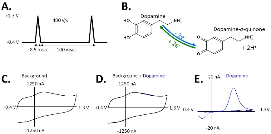
1.2.2.1 Fast-scan Cyclic Voltammetry
flux of electrons is measured as current and is directly proportional to the number of molecules that undergo the electrolysis. Due to the fast scan rate, scanning generates a large capacitive charging current at the electrode surface, which is significantly larger than faradaic currents resulting from redox processes of DA at the microelectrode surface. These background currents are stable over tens of seconds, which allows for their subtraction, revealing the interesting faradaic responses. The resulting background-subtracted cyclic voltammogram (CV) provides information on the identity, redox potentials, reversibility, and electron transfer kinetics of DA. The amplitude of the peaks can be correlated to the concentration of the analyte at the electrode surface with in-vitro calibration, and the shape of the CV can aid DA identification against interferences.
Figure 1.3. Principles of background subtracted FSCV. (A) A triangular potential waveform is applied to a working electrode. (B) The redox reaction of DA will exchange electrons at the electrode surface, generating faradaic current. (C) Application of this waveform generates a stable non-faradaic background current. (D) At low analyte concentrations, the faradaic response is small compared to the background current. (E) The stable non-faradaic background current can be subtracted from the faradaic current arising from the redox reaction. The resulting CV is an electrochemical fingerprint to identify the analyte.
CV can be extracted from a color plot at a specific time (vertical line) to serve as a molecular identifier (Figure 1.4B), or the change of current vs. time can be extracted to study the kinetics associated with dynamic changes (horizontal line) (Figure 1.4C).
Figure 1.4. The color plot. (A) 3-D color plot of DA detection with applied potential on the y-axis, time on the x-axis, and collected current represented in color. (B) CV extracted at time indicated by vertical line aids in analyte identification. (C) Current vs. time extracted at the potential indicated by horizontal line in (A) provides dynamic information.
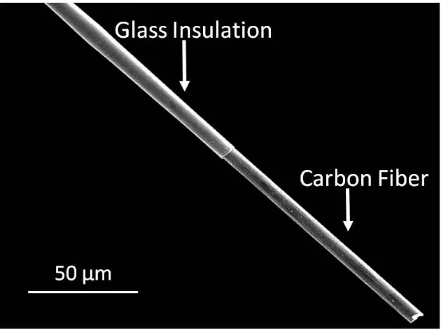
1.3 Carbon-fiber Microelectrodes
The microelectrodes typically used with FCSV are cylindrically shaped carbon-fiber microelectrodes (13). The small size of microelectrodes results in a low time constant that enables high speed measurements. In addition, carbon materials are beneficial as the sensing substrate for in vivo applications due to their wide potential window, biocompatibility, inert chemical nature, and low cost (13). Their surface is rich in oxygen containing functional groups which play a significant role in electron-transfer reactions. Carbon fibers with a
- +
diameter ranging between 5 and 15 µm are commonly employed for the fabrication of microelectrodes (13). A single fiber is aspirated into a glass capillary which works as the insulating material. Using a micropipette puller, the capillary tube containing the fiber is heated and pulled, leaving two glass insulated carbon-fiber microelectrodes. Finally, the fiber length at the sensing tip is adjusted to about 100 µm, using a blade and optical microscope (Figure 1.5). This carbon-fiber electrode is approximately 40-fold shorter and 50-fold smaller in diameter than a typical microdialysis probe, and thus it is particularly well suited to probe brain regions that have gradations in the density of neuronal terminals over these dimensions (16).
Figure 1.5. Representative image of a cylinder carbon-fiber microelectrode. Carbon-fiber microelectrodes are fabricated by aspirating a single fiber into a glass capillary which works as the insulating material. The carbon-fiber of the electrode tip is around 100 µm in length and 7µm in diameter.
In addition to the relatively easy and cost-effective construction, the surface of carbon-fiber microelectrodes can be regenerated by electrochemical oxidation (17). Regeneration of a fresh electrode in real time is advantageous during many electrochemical experiments. It allows for consistency in the electrochemical properties of carbon electrodes and can also help avoid biofouling. Because of all of these advantages, carbon-fiber microelectrodes have enabled real progress in advancing in vivo FSCV for the past 30 years.
1.4 Detecting Dopamine with FSCV in the Brain
Background-subtracted FSCV for the detection of electroactive DA has proven to be sensitive and selective, with detection limits in the nanomolar range for DA when using a cylindrical electrode, approximately 5×100 µm (13). Also, FSCV allows for an assessment of the kinetics of release and uptake of DA in the brain. Thus, this technique has proven to be a valuable tool for studying the dopaminergic system, which is heavily implicated in control of movement (4, 10) and reward-seeking behavior (7, 11, 18-19).
1.4.1 Electrically stimulated DA release in the brain
experimental goals. Typical stimulation parameters are: 60 rectangular pulses, 24 Hz, 150 µA, 2 ms/phase. DA release from a mild stimulation train is highly reproducible, with no significant effects on animals’ behavior. Thus, the effects of a pharmacologic manipulation
on electrically-evoked DA release and uptake can be evaluated by comparing the evoked signal collected before and after drug administration.
Figure 1.6. Stimulating electrode in SN in a schematic representation of the rat brain. Bipolar stimulation electrode can be positioned near dopaminergic cell bodies or dopaminergic axons in a rat’s brain. Application of mild electrical pulses elicits DA release from DA terminals in the striatum. A carbon-fiber microelectrode positioned in the striatum can detect dynamic chemical release events.
1.4.2 Naturally Occurring DA Dynamics in Freely-moving Animals
neurochemical measurement of a naturally occurring DA transient was in the NAc of male rats at their first-time entrance to a novel environment, reported by Rebec et al. (25). Lately, phasic DA signals have been detected and shown to be directly associated with sexual behavior (22), drug-seeking behavior (26) and reward related learning and decision making (27). Thus, transient DA release is now regarded as the functionally relevant mode of DA neurotransmission that mediates the reinforcing properties of drug reward and modulates goal-directed behavior (19).
Figure 1.7. Diagram of experimental set up for behavioral experiments. The animal is placed in the behavioral chamber. It is tethered using the stimulator cable on which the headstage is mounted. The headstage makes voltammetric recordings, while the animal is freely behaving in the operant chamber. The arrows indicate the direction of information flow.
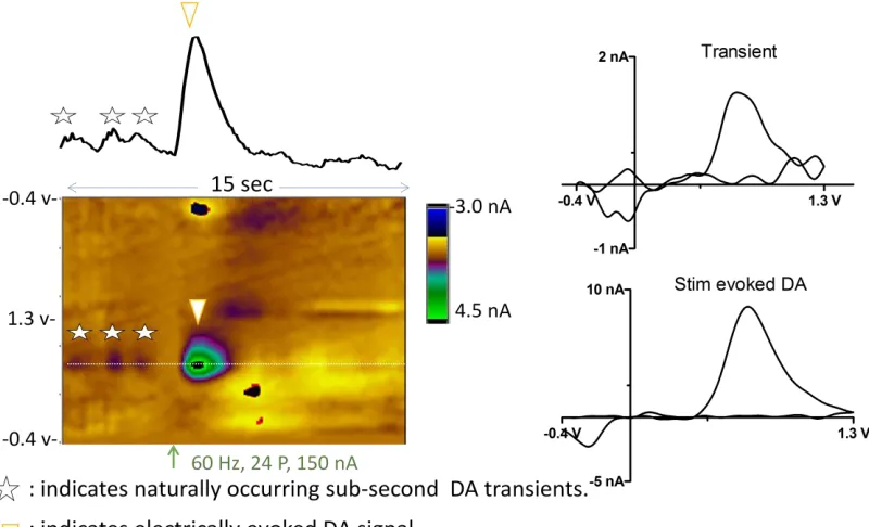
After the rat is placed into the operant box, a micromanipulator containing a retracted microelectrode is locked in place and the working and reference electrodes are connected to the headstage. The electrode is slowly lowered into tissue in small increments until electrically stimulated DA release is repeatedly observed. The electrode is then secured in position by a locking device on the micromanipulator and the experiment is initiated. Figure 1.8 shows an example 15 sec voltammetric recording obtained in the NAc of a resting, freely-moving rat. At this recording site, both electrically evoked and naturally-occurring frequent DA transients were observed. Electrical stimulation of the VTA (red arrow) causes immediate DA release in the NAc (white triangle).The individual background subtracted
Figure 1.8. Representative color plot depicting electrically-stimulated (inverted white triangle) and naturally-occurring (white stars) DA transients measured in an awake rat. The color plot shows a 15s time window of voltammetric data with the current (color) at various applied potentials (y-axis) across time (x-axis). The oxidation potential of DA versus the reference electrode is depicted by the white dotted line. Electrical stimulation (24 pulses, 60Hz, 150 mA) occurred at the time indicated by the orange arrow. Above the color plot, current measured at the oxidation potential of DA is plotted over time and converted to DA concentration via post-experiment electrode calibration. The background-subtracted CVs for DA (right) are identical to those for DA measured in vitro (data not shown).
1.5 Advancing FSCV for longitudinal real-time neurotransmitter measurements in freely- moving animals
animal models or throughout various learning paradigms (27, 31). Additionally, efforts are underway to adopt wireless technology for in vivo measurements and eliminate the cable between the animal and the recording equipment, permitting the animal more unrestricted motion and reducing susceptibility to ambient electrical noise (32-36). However, the amount of data generated and transferred in a typical FSCV experiment can impose a significant hurdle for the continued development and application of long-term data measurements and wireless data transmission (36). Existing FSCV protocols to measure neurotransmitter signalling in the brain were created without consideration for data generation or data transfer rates. Traditionally, 10 cyclic voltammograms are collected per second with 1000 data points per cyclic voltammogram. Therefore, in a single 30 second file, there are 300,000 data points collected. Thus, a chronic experiment lasting several days to months, generates a tremendous amount of data.
1.6 Dissertation overview
This dissertation consists of seven additional chapters that build on the topics discussed in this introduction chapter, with a focus on applying FSCV to investigate unknown neurochemical mechanisms underlying motor disorders and drug addiction in vivo, and advancing FSCV protocols and sensors for longitudinal DA measurements in feely moving animals.
used to assess electrically-evoked DA release and spontaneous H2O2 dynamics in intact and
1.7 References
1. Mark F. Bear, B. W. C., Michael A. Paradiso. (2007) Neuroscience : exploring the brain, 3rd ed., Lippincott Williams & Wilkins, Philadelphia, PA.
2. Schultz, W. (2007) Multiple dopamine functions at different time courses, Annu Rev Neurosci 30, 259-288.
3. Jack R. Cooper, F. E. B., Robert H. Roth (2003) The Biochemical Basis of Neuropharmacology, 8th edition ed., Oxford University Press, New York.
4. Elsworth, J. D., and Roth, R. H. (1997) Dopamine synthesis, uptake, metabolism, and receptors: relevance to gene therapy of Parkinson's disease, Exp Neurol 144, 4-9. 5. Wightman, R. M., and Robinson, D. L. (2002) Transient changes in mesolimbic
dopamine and their association with 'reward', J Neurochem 82, 721-735.
6. Overton, P. G., and Clark, D. (1997) Burst firing in midbrain dopaminergic neurons, Brain Res Brain Res Rev 25, 312-334.
7. Grace, A. A., Floresco, S. B., Goto, Y., and Lodge, D. J. (2007) Regulation of firing of dopaminergic neurons and control of goal-directed behaviors, Trends Neurosci 30, 220-227.
8. Sombers, L. A., Beyene, M., Carelli, R. M., and Wightman, R. M. (2009) Synaptic overflow of dopamine in the nucleus accumbens arises from neuronal activity in the ventral tegmental area, J Neurosci 29, 1735-1742.
10. Kish, S. J., Shannak, K., and Hornykiewicz, O. (1988) Uneven pattern of dopamine loss in the striatum of patients with idiopathic Parkinson's disease. Pathophysiologic and clinical implications, N Engl J Med 318, 876-880.
11. Schultz, W., Dayan, P., and Montague, P. R. (1997) A neural substrate of prediction and reward, Science 275, 1593-1599.
12. Justice, J. B., Jr. (1993) Quantitative microdialysis of neurotransmitters, J Neurosci Methods 48, 263-276.
13. Robinson, D. L., Hermans, A., Seipel, A. T., and Wightman, R. M. (2008) Monitoring rapid chemical communication in the brain, Chem Rev 108, 2554-2584. 14. Heien, M. L., Phillips, P. E., Stuber, G. D., Seipel, A. T., and Wightman, R. M.
(2003) Overoxidation of carbon-fiber microelectrodes enhances dopamine adsorption and increases sensitivity, Analyst 128, 1413-1419.
15. Michael, D. J., Joseph, J. D., Kilpatrick, M. R., Travis, E. R., and Wightman, R. M. (1999) Improving data acquisition for fast-scan cyclic voltammetry, Anal Chem 71, 3941-3947.
16. Arbuthnott, G. W., and Wickens, J. (2007) Space, time and dopamine, Trends Neurosci 30, 62-69.
17. Takmakov, P., Zachek, M. K., Keithley, R. B., Walsh, P. L., Donley, C., McCarty, G. S., and Wightman, R. M. (2010) Carbon microelectrodes with a renewable surface, Anal Chem 82, 2020-2028.
19. Wise, R. A. (2004) Dopamine, learning and motivation, Nat Rev Neurosci 5, 483-494. 20. Kuhr, W. G., Wightman, R. M., and Rebec, G. V. (1987) Dopaminergic neurons: simultaneous measurements of dopamine release and single-unit activity during stimulation of the medial forebrain bundle, Brain Res 418, 122-128.
21. Wightman, R. M., Amatore, C., Engstrom, R. C., Hale, P. D., Kristensen, E. W., Kuhr, W. G., and May, L. J. (1988) Real-time characterization of dopamine overflow and uptake in the rat striatum, Neuroscience 25, 513-523.
22. Robinson, D. L., Phillips, P. E., Budygin, E. A., Trafton, B. J., Garris, P. A., and Wightman, R. M. (2001) Sub-second changes in accumbal dopamine during sexual behavior in male rats, Neuroreport 12, 2549-2552.
23. Robinson, D. L., Heien, M. L., and Wightman, R. M. (2002) Frequency of dopamine concentration transients increases in dorsal and ventral striatum of male rats during introduction of conspecifics, J Neurosci 22, 10477-10486.
24. Robinson, D. L., Venton, B. J., Heien, M. L., and Wightman, R. M. (2003) Detecting subsecond dopamine release with fast-scan cyclic voltammetry in vivo, Clin Chem 49, 1763-1773.
25. Rebec, G. V., Christensen, J. R., Guerra, C., and Bardo, M. T. (1997) Regional and temporal differences in real-time dopamine efflux in the nucleus accumbens during free-choice novelty, Brain Res 776, 61-67.
27. Clark, J. J., Hollon, N. G., and Phillips, P. E. (2012) Pavlovian valuation systems in learning and decision making, Curr Opin Neurobiol 22, 1054-1061.
28. Wightman, R. M., Heien, M. L., Wassum, K. M., Sombers, L. A., Aragona, B. J., Khan, A. S., Ariansen, J. L., Cheer, J. F., Phillips, P. E., and Carelli, R. M. (2007) Dopamine release is heterogeneous within microenvironments of the rat nucleus accumbens, Eur J Neurosci 26, 2046-2054.
29. Stuber, G. D., Roitman, M. F., Phillips, P. E., Carelli, R. M., and Wightman, R. M. (2005) Rapid dopamine signaling in the nucleus accumbens during contingent and noncontingent cocaine administration, Neuropsychopharmacology 30, 853-863. 30. Roitman, M. F., Stuber, G. D., Phillips, P. E., Wightman, R. M., and Carelli, R. M.
(2004) Dopamine operates as a subsecond modulator of food seeking, J Neurosci 24, 1265-1271.
31. Clark, J. J., Sandberg, S. G., Wanat, M. J., Gan, J. O., Horne, E. A., Hart, A. S., Akers, C. A., Parker, J. G., Willuhn, I., Martinez, V., Evans, S. B., Stella, N., and Phillips, P. E. (2010) Chronic microsensors for longitudinal, subsecond dopamine detection in behaving animals, Nat Methods 7, 126-129.
33. Agnesi, F., Tye, S. J., Bledsoe, J. M., Griessenauer, C. J., Kimble, C. J., Sieck, G. C., Bennet, K. E., Garris, P. A., Blaha, C. D., and Lee, K. H. (2009) Wireless Instantaneous Neurotransmitter Concentration System-based amperometric detection of dopamine, adenosine, and glutamate for intraoperative neurochemical monitoring, J Neurosurg 111, 701-711.
34. Kimble, C. J., Johnson, D. M., Winter, B. A., Whitlock, S. V., Kressin, K. R., Horne, A. E., Robinson, J. C., Bledsoe, J. M., Tye, S. J., Chang, S. Y., Agnesi, F., Griessenauer, C. J., Covey, D., Shon, Y. M., Bennet, K. E., Garris, P. A., and Lee, K. H. (2009) Wireless Instantaneous Neurotransmitter Concentration Sensing System (WINCS) for intraoperative neurochemical monitoring, Conf Proc IEEE Eng Med Biol Soc 2009, 4856-4859.
35. Griessenauer, C. J., Chang, S. Y., Tye, S. J., Kimble, C. J., Bennet, K. E., Garris, P. A., and Lee, K. H. (2010) Wireless Instantaneous Neurotransmitter Concentration System: electrochemical monitoring of serotonin using fast-scan cyclic voltammetry--a proof-of-principle study, J Neurosurg 113, 656-665.
CHAPTER 2
Nafion-coated Carbon-fiber Microelectrodes for Rapid Detection of L-DOPA’s Effects on Dopamine Signalling in Rat Brain
This work was completed in collaboration with: Stephanie Hughes and Leslie A. Sombers. The author designed the experiments, collected, and analyzed the data presented in this chapter.
2.1 Introduction
Parkinson’s disease (PD) is a neurodegenerative disorder that affects more than a
million people in the United States (1). It is characterized by motor deficits including bradykinesia, rigidity, and resting tremor, resulting from a progressive loss of nigrostriatal dopamine (DA) neurons (2-3). DA does not readily cross the blood-brain barrier, thus its use for treating the symptoms of PD is precluded (4). 3, 4-dihydroxyphenyl-L-alanine (L-DOPA), the metabolic precursor to DA, has routinely been used for symptomatic treatment of PD since the late 1960s (3, 5-6). However, the efficacy of prolonged L-DOPA treatment wanes over time and patients develop serious motor complications (3, 7-8). Despite common use in the therapeutic management of PD, remarkably little is known about how L-DOPA replacement therapy alters the dynamics of pulsatile DA fluctuations that occur on a fast (seconds) timescale.
method that is well suited to examine steady-state or slowly changing levels of analytes in the extracellular fluid (13-15). Thus, these studies have significantly advanced our knowledge on how L-DOPA serves to gradually increase striatal DA levels (9-12). However, quantification of the effects of L-DOPA on rapid DA dynamics is also important, because rapid DA signalling in both the ventral medial and sensorimotor striatum have been shown to play keys roles in controlling specific aspects of behaviour (15-17). Over the past twenty years, electrochemical techniques have proven to be particularly useful for monitoring rapid changes in DA concentration resulting from discrete neurochemical events. However, electrochemical studies investigating the effects of L-DOPA administration on electrically-evoked DA release in intact animals have reported conflicting results with significant variability (18-21). Clarifying this question is critical to developing improved therapies for the treatment of PD.
Figure 2.1. DA polymerization pathway.
(40). We have systematically optimized the procedure for Nafion coating cylindrical carbon-fiber microelectrodes for in vivo applications. We use fast-scan cyclic voltammetry (FSCV) to demonstrate that generation of a Nafion membrane after electrochemical conditioning reliably produces a uniform film on the sensor surface that preserves a rapid response to DA while preventing fouling. We then use this approach to assess the effects of various concentrations of systemic L-DOPA administration on DA dynamics measured in the striatum with sub-second temporal resolution. In addition to providing chemical information that is highly relevant to the most common therapy used in the treatment of PD, this work serves to benefit a wide variety of studies plagued by electrode fouling because Nafion is so commonly used in electroanalytical chemistry.
2.2 Experimental section 2.2.1 Chemicals
See Appendix A.
2.2.2 Carbon-Fiber Microelectrode Fabrication
See Appendix A.
2.2.3 Data Acquisition
frequency of 10 Hz, as described previously (43). More details about data acquisition process are described in Appendix.
2.2.4 Flow Injection Apparatus
See Appendix A.
2.2.5 Nafion-Coating Protocols
2.2.6 Surgery
See Appendix A.
2.2.7 In-vivo Experimental Design
Following electrode implantation, carbon-fiber microelectrodes were cycled at 10 Hz for 15 min to stabilize the background current. L-DOPA methyl-ester and benserazide-hydrochloride, a peripheral DOPA decarboxylase inhibitor (46), were dissolved together in physiological saline. After baseline data collection, each animal received an acute treatment with L-DOPA methyl-ester/benserazide cocktail at a clinically-relevant dose (6 mg/kg + 10 mg/kg benserazide, i.p.), followed by a higher dose (250 mg/kg + 400 mg/kg benserazide, i.p.). These doses of DOPA methyl-ester are equivalent to 5 mg/kg and 200 mg/kg of L-DOPA, respectively (21). Data was collected for one hour after each drug administration.
2.2.8 Statistics
See Appendix A.
2.3 Results and Discussion
2.3.1 Microelectrode Fouling by L-DOPA Administration
Figure 2.2. L-DOPA decreases electrically-evoked DA release recorded at bare carbon-fiber microelectrodes. (A) Representative data collected at a bare carbon-fiber microelectrode before L-DOPA administration (left column), 20 min after administration of L-DOPA (5 mg/kg, i.p., middle), and 20 min after administration of a higher dose (200 mg/kg, i.p., right). Top: Color plots depicting all changes in current (false color) collected over a 15-s window (x-axis) across all potentials (y-axis). Electrical stimulation of the MFB is indicated by the black arrow. Middle: Current vs. time traces extracted at the oxidation potential of DA (+0.6V). Bottom: Cyclic voltammograms extracted from the data. (B) Across the entire data set, there is a significant decrease in electrically-evoked DA release upon acute L-DOPA treatment (n = 5, *p < 0.05, **p<0.01, ***p<0.001, one-way repeated measures ANOVA).
2.3A); however, 50 µM L-DOPA significantly attenuated sensitivity (Figure 2.3B, n = 6, *** p<0.001). Indeed, Spina and Cohen have reported a peak value of 50 µM L-DOPA in the rat striatum after acute i.p. administration of 100 mg/kg L-DOPA (58). Thus, the decrease in evoked DA detected at bare carbon-fiber microelectrodes after a 200 mg/kg L-DOPA treatment suggests fouling of the electrode surface. If not taken into account, this significantly skews interpretation and quantification of in vivo data.
Figure 2.3. L-DOPA fouls carbon-fiber microelectrodes. Normalized response of bare carbon-fiber microelectrodes to 1 μM DA in the absence (black) and presence (grey) of (A) 1 µM or (B) 50 µM L-DOPA in vitro. Sensitivity was significantly attenuated in the presence of 50 µM L-DOPA (n = 6, paired t- test,*** p<0.001).
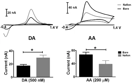
2.3.2 Systematic Characterization of Nafion-Coating Procedure
collected in vitro using bare and Nafion-coated carbon-fiber microelectrodes. The entire data set is summarized in the lower panel. Nafion-coated electrodes provided a 71% increase in sensitivity to DA, and decreased sensitivity to AA to 57% of the value inherent to uncoated electrodes. Thus, the presence of a Nafion layer can be confirmed by reporting a sensitivity ratio for these species (the quotient of the sensitivities measured before and after coating). A large deviation from unity indicates robust Nafion membrane performance.
Figure 2.5. Systematic characterization of Nafion-coating procedures. (A) Dip-coating versus electrodeposition, (B) Electrochemical pretreatment before vs. after Nafion electrodeposition, (C) Various electrodeposition potentials, (D) Various electrodeposition times. Data are presented as normalized sensitivity ratio (n = 6 –17 electrodes per protocol, one way ANOVA, * p<0.05; ** p< 0.01; *** p<0.001).
than ~1.3V can chemically alter, or even etch, the electrode surface (34, 65, 72). Thus, we hypothesized that electrochemical conditioning with a commonly-used waveform (triangular, ranging from -0.4 V to 1.4 V vs. Ag/AgCl) would exacerbate the generally inconsistent adhesion of Nafion to the electrode surface. We quantitatively compared the performance of Nafion-coated carbon-fiber microelectrodes that were electrochemically pre-treated before Nafion deposition with those that were conditioned immediately prior to measurement. The data unequivocally demonstrate that Nafion eletrodeposition was significantly more effective when electrodes were conditioned prior to the deposition procedure (Figure 2.5B). Based on this, all subsequent protocols included electrochemical pretreatment of bare carbon-fiber microelectrodes prior to electrodeposition of Nafion.
Next, the potentials employed to electrodeposit the Nafion membrane were systematically investigated. Three potentials were selected: +0.5, +1.0 and +1.5 V vs. Ag/AgCl, as these have all been previously reported in the literature (38, 41, 73-74). The results indicate that +1.0 and +1.5 V were more effective than +0.5 V in generating a reliable membrane (Figure 2.5C). However, as described above, the application of positive potentials can modify the surface of the carbon fiber, making it highly absorptive and potentially slowing electrode response time, convoluting electrochemical performance (63, 65). Thus, 1.0 V (vs. Ag/AgCl) was selected as the most appropriate potential for the electrodeposition of Nafion.
Figure 2.7. Representative DA concentration traces upon L-DOPA treatment recorded at different Nafion-modified electrodes. Electrical stimulation was used to evoke striatal DA release every 5 minutes. X-axis shows the 2- h window of data collection. Y-axis represents the normalized amplitude of electrically-evoked DA release. Arrows indicate the time at which L-DOPA was administrated (orange: 5 mg/kg, red: 200 mg/kg).
2.3.3 The Effects of L-DOPA on Striatal DA Dynamics
administration (n = 4, *p < 0.05, **p < 0.01, ***p<0.001). This suggests that following acute administration of a clinically relevant dose of L-DOPA (5 mg/kg), the regulatory mechanisms of the intact animals are capable of regulating extracellular DA very efficiently. However, administration of a higher dose of L-DOPA (200 mg/kg) is capable of enhancing DA overflow. These data are consistent with previous reports using microdialysis in which striatal DA was increased by ~200% in intact animals following a 200 mg/kg or higher dose of L-DOPA (10, 20, 76).
Figure 2.8. L-DOPA increases electrically-evoked DA release. (A) Representative data collected at a Nafion-coated carbon-fiber microelectrode in an intact animal before L-DOPA administration (left column), 20 min after administration of L-DOPA (5mg/kg, i.p (middle), and 20 min after administration of a higher dose of L-DOPA (200 mg/kg, i.p.right). (B) Across all animals, the amplitude of DA release was increased, but was not significantly increased until 200 mg/kg was administered (red, n = 4, *p < 0.05, **p<0.01, one-way repeated measures ANOVA).
to successfully prevent the fouling caused by L-DOPA at carbon-fiber microelectrodes, and to accurately report the effects of this treatment on DA dynamics in vivo when using electrochemical measurements.
Figure 2.9. Direct comparison of bare vs. Nafion-coated carbon-fiber microelectrodes in vivo. Nafion-coated carbon-fiber microelectrodes (blue, n =4), recorded an increase in evoked DA release in response to acute L-DOPA treatment in intact animals, in direct contrast to the results collected at bare electrodes (black, n = 5, repeated measures two-way ANOVA , *p < 0.05 **p<0.01, ***p< 0.001).
2.4 Conclusions
2.5 References
1. Sutachan, J. J., Casas, Z., Albarracin, S. L., Stab, B. R., 2nd, Samudio, I., Gonzalez, J., Morales, L., and Barreto, G. E. (2012) Cellular and molecular mechanisms of antioxidants in Parkinson's disease, Nutr Neurosci 15, 120-126.
2. Jankovic, J. (2008) Parkinson's disease: clinical features and diagnosis, J Neurol Neurosur Ps 79, 368-376.
3. Fahn, S. (2006) Levodopa in the treatment of Parkinson's disease, J Neural Transm Suppl, 1-15.
4. Mark F. Bear, B. W. C., Michael A. Paradiso. (2007) Neuroscience : exploring the brain, 3rd ed., Lippincott Williams & Wilkins, Philadelphia, PA.
5. Cotzias, G. C., Van Woert, M. H., and Schiffer, L. M. (1967) Aromatic amino acids and modification of parkinsonism, N Engl J Med 276, 374-379.
6. Hornykiewicz, O. (2002) L-DOPA: from a biologically inactive amino acid to a successful therapeutic agent, Amino Acids 23, 65-70.
7. Ahlskog, J. E., and Muenter, M. D. (2001) Frequency of levodopa-related dyskinesias and motor fluctuations as estimated from the cumulative literature, Movement Disord 16, 448-458.
8. Nutt, J. G. (1992) Dyskinesia induced by levodopa and dopamine agonists in patients with Parkinson’s disease, in Drug-Induced Movement Disorders, Futura Publishing
Co. Inc., Mount Kisko.
rat model of Parkinson's disease: temporal and quantitative relationship to the expression of dyskinesia, J. Neurochem. 112, 1465-1476.
10. Abercrombie, E. D., Bonatz, A. E., and Zigmond, M. J. (1990) Effects of L-dopa on extracellular dopamine in striatum of normal and 6-hydroxydopamine-treated rats, Brain Res 525, 36-44.
11. Robinson, T. E., Mocsary, Z., Camp, D. M., and Whishaw, I. Q. (1994) Time course of recovery of extracellular dopamine following partial damage to the nigrostriatal dopamine system, J. Neurosci. 14, 2687-2696.
12. Zhang, W. Q., Tilson, H. A., Nanry, K. P., Hudson, P. M., Hong, J. S., and Stachowiak, M. K. (1988) Increased dopamine release from striata of rats after unilateral nigrostriatal bundle damage, Brain Res. 461, 335-342.
13. Lu, Y., Peters, J. L., and Michael, A. C. (1998) Direct comparison of the response of voltammetry and microdialysis to electrically evoked release of striatal dopamine, J Neurochem 70, 584-593.
14. Khan, A. S., and Michael, A. C. (2003) Invasive consequences of using micro-electrodes and microdialysis probes in the brain, Trac-Trend Anal Chem 22, 503-508. 15. Robinson, D. L., Hermans, A., Seipel, A. T., and Wightman, R. M. (2008)
Monitoring rapid chemical communication in the brain, Chem Rev 108, 2554-2584. 16. Phillips, P. E., Stuber, G. D., Heien, M. L., Wightman, R. M., and Carelli, R. M.
(2003) Subsecond dopamine release promotes cocaine seeking, Nature 422, 614-618. 17. Shnitko, T. A., and Robinson, D. L. (2014) Regional variation in phasic dopamine
18. May, L. J., Kuhr, W. G., and Wightman, R. M. (1988) Differentiation of dopamine overflow and uptake processes in the extracellular fluid of the rat caudate nucleus with fast-scan in vivo voltammetry, J Neurochem 51, 1060-1069.
19. Garris, P. A., Ciolkowski, E. L., Pastore, P., and Wightman, R. M. (1994) Efflux of dopamine from the synaptic cleft in the nucleus accumbens of the rat brain, J Neurosci 14, 6084-6093.
20. Rodriguez, M., Morales, I., Gonzalez-Mora, J. L., Gomez, I., Sabate, M., Dopico, J. G., Rodriguez-Oroz, M. C., and Obeso, J. A. (2007) Different levodopa actions on the extracellular dopamine pools in the rat striatum, Synapse 61, 61-71.
21. Lundblad, M., af Bjerken, S., Cenci, M. A., Pomerleau, F., Gerhardt, G. A., and Stromberg, I. (2009) Chronic intermittent L-DOPA treatment induces changes in dopamine release, J. Neurochem. 108, 998-1008.
22. Yu, M. E., Hwang, J. Y., and Deming, T. J. (1999) Role of L-3,4-dihydroxyphenylalanine in mussel adhesive proteins, J Am Chem Soc 121, 5825-5826.
23. Tse, D. C., McCreery, R. L., and Adams, R. N. (1976) Potential oxidative pathways of brain catecholamines, J Med Chem 19, 37-40.
24. Harreither, W., Trouillon, R., Poulin, P., Neri, W., Ewing, A. G., and Safina, G. (2013) Carbon nanotube fiber microelectrodes show a higher resistance to dopamine fouling, Anal Chem 85, 7447-7453.
26. Azari, S., and Zou, L. D. (2012) Using zwitterionic amino acid L-DOPA to modify the surface of thin film composite polyamide reverse osmosis membranes to increase their fouling resistance, J Membrane Sci 401, 68-75.
27. Xi, Z. Y., Xu, Y. Y., Zhu, L. P., Wang, Y., and Zhu, B. K. (2009) A facile method of surface modification for hydrophobic polymer membranes based on the adhesive behavior of poly(DOPA) and poly(dopamine), J Membrane Sci 327, 244-253.
28. Koile, R. C., and Johnson, D. C. (1979) Electrochemical Removal of Phenolic Films from a Platinum Anode, Analytical Chemistry 51, 741-744.
29. Adams RN, M. C. (1982) Electrochemical detection methods for monoamine measurements in vitro and in vivo, Plenum Press.
30. Stamford, J. A. (1986) In vivo voltammetry: some methodological considerations, J Neurosci Methods 17, 1-29.
31. Cahill, P. S., Walker, Q. D., Finnegan, J. M., Mickelson, G. E., Travis, E. R., and Wightman, R. M. (1996) Microelectrodes for the measurement of catecholamines in biological systems, Anal Chem 68, 3180-3186.
32. Burmeister, J. J., Pomerleau, F., Huettl, P., Gash, C. R., Werner, C. E., Bruno, J. P., and Gerhardt, G. A. (2008) Ceramic-based multisite microelectrode arrays for simultaneous measures of choline and acetylcholine in CNS, Biosens Bioelectron 23, 1382-1389.
34. Roberts, J. G., Moody, B. P., McCarty, G. S., and Sombers, L. A. (2010) Specific oxygen-containing functional groups on the carbon surface underlie an enhanced sensitivity to dopamine at electrochemically pretreated carbon fiber microelectrodes, Langmuir 26, 9116-9122.
35. Gerhardt, G. A., Oke, A. F., Nagy, G., Moghaddam, B., and Adams, R. N. (1984) Nafion-coated electrodes with high selectivity for CNS electrochemistry, Brain Res 290, 390-395.
36. Rice, M. E., Oke, A. F., Bradberry, C. W., and Adams, R. N. (1985) Simultaneous voltammetric and chemical monitoring of dopamine release in situ, Brain Res 340, 151-155.
37. Singh, Y. S., Sawarynski, L. E., Dabiri, P. D., Choi, W. R., and Andrews, A. M. (2011) Head-to-head comparisons of carbon fiber microelectrode coatings for sensitive and selective neurotransmitter detection by voltammetry, Anal Chem 83, 6658-6666.
38. Hashemi, P., Dankoski, E. C., Petrovic, J., Keithley, R. B., and Wightman, R. M. (2009) Voltammetric detection of 5-hydroxytryptamine release in the rat brain, Anal Chem 81, 9462-9471.
39. Valdes, T. I., and Moussy, F. (2000) In vitro and in vivo degradation of glucose oxidase enzyme used for an implantable glucose biosensor, Diabetes Technol Ther 2, 367-376.
41. Capella, P., Ghasemzadeh, B., Mitchell, K., and Adams, R. N. (1990) Nafion-Coated Carbon-Fiber Electrodes for Neurochemical Studies in Brain-Tissue, Electroanal 2, 175-182.
42. Ross, A. E., and Venton, B. J. (2012) Nafion-CNT coated carbon-fiber microelectrodes for enhanced detection of adenosine, Analyst 137, 3045-3051.
43. Roberts, J. G., Lugo-Morales, L. Z., Loziuk, P. L., and Sombers, L. A. (2013) Real-time chemical measurements of dopamine release in the brain, Methods Mol Biol 964, 275-294.
44. Bath, B. D., Michael, D. J., Trafton, B. J., Joseph, J. D., Runnels, P. L., and Wightman, R. M. (2000) Subsecond adsorption and desorption of dopamine at carbon-fiber microelectrodes, Anal Chem 72, 5994-6002.
45. Paxinos, G. W., C. (1986) The rat brain in stereotaxic coordinates, 2nd ed. ed., Academic Press, Orlando, FL,.
46. Silva, M. A., Mattern, C., Hacker, R., Tomaz, C., Huston, J. P., and Schwarting, R. K. (1997) Increased neostriatal dopamine activity after intraperitoneal or intranasal administration of L-DOPA: on the role of benserazide pretreatment, Synapse 27, 294-302.
47. Michael, D., Travis, E. R., and Wightman, R. M. (1998) Color images for fast-scan CV measurements in biological systems, Anal Chem 70, 586A-592A.
49. Marburger, A., Sohr, R., Reum, T., and Morgenstern, R. (2000) Comparison by microdialysis of striatal L-DOPA after its systemic administration in rats with probes implanted acutely or through a guide cannula, J Neurosci Meth 102, 127-132.
50. Zhang, J., Qu, F. R., Nakatsuka, A., Nomura, T., Nagai, M., and Nomoto, M. (2003) Pharmacokinetics of L-dopa in plasma and extracellular fluid of striatum in common marmosets, Brain Res 993, 54-58.
51. Eslami, M., Zare, H. R., and Namazian, M. (2012) Thermodynamic parameters of electrochemical oxidation of L-DOPA: experimental and theoretical studies, J Phys Chem B 116, 12552-12557.
52. Aslanoglu, M., Kutluay, A., Goktas, S., and Karabulut, S. (2009) Voltammetric behaviour of levodopa and its quantification in pharmaceuticals using a beta-cyclodextrine doped poly(2,5-diaminobenzenesulfonic acid) modified electrode, J Chem Sci 121, 209-215.
53. Cabanes, J., Garciacanovas, F., Lozano, J. A., and Garciacarmona, F. (1987) A Kinetic-Study of the Melanization Pathway between L-Tyrosine and Dopachrome, Biochim Biophys Acta 923, 187-195.
54. Robinson, G. M., and Smyth, M. R. (1997) Simultaneous determination of products and intermediates of L-dopa oxidation using capillary electrophoresis with diode-array detection, Analyst 122, 797-802.
55. Prota, G. (1992) Melanins and Melanogenesis, Academic Press, New York.
57. Nicolai, M., Goncalves, G., Natalio, F., and Humanes, M. (2011) Biocatalytic formation of synthetic melanin: The role of vanadium haloperoxidases, L-DOPA and iodide, J Inorg Biochem 105, 887-893.
58. Spina, M. B., and Cohen, G. (1988) Exposure of School Synaptosomes to L-Dopa Increases Levels of Oxidized Glutathione, J Pharmacol Exp Ther 247, 502-507. 59. Jackson, B. P., Dietz, S. M., and Wightman, R. M. (1995) Fast-scan cyclic
voltammetry of 5-hydroxytryptamine, Anal Chem 67, 1115-1120.
60. Pihel, K., Walker, Q. D., and Wightman, R. M. (1996) Overoxidized polypyrrole-coated carbon fiber microelectrodes for dopamine measurements with fast-scan cyclic voltammetry, Anal Chem 68, 2084-2089.
61. Engstrom, R. C. (1982) Electrochemical Pretreatment of Glassy-Carbon Electrodes, Analytical Chemistry 54, 2310-2314.
62. Engstrom, R. C., and Strasser, V. A. (1984) Characterization of Electrochemically Pretreated Glassy-Carbon Electrodes, Analytical Chemistry 56, 136-141.
63. Gonon, F. G., Fombarlet, C. M., Buda, M. J., and Pujol, J. F. (1981) Electrochemical Treatment of Pyrolytic Carbon-Fiber Electrodes, Analytical Chemistry 53, 1386-1389.
64. Saraceno, R. A., and Ewing, A. G. (1988) Electron-Transfer Reactions of Catechols at Ultrasmall Carbon Ring Electrodes, Analytical Chemistry 60, 2016-2020.
66. Langley, L. A., Villanueva, D. E., and Fairbrother, D. H. (2006) Quantification of surface oxides on carbonaceous materials, Chem Mater 18, 169-178.
67. Fagan, D. T., and Kuwana, T. (1989) Thermal-Desorption Mass Analysis of Carbon-Fiber Surfaces - Surface Oxygen Complexes, Analytical Chemistry 61, 1017-1023. 68. Pantano, P., and Kuhr, W. G. (1993) Characterization of the Chemical Architecture of
Carbon-Fiber Microelectrodes .2. Correlation of Carboxylate Distribution with Electron-Transfer Properties, Analytical Chemistry 65, 2452-2458.
69. Pantano, P., and Kuhr, W. G. (1993) Dehydrogenase-Modified Carbon-Fiber Microelectrodes for the Measurement of Neurotransmitter Dynamics .2. Covalent Modification Utilizing Avidin Biotin Technology, Analytical Chemistry 65, 623-630. 70. Pantano, P., and Kuhr, W. G. (1991) Characterization of the Chemical Architecture of
Carbon-Fiber Microelectrodes .1. Carboxylates, Analytical Chemistry 63, 1413-1418. 71. Ray, K. R., and McCreery, R. L. (1999) Characterization of the surface carbonyl and
hydroxyl coverage on glassy carbon electrodes using Raman spectroscopy, J Electroanal Chem 469, 150-158.
72. Takmakov, P., Zachek, M. K., Keithley, R. B., Walsh, P. L., Donley, C., McCarty, G. S., and Wightman, R. M. (2010) Carbon microelectrodes with a renewable surface, Anal Chem 82, 2020-2028.
74. Crespi, F., Martin, K. F., and Marsden, C. A. (1988) Measurement of extracellular basal levels of serotonin in vivo using nafion-coated carbon fibre electrodes combined with differential pulse voltammetry, Neuroscience 27, 885-896.
75. Kawagoe, K. T., and Wightman, R. M. (1994) Characterization of amperometry for in vivo measurement of dopamine dynamics in the rat brain, Talanta 41, 865-874. 76. Di Monte, D. A., DeLanney, L. E., Irwin, I., Royland, J. E., Chan, P., Jakowec, M.
W., and Langston, J. W. (1996) Monoamine oxidase-dependent metabolism of dopamine in the striatum and substantia nigra of L-DOPA-treated monkeys, Brain Res 738, 53-59.
77. Zigmond, M. J., Abercrombie, E. D., Berger, T. W., Grace, A. A., and Stricker, E. M. (1990) Compensations after lesions of central dopaminergic neurons: some clinical and basic implications, Trends Neurosci 13, 290-296.
CHAPTER 3
Simultaneously Monitoring the Effects of Levodopa Treatment on Dopamine and H2O2
Dynamics in Intact and 6-OHDA Lesioned Striatal Tissue Using Fast-scan Cyclic Voltammetry
This work was conducted in collaboration with: Christie Lee, Kristen Blanton, Leslie Wilson and Leslie Sombers.
3.1 Introduction
1-methyl-4-phenyl-1,2,3,6-terahydropyridine (MPTP) and 6-hydroxydopamine (6-OHDA) can cause motor dysfunction in animals (9-13). However, direct evidence demonstrating a role for ROS in PD is still lacking.
Among all of the available clinical therapies for PD, treatment with 3, 4-dihydroxyphenyl-L-alanine (L-DOPA), the metabolic precursor to dopamine (DA), has been the most efficacious in relieving Parkinsonian symptoms since the late 1960s (4, 14-16). However, the efficacy of prolonged L-DOPA treatment wanes over time and patients develop serious motor complications (4, 17-18). Despite common use in the therapeutic management of PD, remarkably little is known about how L-DOPA alters the real-time DA dynamics in the brain of animals. Additionally, reports on the effects of L-DOPA on oxidative stress are contradictory (4, 19). Some studies have suggested that L-DOPA treatment may damage dopaminergic neurons, since free radical and quinone metabolites of L-DOPA are toxic (20-23). Other studies indicate that L-DOPA may act as a neuroprotectant in animal models of PD, as it can decrease lipid peroxidation and reduce tissue content of ROS in striatum (24-25). The reason for these opposing results may be partially attributed to the static nature of the techniques used to measure ROS. These measurements are accomplished utilizing microdialysis sampling coupled with a trapping compound, or a fluorescence imaging approach (26-29).