University of Windsor University of Windsor
Scholarship at UWindsor
Scholarship at UWindsor
Electronic Theses and Dissertations Theses, Dissertations, and Major Papers
2009
Applications of gold nanoparticles to thiol and nitric oxide
Applications of gold nanoparticles to thiol and nitric oxide
chemistry
chemistry
Suzanne Durocher University of Windsor
Follow this and additional works at: https://scholar.uwindsor.ca/etd
Recommended Citation Recommended Citation
Durocher, Suzanne, "Applications of gold nanoparticles to thiol and nitric oxide chemistry" (2009). Electronic Theses and Dissertations. 7974.
https://scholar.uwindsor.ca/etd/7974
This online database contains the full-text of PhD dissertations and Masters’ theses of University of Windsor students from 1954 forward. These documents are made available for personal study and research purposes only, in accordance with the Canadian Copyright Act and the Creative Commons license—CC BY-NC-ND (Attribution, Non-Commercial, No Derivative Works). Under this license, works must always be attributed to the copyright holder (original author), cannot be used for any commercial purposes, and may not be altered. Any other use would require the permission of the copyright holder. Students may inquire about withdrawing their dissertation and/or thesis from this database. For additional inquiries, please contact the repository administrator via email
APPLICATIONS OF GOLD NANOPARTICLES TO THIOL AND NITRIC OXIDE CHEMISTRY
By
Suzanne Durocher
A Thesis
Submitted to the Faculty of Graduate Studies Through the Department of Chemistry and Biochemistry
In Partial Fulfillment of the Requirements for The Degree of Master of Science at the
University of Windsor
Windsor, Ontario, Canada 2009
1*1
Library and Archives CanadaPublished Heritage Branch
395 Wellington Street OttawaONK1A0N4 Canada
Bibliotheque et Archives Canada
Direction du
Patrimoine de I'edition
395, rue Wellington Ottawa ON K1A 0N4 Canada
Your file Votre r&ference ISBN: 978-0-494-57640-3 Our file Notre reference ISBN: 978-0-494-57640-3
NOTICE: AVIS:
The author has granted a
non-exclusive license allowing Library and Archives Canada to reproduce, publish, archive, preserve, conserve, communicate to the public by
telecommunication or on the Internet, loan, distribute and sell theses
worldwide, for commercial or non-commercial purposes, in microform, paper, electronic and/or any other formats.
L'auteur a accorde une licence non exclusive permettant a la Bibliotheque et Archives Canada de reproduire, publier, archiver, sauvegarder, conserver, transmettre au public par telecommunication ou par Plnternet, preter, distribuer et vendre des theses partout dans le monde, a des fins commerciales ou autres, sur support microforme, papier, electronique et/ou autres formats.
The author retains copyright ownership and moral rights in this thesis. Neither the thesis nor substantial extracts from it may be printed or otherwise reproduced without the author's permission.
L'auteur conserve la propriete du droit d'auteur et des droits moraux qui protege cette these. Ni la these ni des extra its substantiels de celle-ci ne doivent etre imprimes ou autrement
reproduits sans son autorisation.
In compliance with the Canadian Privacy Act some supporting forms may have been removed from this thesis.
Conformement a la loi canadienne sur la protection de la vie privee, quelques
formulaires secondaires ont ete enleves de cette these.
While these forms may be included in the document page count, their removal does not represent any loss of content from the thesis.
Bien que ces formulaires aient inclus dans la pagination, il n'y aura aucun contenu manquant.
DECLARATION OF CO-AUTHORSHIP/PREVIOUS PUBLICATION
I. Declaration of Previous Publication
This thesis includes two original papers that have been previously published in peer reviewed journals, as follows:
Chapter 2
Chapter 3
Mikula, I., Durocher, S., Martasek, P., Mutus, B. and Slama-Schwok, A. Isoform-specific differences in the nitrite reductase activity of nitric oxide synthases under hypoxia (2009). Biochem
J. 418(3):673-82.
Durocher, S., Rezaee, A., Hamm, C , Rangan, C , Mittler, S. and Mutus, B. (2009). Disulfide-linked, gold nanoparticle-based reagent for detecting small molecular weight thiols. J Am Chem
Soc. 131(7):2475-7.
Published
Published
I certify that I have obtained a written permission from the copyright owners to include the above published materials in my thesis. I certify that the above material describes work completed during my registration as graduate student at the University of Windsor.
I declare that, to the best of my knowledge, my thesis does not infringe upon anyone's copyright nor violate any proprietary rights and that any ideas, techniques, quotations, or any other material from the work of other people included in my thesis, published or otherwise, are fully acknowledged in accordance with the standard referencing practices. Furthermore, to the extent that I have included copyrighted material that surpasses the bounds of fair dealing within the meaning of the Canada Copyright Act, I certify that I have obtained a written permission from the copyright owners to include such materials in my thesis.
I declare that this is a true copy of my thesis, including any final revisions, as approved by my thesis committee and the Graduate Studies office, and that this thesis has not been submitted for a higher degree to any other University or Institution.
II. Co-Authorship Declaration
Chapter 3 was also the result of joint research. In all cases the experimental design, data and data interpretation were performed by the author under the supervision of Dr. Bulent Mutus. The collaborators, Dr. Chitra Rangan of the University of Windsor, Dr. Caroline Hamm of the Windsor Regional Cancer Centre in Windsor, Ontario, Asad Rezaee and Dr. Silvia Mittler of the University of Western Ontario in London, Ontario, contributed through funding and through discussion of experimental design and data interpretation.
I am aware of the University of Windsor Senate Policy on Authorship and I certify that I have properly acknowledged the contribution of other researchers to my thesis, and have obtained written permission from each of the co-authors to include the above materials in my thesis.
ABSTRACT
The first project studied the three nitric oxide synthases under different oxygen
tensions. We found that eNOS can reduce NO2" to NO in hypoxia and anoxia, using
endothelial cells and isolated enzymes. This suggests a new role for eNOS in times of
restricted oxygen.
In the next project, gold nanoparticles were joined with a disulfide bond-bearing
crosslinker to create a blue probe that turned red upon reduction with low molecular
weight thiols. The rate depended on the size of the molecule. Since small thiols help
control the redox of proteins their quantification helps identify oxidative stress.
In the third project, seven proteins isolated by gold nanoparticles were identified;
these may have bound by their surface thiols. Additionally, gold nanoparticles were used
to S-denitrosylate GSNO, and a flow device was designed that would automate this
method. The prototype model is manually operated but shows potential to be developed
DEDICATION
ACKNOWLEDGEMENTS
I am much indebted to Dr. Bulent Mutus for his mentorship during my time as a
Master's student, both for his supervision over my research, and for his personal interest
in the well-being of myself and his other students. He has been an invaluable person in
my life.
My fellow graduate students, past and present, have greatly enhanced my
experience as well. Dr. Arun Raturi, Dr. Inga Sliskovic, Rebecca Heeney, Adam
Faccenda, Vasantha Kallakunta, Harmanpreet Kaur, and Ruchi Chaube have made the
past 2 years very enjoyable, through sharing lunches, advice, and academic discussions. I
wish everyone great success in their scientific careers.
I also wish to thank my friends and family for helping me grow as a person during
this time. The support and pride of my mother, father, sister, and brother have been
greatly motivating. Fun evenings out or in with my friends, especially Christine Koncan,
Karl Leboeuf, and Erica Lee, have given me many wonderful memories. Thanks also to
Christine for assistance in editing this thesis.
Finally, I would like to extend my gratitude to Arvin Singla. I could not have done
TABLE OF CONTENTS
DECLARATION OF CO-AUTHORSHIP/PREVIOUS PUBLICATION iii
ABSTRACT v DEDICATION vi ACKNOWLEDGEMENTS vii
LIST OF TABLES xi LIST OF FIGURES xii LIST OF SCHEMES xiii LIST OF ABBREVIATIONS xiv
CHAPTER 1 1
1.1 Nitric Oxide 2
1.1.1 Introduction 2 1.1.2 Biosynthesis 2
1.1.2.1 Nitric oxide synthase 2 1.1.2.2 De novo NO synthesis 4
1.1.3 Physical properties 6 1.1.4 Different forms 6 1.1.5 Reactions 8 1.1.6 Transport 12 1.1.7 Regulation 13 1.1.8 Detection methods 14
1.2 Gold Nanoparticles 17
1.2.1 History 17 1.2.2 Synthesis 17 1.2.3 PDMS and synthesis of surface gold nanoparticles 18
1.2.4 Localized surface plasmon resonance... 20
1.2.5 Applications of AuNP 22
CHAPTER 2 24
2.1 Introduction 25
2.2 Materials and Equipment 25
2.2.1 Materials 25 2.2.2 Equipment 26
2.3 Methods 27 2.3.1 Cell culture 27
2.3.2 Treatment of cells 27 2.3.3 Inhibitor treatment of cells 28
2.3.5 Nitric oxide detection from in vitro NOS isoforms 28
2.4 Results 30
2.4.1 NO release by endothelial cells under normoxia, hypoxia or anoxia 30 2.4.2 Effect of inhibitors on NO release by endothelial cells under anoxia 32 2.4.3 NO formed by in vitro NOS or NOS reductase domain under anoxia 34
2.5 Discussion 36
CHAPTER 3 41
3.1 Introduction 42
3.2 Materials and Equipment 43
3.2.1 Materials 43 3.2.2 Equipment 43
3.3 Methods 44
3.3.1 Synthesis of gold nanoparticles 44 3.3.2 Synthesis of the gold nanoparticle-based reagent 44
3.3.2.1 Coating with DSP 44 3.3.2.2 Crosslinking with GSSG 45 3.3.3 Transmission Electron Microscopy 45 3.3.4 Purification of recombinant human PDI 45 3.3.5 Reduction of the reagent with small thiols 46 3.3.6 Synthesis of disulfide-linked AuNP with a larger spacer ligand 46
3.3.6.1 Oxidation of peptides 46 3.3.6.2 Synthesis of the reagent 47 3.3.6.3 Reduction of the larger reagent 47
3.4 Results 48
3.4.1 Gold nanoparticle synthesis 48 3.4.2 Au-DSP-GSSG synthesis 48
3.4.2.1 Optimization of DSP concentration 48
3.4.2.2 Final product formation 51
3.4.3 TEM of Au-DSP-GSSG 51 3.4.4 Reaction with thiols ...55
3.4.5 Effects of size and concentration on reduction rates 58 3.4.6 Synthesis of disulfide-linked AuNP with a larger spacer ligand 62
3.5 Discussion 65
CHAPTER 4 70
4.2 Materials and Equipment 72
4.2.1 Materials 72 4.2.2 Equipment 73
4.3 Methods 74
4.3.1 Synthesis of gold nanoparticles 74 4.3.2 Synthesis of PDMS-bound gold nanoparticles 74
4.3.3 Isolation of plasma from whole blood 74 4.3.4 Isolation of proteins from serum 74
4.3.4.1 Using aqueous AuNP 74 4.3.4.2 Using PDMS-AuNP 75 4.3.5 Identification of the isolated proteins 77
4.3.5.1 SDS-PAGE and staining 77
4.3.5.2 Analysis of gels 77 4.3.5.3 Mass Spectrometry 77 4.3.5.4 Western blot 78 4.3.6 Synthesis of GSNO 78 4.3.7 S-denitrosylation of GSNO by AuNP 79
4.3.7.1 Aqueous AuNP 79 4.3.7.2 PDMS-AuNP and PDMS 79
4.3.8 Flow device design 80 4.3.9 S-denitrosylation of GSNO using the flow device 80
4.4 Results 81
4.4.1 Synthesis of PDMS-AuNP , 81 4.4.2 Identification of AuNP-bound plasma proteins 81
4.4.3 Properties of identified proteins 88 4.4.4 AuNP can S-denitrosylate GSNO 92 4.4.5 PDMS-AuNP can S-denitrosylate GSNO 94
4.4.6 S-denitrosylation of GSNO using the flow device 96
4.5 Discussion 99
5.1 Conclusions 105
REFERENCES 107
LIST OF TABLES
LIST OF FIGURES
Figure 1.1 Structure of the active NOS 5 Figure 2.1 Detection of NO release from nitrite under normoxia, hypoxia and anoxia..
31
Figure 2.2 Effect of inhibitors on nitrite reduction under anoxia 33 Figure 2.3 Detection of NO released by nNOS and eNOS under argon 35
Figure 3.1 Visualization of newly synthesized AuNP 49 Figure 3.2 DSP concentration optimization based on hydrolysis rates 50
Figure 3.3 Synthesis of Au-DSP-GSSG 53 Figure 3.4 Overnight stability of Au-DSP-GSSG after synthesis 54
Figure 3.5 Reduction of Au-DSP-GSSG by 1 [xM thiol 56
Figure 3.6 Effect of size on reduction rate 60 Figure 3.7 Effect of concentration on reduction 61
Figure 3.8 Oxidation of GGGGC 63
Figure 3.9 Synthesis of Au-DSP-G4CCG4 64
Figure 4.1 AuNP-bound proteins 83 Figure 4.2 Histograms of the protein banding of pre-mass spectrometry gels 85
Figure 4.3 Identification of PDI and HSP70 by Western blot 87 Figure 4.4 Structures of identified possible thiol proteome members 90
Figure 4.5 S-denitrosylation of GSNO by AuNP 93 Figure 4.6 5-denitrosylation of GSNO by PDMS and PDMS-AuNP 95
LIST OF SCHEMES
Scheme 1 Synthesis process of Au-DSP-GSSG 52 Scheme 2 Experimental design for the isolation of serum proteins using
LIST OF ABBREVIATIONS 7-NI p-ME Au-DSP-GSSG AuNP BH4 cGMP Cys DMSO DSP DTNB DTT ECM eNOS eNOSred FAD FMN GSH GSNO GSSG HDMEC HMWT HSP70 iNOS LMWT L-NMMA LSPR MALDI-TOF NADPH NaSH nNOS nNOSred NO N02
- NO3-NOA NOS NOx PDI PDMS PDMS-AuNP PNGase F PRP RSNO SDS-PAGE TEM 3-bromo-7-nitroindazole P-mercaptoethanol
Disulfide-linked, gold nanoparticle-based reagent Gold nanoparticles
Tetrahydrobiopterin
Cyclic guanosine monophosphate L-cysteine
Dimethyl sulfoxide
Dithiobis(succinimidyl propionate) 5,5' -dithiobis(2-nitrobenzoic acid) L-dithiothreitol
Endothelial cell medium
Endothelial nitric oxide synthase
Endothelial nitric oxide synthase reductase domain Flavin adenine dinucleotide
Flavin mononucleotide Glutathione (reduced) 5-nitrosglutathione Glutathione (oxidized)
Human dermal microvascular endothelial cells High molecular weight thiol
Heat-shock protein 70
Inducible nitric oxide synthase Low molecular weight thiol NG-monomethyl-L-arginine
Localized surface plasmon resonance
Matrix-assisted laser desorption ionization - time of flight Nicotinamide adenine dinucleotide phosphate (reduced) Sodium hydrosulfide
Neuronal nitric oxide synthase
Neuronal nitric oxide synthase reductase domain Nitric oxide
Nitrite Nitrate
Nitric oxide analyzer Nitric oxide synthase
Nitric oxide and its related species Protein disulfide isomerase
Poly(dimethylsiloxane)
Poly(dimethylsiloxane)-bound gold nanoparticles Peptide N-glycosidase F
Platelet-rich plasma 5-nitrosothiol
CHAPTER 1
1.1 Nitric Oxide
1.1.1 Introduction
Most people are aware of some effects of nitric oxide (NO) with no prior
knowledge of the molecule itself. It is a component of air pollution, used to treat heart
disease in the form of nitroglycerin, and the active ingredient of Viagra. Similarly, one of
the main physiological roles of NO was known before the molecule itself was identified
as the causative agent: that is the promotion of smooth muscle relaxation (1). The
diversity of the effects of NO has resulted in interest from different groups of researchers
such as athletes, doctors and academic scientists.
There was interest in the 1980s in a molecule dubbed endothelium-derived
relaxing factor (EDRF). As more was learned about EDRF and NO, Ignarro (2), Palmer et
al (3) and others independently discovered that they were the same molecule. This was
only a few years after NO was found to activate the enzyme guanylate cyclase, which was
already known to promote smooth muscle relaxation through its product cyclic guanosine
monophosphate (cGMP) (1). Since then, NO research has held immense interest and
scientists continue to learn more about its importance, underscored by the wide diversity
of its roles and physiological effects such as platelet aggregation inhibition (4),
angiogenesis (5), and immune response (6), among others.
This brief survey will attempt to impart an understanding of the fundamentals of
NO. Beginning with synthesis and properties, it will then explain the reactions NO readily
undergoes, touching on the influence this has in health and disease. Finally, current
detection methods will be described as they are vital to NO research.
1.1.2 Biosynthesis
Nitric oxide synthase (NOS) is the enzyme responsible for the biosynthesis of NO.
There are 3 isoforms, named for their role or location of first discovery: neuronal NOS
(nNOS), also known as NOS I, which was first isolated in 1990 (7); inducible NOS
(iNOS), originally discovered in murine macrophages and known as NOS II (8); and
endothelial NOS (eNOS), also known as NOS III (9). nNOS is further divided into
sub-types [i, a, p\ or y depending on the specific tissue in which it is expressed. eNOS and
nNOS are constitutively expressed, producing low levels of NO upon receiving a signal.
iNOS, however, is not expressed until it is activated (1). It can be activated in many tissue
types, producing high amounts of NO until no longer needed.
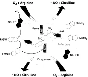
The 3 isozymes share basic structural similarities. NOS is a homodimer, each
monomer containing an N-terminal oxygenase domain and a C-terminal reductase domain
(10). The oxygenase domains bind to heme and tetrahydrobiopterin (BH4), while the
reductase domains contain an electron transport system comprised of nicotinamide
adenine dinucleotide phosphate (NADPH), flavin adenine dinucleotide (FAD), and flavin
mononucleotide (FMN). The calmodulin and zinc cofactors are located at the interface
between monomers, with two cysteine residues from each monomer coordinated to the
zinc. While eNOS and nNOS bind calmodulin reversibly, its binding to iNOS is
irreversible. The NOS active sites are highly conserved (9) as they all catalyze the same
reaction to produce NO from arginine and oxygen (6). An image of NOS can be found in
Figure 1.1.
Aside from these fundamental similarities, the NOS isozymes have important
differences to accommodate their varied roles in the body. They only share 50-60%
homology with one another and are somewhat different in size (11). eNOS and nNOS are
regulation also varies and will be discussed later. These and other variations between the
3 isozymes allow them to maintain their separate roles based on the location of their
expression.
1.1.2.2 De novo NO synthesis
NO is a product of the 5-electron oxidation reaction of arginine to citrulline (6). It
is a multi-step mechanism involving the electron transport system of the reductase
domain. Electron flow begins with NADPH, through FAD to FMN, which passes one
electron at a time to the heme iron in the opposite monomer's oxygenase domain, as
depicted in Figure 1.1. Once the iron is reduced, it is able to bind a molecule of oxygen
and begin catalysis (13). The first main step is the formation of one mole of NG
-hydroxy-L-arginine (NOHA) from one mole of arginine, NADPH and O2. Next, one electron (i.e.,
0.5 moles of NADPH consumed) is transferred to heme where one more mole of O2 is
consumed to convert NOHA to citrulline, releasing NO. The overall stoichiometry of the
reaction is 2 moles of O2 and 1.5 moles of NADPH consumed per mole of arginine to
produce one mole of NO and one of citrulline (14). Since the reaction occurs at the
terminal amine of L-arginine, NG-monomethyl-L-arginine (L-NMMA) is a potent
NO + Citrulline
FADH"<
, A , J FMNH2
FADH,
F M N H - l ^ y ^ N A D P H
• NO + Citrulline O2 + Arginine
Figure 1.1 Structure of the active NOS
While maintaining differences in sequence and regulation, all 3 of the NOS
isozymes follow the common, basic structure depicted above. In the catalytically active
form, it is a homodimer with each monomer comprised of an oxygenase domain, shown
in gray, and a reductase domain, depicted in pink. Electron flow is shown on the left-hand
side, where an electron is transferred from NADPH to FAD, then to FMN, and from there
to the heme of the opposite monomer. Oxygen can bind to the reduced heme and this
begins the conversion of arginine to citrulline and NO via the NOHA intermediate. The
1.1.3 Physical properties
NO's small size and hydrophobic nature are responsible for its opportunities for
reactivity inside the body (1). It is slightly more soluble in water than O2 and it is 9 times
more soluble in organic solvents than in water (16). This means it can freely diffuse
across cell membranes as well as react inside them. Since the cell membrane occupies
only 3% of a cell's volume, it serves to concentrate existing NO into a smaller area,
enabling faster reactions (1).
More powerful, however, is the diffusibility of NO. It has a diffusion constant of
3300 nmV1 in water (17) and takes 2 ms to diffuse out of a 5-u.m cell; therefore, given a
half-life of 5-10 s, NO is a very effective signaling molecule capable of reaching several
hundred thousand cells surrounding that in which it was produced (18). To underscore
this statement, it has been theoretically shown that since NO diffuses more rapidly than it
reacts, in reactions occurring within NO-producing cells, the NO is more likely to have
come from a different cell than itself (19). These two properties of solubility and
diffusibility are thus responsible for the rapid reaction rates and the paracrine nature of
NO signaling.
1.1.4 Different forms
NO has 11 valence electrons, making it an uncharged, paramagnetic, free radical
and consequently highly reactive (20). Its reactivity is the reason it has a half-life of only
a few seconds in the body. The nitrogen-oxygen bond itself is a double bond, N=0. While
it is capable of forming a dimer, 0=N—N=0, the N—N bond is weak and not
energetically favourable (21), supporting the hypothesis that dimerization is transient at
Although it does not readily dimerize, NO reacts with O2 which can be considered
a biradical given its two unpaired electrons. »N02, another free radical, can be formed
from NO and O2 in a multi-step reaction (22). This is a strong oxidant which is able to
abstract hydrogen atoms, add to unsaturated bonds, or react with anions (23). It is also
able to react with other radicals, including itself and NO. It dimerizes much more readily
than NO, producing N204 which is a good NO donor to thiols. However, it is unstable and
dissociates in water to nitrite (NO2) and nitrate (NO3). In addition, »N02 will react with
NO to form the NO donor N2O3. In water this will remain in equilibrium with two
equivalents of N 02\ but in a hydrophobic environment it is more stable. This includes not
only the cell membrane but interior hydrophobic pockets of proteins such as protein
disulfide isomerase (PDI) (24). NO2" can also be formed more directly through a reaction
between NO, 02, and H20 (25).
Oxygen can gain one electron to become superoxide (O2*). It is negatively
charged and still a free radical because one electron remains unpaired; therefore, it is very
highly reactive. One common reaction occurs between O2*" and NO, resulting in
peroxynitrite (ONOO) (26). In aqueous solution it is in equilibrium with its acid,
ONOOH, and is a strong oxidizing agent and nucleophile. Its most common reaction
occurs with CO2 to ultimately yield NO3" and bicarbonate (HCO3) (27). However, it also
reacts with 02" to produce 02, #N02 and H20, or with NO to form »N02 and N02" (28).
It had previously been thought that that NO2" and NO3" are inert end products of
NO metabolism; however, recent evidence shows that they can exert the same influence
on physiology as NO (29). For example, ingestion of NO2" relaxes smooth muscle and
leads to an increase in Hb-Fe-NO, similarly to when NO is synthesized (30). Furthermore,
can be chemically reduced to NO, the reaction is prohibitively slow except in acidic
conditions such as the stomach (31). In recent years several mammalian enzymes have
been identified as being capable of catalyzing this reduction in addition to their main
functions. These include xanthine oxidase, cytochrome c oxidase, and deoxyhemoglobin
(30). Interestingly, this nitrite reduction is observed only when there is a lack of oxygen:
xanthine oxidase is up-regulated in hypoxia, and deoxyhemoglobin by definition has no
bound O2 molecules. While mammals have no such enzyme, commensal bacteria are able
to reduce NO3" to NO2", making ingested nitrate protective under hypoxia as well.
In addition to functional similarities, several NO derivatives described above are
also potent oxidizing agents and generally highly reactive, especially N2O3 and »N02. It is
this property that allows NO to affect the cell physiology as it does, through reactions
with lipids, thiols, and other essential components as discussed in the next section.
1.1.5 Reactions
Unsaturated fatty acid side chains are subject to hydrogen abstraction, either by
enzymes or by radicals, resulting in a lipid free radical denoted as L» (32). Oxygen binds
to it to yield LOO*; this is known as lipid peroxidation. The formation of one LOO*
begins a chain of reactions whereby affected fatty acids oxidize their neighbours, and so
on. This regularly occurs in a controlled manner since LOO* serve as signaling
molecules, and the peroxidation reactions cease upon low oxygen availability. However,
in a situation of oxidative stress lipid peroxidation becomes uncontrolled, undergoing
what is known as propagation reactions.
NO can either initiate or quench these propagation reactions depending on the
circumstances in the cell. ONOO" is known to directly induce it by abstracting H atoms
lesser extent (32). Therefore, in cells where O2" is available, the chance of lipid
peroxidation by NO-derived species becomes greater.
Conversely, since NO is a free radical it is able to react with LOO* to form
LOONO, thus quenching the propagation (34). This may stay as is or hydrolyze to form
LOOH and N02 . By eliminating the lipid free radical, NO can terminate the reaction
from propagating further. This is common in conditions where H2O2, transition metals or
other sources initiate the propagation reaction rather than NOx. NO can also act as a
quenching agent in the presence of O2"" as long as it is in excess (32). In addition, NO can
scavenge O2*" and other reactive oxygen species directly, reducing the chance of ONOO"
initiating lipid peroxidation (35).
Low molecular weight thiols (LMWT) such as cysteine, N-acetylcysteine,
glutathione, and others circulate through the blood where they perform a variety of
functions. They may be reduced, bearing one or more free SH group, or oxidized,
connected through disulfide (S—S) bonds (36). LMWT maintain the redox status of
proteins and other high molecular weight thiols (HMWT) through the interchangeability
of these two forms. For example, one function of PDI is to reduce disulfide bonds of other
proteins; in the process, PDI becomes oxidized (37). It must return to its original reduced
state to be able to perform the same reaction again, and LMWT allow this to occur. When
there is a state of oxidative stress the LMWT reduce proteins to enable them to continue
their functions, absorbing the oxidized state (36). Reduced LMWT can be replenished
through de novo synthesis or via reduction by enzymes such as glutathione reductase.
NO is known to become bound to thiols which are then called S-nitrosothiols,
denoted as RSNO (38). These can be LMWT and HMWT alike. This formation has
peroxidation, the ability of a thiol to become S-nitrosylated depends on its environment,
specifically the local pH and the presence of oxygen and metals.
A mechanism was proposed in 1997 wherein NO directly reacts with a free thiol
to produce an intermediate RSN»-OH, which donates an electron to an acceptor to yield
RSNO (39). However, given NO's relative inability to react with non-radicals, and the
fact that only specific RSNOs form in vivo, it is not likely that the reaction occurs
spontaneously (1,40). Since then, copper (Cu2+) has been shown to act as a catalyst for the
direct 5-nitrosylation by NO (41). Cu2+ can oxidize NO to NO+, as well as oxidize R-SH
to thiolate, RS". When it performs one or both of the oxidations, the two species can
directly bind to form RSNO.
5-nitrosothiols are mainly formed through the reaction between a thiol and an NO
donor rather than directly (38). Transnitrosylation occurs when an RSNO donates its NO
group to another thiol. LMWT in the blood such as glutathione (GSH) act as NO
reservoirs, and upon a stimulus will donate the NO to proteins; this is an important signal
transduction mechanism. As mentioned above, N2O3 and N2O4 are excellent NO donors
as well (42). These will react with thiols directly to form RSNO and N02" or N03"
respectively. In addition, ONOOH - the acid of peroxynitrite - will react with thiolates
and release peroxide (HOO) along with RSNO; however, this occurs less readily due to
competition from CO2 (43).
Although S-nitrosothiols are important carriers of NO, which extend its lifetime
and mediate signal transduction, they are susceptible to decomposition. While Cu + can
oxidize NO to NO+, allowing it to bind to thiolates, Cu+ is effective at reducing RSNO
and RSNO is light sensitive. Thus, the occurrence of S-nitrosothiols is highly regulated
via its formation and decomposition pathways.
Finally, NO will react with metal centres, with the notable example of reaction
with iron inside heme groups. The M—NO bond is usually linear but may be bent if NO
acts as an electron acceptor from the metal (1). There are three main reactions that may
undergo between NO and a hemoprotein such as hemoglobin (45). In one, NO reacts with
deoxyhemoglobin (Hb-Fe(II)) to form Hb-Fe(II)-NO. Depending on the environment, this
may then transfer an NO equivalent to a thiol, serving as an NO donor for S-nitrosothiol
formation (46). NO may also react with oxyhemoglobin (Hb-Fe(II)-02), reducing the
heme to Hb-Fe(III) and itself becoming oxidized to NO3 in the process by the released O2
(45). Thirdly, NO may react with Hb-Fe(III) (methemoglobin), resulting in its oxidation
while methemoglobin becomes Hb-Fe(III)-OH2.
Many proteins contain a heme group and therefore regulate, or are regulated by,
NO. Hemoglobin, myoglobin, cytochrome P450, and xanthine oxidase are a handful of
examples. Importantly, NO activates guanylate cyclase by binding to its heme group,
culminating in smooth muscle relaxation (47). Binding leads to a conformational change
in which the weak bond between Fe(II) and its axial ligand, histidine 105, is broken (48).
The active site then becomes exposed, substantially improving the affinity for its
substrate, guanosine triphosphate (GTP). The product, cyclic guanosine monophosphate
(cGMP), activates cGMP-dependent protein kinases and phosphodiesterases (PDE);
PDE3 stimulates the sarcoplasmic reticulum calcium pump (SERCA2) to sequester
calcium, leading to the relaxation of smooth muscle (49). This example highlights the
Overall, the reactivity of NO can be either positive or detrimental to the cell,
depending in part on its concentration (50). Under normal circumstances the amount of
NO is sufficient to reversibly activate certain enzymes, such as guanylate cyclase (47), or
inhibit others through S-nitrosylation, such as PDI (24). In the event of high
concentrations of NO, however, nitrosative stress can occur in which excessive levels of
ONOO", N203 and other highly reactive species are produced. These can react with
proteins unnecessarily, initiate lipid peroxidation, or damage DNA when unchecked (1).
Therefore, it is critical that the body strictly regulates its NO supply to ensure that it can
perform its functions without extraneous and damaging reactions occurring.
1.1.6 Transport
As stated above, NO is able to freely diffuse due to its small size and
hydrophobicity. However, free NO has a short half-life and is more likely to react than to
diffuse for very long (19). RSNOs therefore act as vehicles for the transport of NO from
cells into serum, through the circulatory system, and to specific tissues.
PDI has been shown to S-denitrosylate RSNOs such as GSNO or PDI-SNO (24).
This can produce free NO or PDI itself may become 5-nitrosylated in the process. In
addition, N2O3 formed in a hydrophobic region inside PDI may directly S-nitrosylate
PDI's active site thiols. Since PDI is known to be excreted to the cell surface in low
amounts, and may cycle back in as well (51), it is likely that PDI carries NO across the
cell membrane where it is then released by free PDI. This would serve as an important
mechanism to bring NO from a location of high concentration - inside an NO-producing
cell or platelet - to a site of low concentration, the cell exterior (24).
5-nitrosoalbumin (Alb-SNO) is one of the primary reservoirs of NO in human
of Alb-SNO may be as high as 5 uM due to the abundance of albumin circulating (1).
Studies have shown that hydrophobic pockets in albumin facilitate the formation of N2O3,
which can then S-nitrosylate the same albumin (53). Additionally, transnitrosylation
reactions occur readily from low molecular weight RSNOs, especially GSNO and
S-nitrosocysteine (CysNO), to albumin (52). Alb-SNO may thus be a transport vessel to
circulate NO in the blood while it is released in places where it is needed. Hemoglobin
binds NO both in its heme and by S-nitrosylation of cys93 of the P subunit, but its role in
the possible transport of NO remains unclear (54).
1.1.7 Regulation
The formation of RSNOs and metal-NO complexes regulates the levels of free
NO, and facilitates its transport to tissues where its activity is needed. However, most of
the regulation of the amount of NO or NOx occur at the level of the enzyme NOS. eNOS
and nNOS are the most similar to one another and consequently are regulated in similar
manners (1). Before it is catalytically active, e/nNOS must form a dimer and bind all of
its cofactors; the cofactor which has the most influence on this process is calmodulin (55).
e/nNOS exists as a monomer before its activation by Ca2+. When NO is required, Ca2+ is
released to the cytosol where it binds to calmodulin. This induces a conformational
change in calmodulin, allowing it to bind to specific consensus sites on certain proteins.
On NOS the site is located such that two calmodulins bind to two oxygenase domains,
bringing them together (Figure 1.1). Dimerization is further stabilized by the cofactor
BH4. Holoenzyme assembly is thus the main temporal regulation of the amount of NO
produced by those two isozymes.
eNOS and nNOS are also regulated spatially. These isozymes are active when
myristoylated and palmitoylated, and while it is there it is constitutively phosphorylated
at serine 1179, making it active (56). It is inactive in the cytosol when it lacks
phosphorylation at that site. nNOS is also active at the plasma membrane where it
localizes through protein-protein interactions (57). The last major sources of e/nNOS
regulation are substrate availability, namely arginine and O2 (58), and feedback inhibition
by NO (59).
iNOS has its own forms of regulation. Calmodulin binding to iNOS is so tight that
it is considered a subunit more so than a cofactor, and consequently Ca + availability has
no effect on induction of NO synthesis (55). Rather, the synthesis and inhibition of iNOS
itself are the main sources of regulation (1). In most cell types, iNOS is not expressed
until a stimulus induces it. Effective stimulants include lipopolysaccharide (LPS) (60),
cytokines such as interleukin 1 or tumour necrosis factor (TNF) (61), and endotoxins.
Once activated, iNOS synthesizes copious amounts of NO continuously until it is
stopped. One method of down-regulation is feedback inhibition by NO itself through its
ability to bind to the heme group, which is the site of catalysis (62). iNOS is also limited
by arginine availability (58); arginase is co-induced along with iNOS, ensuring that
arginine will deplete so that NO levels will no longer increase after a certain length of
time (63). iNOS is eventually degraded by the usual cellular mechanism and once the
induction signal is no longer present, it will stop being produced by the cell.
1.1.8 Detection methods
Several detection methods exist that are specific for one form of NO; others are
more general and simply detect NOx. 4,5-diaminofluorescein diacetate (DAF-2-DA) is
commonly used on cells for the purpose of detection (64). The acetate groups allow it to
forms - reacts with DAF-2 in the presence of oxygen, resulting in the formation of a
fluorescent product that can be quantified with a microscope. As a probe that only
requires 30 minutes to finish reacting, this makes it a convenient detector for changes in
NO concentrations in the cell after a stimulus such as lipopolysaccharide.
Nitric oxide analyzers (NOA) are available from Sievers (General Electric) and
can be used to detect either NO or NO2" by chemiluminescence (65). The purge vessel can
be filled with acetic acid and 50 mg of sodium iodide. The iodide ions reduce injected
N02" to NO, with the acid providing protons for the reaction, while the injected solution
is carried to the NOA. Inside, the NO is mixed with ozone, O3, and they react to form an
excited NO2* which releases a photon to become N02#. The photon is detected in the
photomultiplier tube and displayed in the software as a peak; the area under the peak is
directly proportional to the number of photons detected, which in turn is directly
proportional to the number of NO molecules produced. Therefore, by taking the integral
of the peak area and interpolating it into an NO2" standard curve, one can calculate the
concentration of NO2 originally injected. If NO is being measured directly instead of
N02" then acid and iodide are not needed; simply water or buffer can be used to fill the
purge vessel so that only NO itself will be detected. This can be used for a variety of
solutions including cell culture media, exhaled breath, or serum.
The biotin switch assay is a commonly used method to detect 5-nitrosylated
proteins, developed in 2001 by Jaffrey et al (66). In the first step, methyl
methanethiosulfonate (MMTS) is added to a sample of S-nitrosoprotein(s) to block free
thiols with a methyl group. Once excess MMTS is removed, ascorbate is added to reduce
the S-NO bond, leaving newly formed free thiols. Finally, biotin is conjugated to the
process. In short, biotin is conjugated to proteins only at sites that were S-nitrosylated,
making S-nitrosoproteins detectable by Western blot through anti-biotin primary
antibodies. This method is useful for determining whether a particular protein is able to
be S-nitrosylated, or for determining the proteome of S-nitrosoproteins in serum, among
other applications.
These are the most commonly used methods for their respective applications.
While they are for the most part effective, they still all have their drawbacks. DAF reacts
with radicals inside the cell which are not necessarily NOx (67), and the biotin switch
assay is convoluted and has been known to produce false positives (68). Therefore, as NO
1.2 Gold Nanoparticles
1.2.1 History
Gold nanoparticles (AuNP), also referred to as colloidal gold, have been used for
millennia, but it is only in the last -150 years that they have been extensively
characterized and studied (69). The first known cultures to make use of AuNP were
ancient Egypt and China (400-500 years B.C.E.) which used the wine-red solution as a
dye for glass and ceramic due to its brilliant colour; this is still in practice today. In
addition, solutions of AuNP were thought to be medicinal and were drank to cure a
variety of ailments throughout the middle ages and up until the 19th century (70).
In 1857 Michael Faraday published the first scientific investigation into the
properties of the gold nanoparticles (71). While he was not the first to speculate that the
particles were too small to view with the naked eye (69), he was able to estimate them to
be in the 10-nm range. He also realized that he could produce them by reducing the
AuCU" ion. Since then, a variety of methods have been established to synthesize AuNP
which will be briefly outlined below.
1.2.2 Synthesis
Many types of nonmetal and metal nanoparticles exist, including iron, copper,
silver, cadmium, and platinum (72); however, for the purpose of brevity, only synthesis
methods of AuNP will be discussed here. As mentioned above, Faraday was the first
person to synthesize AuNP by reducing AuCLf with phosphorus in CS2 (71). Currently
used methods are able to produce nanoparticles of specific sizes and shapes. The
Turkevich method (73), as modified by Frens (74), uses trisodium citrate to both reduce
aqueous AuCU" and stabilize the resultant spherical AuNP. While Turkevich's protocol
and citrate to result in nanoparticles ranging from 16 to 147 nm. The method has been
further modified by various groups including Grabar et al (75) to produce smaller
spherical AuNP - in the case of Grabar's group, 12 nm in diameter.
The seeded growth method is used to control the shape of the AuNP (76). In this
protocol, AuCLf is mixed with trisodium citrate and the resultant "seed" is mixed in
specific proportions with cetyltrimethylammonium bromide (CTAB), HAuCU, and
ascorbic acid. Simply by adjusting the concentrations of each reagent as well as the
timing and sequence of each reagent's addition, one can obtain nanoparticles shaped like
triangles, rods of specific aspect ratios, hexagons, cubes, and others (77).
Gold nanoparticles can be produced using reducing agents other than trisodium
citrate. The Brust-Schiffrin method utilizes tetraoctylammonium bromide to transfer
AuCLf to toluene where it is reduced by NaBHU (78). The nanoparticles are commonly
protected by the subsequent addition of a monolayer of thiol. Alternatively, top-down
methods can be used for synthesis in which a larger material is effectively whittled down
to the nanoparticles; these include photolithography and electron beam lithography
(79-80).
While this description of AuNP synthesis is not exhaustive, these are some of the
most commonly used methods which have been refined over the past 1-2 decades. It is
clear that just by using the methods discussed above, one can obtain nanoparticles of any
desired size and shape and consequently develop a variety of applications.
1.2.3 PDMS and synthesis of surface gold nanoparticles
Poly(dimethylsiloxane) (PDMS) is a type of silicone used in many household and
industrial products, such as contact lenses and microfluidic devices (81). Its notable
inexpensive (81), and it is chemically inert, though it can be made reactive through
oxidation if needed. PDMS is prepared by mixing the vinyl-containing, silicon-based
monomer with a crosslinker that includes silyl (SiH) groups in the presence of a platinum
catalyst and heat (82). It forms a three-dimensional network based on two reactions. The
primary reaction occurs between the SiH of the crosslinker and the vinyl group of the
monomer, shown in Reaction 1:
—Si-H + CH2=CH-Si— -> — Si-CH2-CH2-Si— (1)
The secondary reaction is favoured in a high excess of crosslinker. The Si-H group is
hydrolyzed to become Si-OH and this reacts with either another Si-OH or with Si-H to
form Si-O-Si chains, as seen in Reactions 2 and 3:
—Si-H + HO-Si— -> — Si-O-Si— + H2 (2)
—Si-OH + HO-Si— <-> —Si-O-Si— + H20 (3)
Changing the ratio of monomer to curing agent changes the polymer's properties: for
example, adding more crosslinker makes PDMS more rigid. Although it was omitted
from Reactions 1-3 for simplicity, each silicon atom is bonded to methyl groups or
hydrogen atoms wherever a bond is not shown, making PDMS very hydrophobic and thus
inert.
In 2007, Zhang et al (83) published a new technique which allows AuNP to be
formed on the surface of PDMS. Simply by incubating a 0.5% (w/v) solution of AuCLf
on the surface of the pre-formed polymer for 24-48 hours, they were able to obtain a pink
layer of nanoparticles on the surface. Their explanation is that excess SiH groups acted as
the reducing agent, and the nanoparticles embedded into the pores between crosslinker
morphology and size variation; for example, a ratio of 0.06 led to the formation of
spherical nanoparticles with an average diameter of 7 nm, while a ratio of 0.1 resulted in
variably-shaped nanoparticles averaging 13 nm in diameter.
The Mutus lab recently optimized this process to achieve a greater density of
AuNP on the surface of the PDMS (unpublished observations). PDMS was polymerized
using a ratio of either 0.02 or 0.04 and then AuCLf and trisodium citrate were
simultaneously added to the surface. With this method the citrate acts as the reducing
agent and the large pores caused by the low r\ gives the nanoparticles a site to embed. The
colour of the solution is a darker red compared to when there is a lack of citrate,
indicating that a higher density of nanoparticles is present on the PDMS surface.
1.2.4 Localized surface plasmon resonance
The characteristic brilliant colour of gold nanoparticles is a property unique to
materials in the 1-100 nm range (84). They behave neither as bulk objects nor as single
molecules. At this point, their identities becomes negligible and the only factors affecting
their colour are size, shape, and the dielectric constants of the nanoparticles and the
surrounding media (85). Gold has d-orbital electrons that travel more freely than those of
lower orbitals. Since the AuNP are smaller than the wavelength of light, passing light
waves are able to influence the positions of all the d electrons of a nanoparticle, rather
than individual atoms. The light causes them to cohesively oscillate between ends of the
nanoparticles, creating a dipole (84). This is known as localized surface plasmon
resonance (LSPR).
The frequency of the oscillation lies in the visible region of the light spectrum and
absorbs strongly at characteristic wavelengths. In the case of unmodified 11-nm spherical
does not greatly affect this peak. However, if the nanoparticles were rod-shaped then the
resonance is more complicated, as there is one transverse and one longitudinal plasmon
resonance, each with their own absorbance peak (86). Altering the aspect ratio greatly
influences the overall oscillation frequency and therefore the colour. Thus, each shape of
AuNP has its own characteristic LSPR absorption.
The presence of other molecules on the surface is the factor which has the greatest
effect on the LSPR absorption for nanoparticles of given size and shape. Sulfur has a
strong attraction to AuNP. It has been shown with x-ray crystallography that while the
distance between gold atoms within a nanoparticle is 2.8-3.1 A, the distance between gold
and sulfur atoms is 2.2-2.6 A (87). The bond is not covalent, but it is far stronger than
electrostatic, so that thiol-containing molecules are stably attached to the surface. Each
sulfur atom binds between two gold atoms of the nanoparticle. The presence of bonded
molecules changes the electron density on the surface of the nanoparticles and
consequently a small red-shift in absorbance peak is seen (84). Many of the applications
of AuNP involve coating the surface with molecules via their thiols, so this phenomenon
is frequently observed.
When molecules are added to AuNP such that they become linked together into
3-dimensional networks, the LSPR absorption undergoes a more extensive shift (88). The
crosslinking molecules act as conductors for the electrons, with interparticle distance
(determined by crosslinker length) controlling the conductivity (89). Consequently, the
plasmon resonance becomes synced for all of the connected AuNP, leading to a new
LSPR absorption. Specifically, the absorbance peak will broaden and red-shift, though the
extent will depend on the nature and length of the connecting molecules. This colour
1.2.5 Applications of AuNP
Gold nanoparticles are brightly coloured, are able to undergo colour change when
crosslinked, and are visible in transmission electron microscopy (TEM). They can be
synthesized in solution or on a surface, are dense enough to centrifuge, and enhance the
fluorescence of nearby molecules (70,90). They are similar in size to some proteins and
are non-cytotoxic (91). These properties make them useful in a large variety of
applications. Here, only a selection will be discussed with the intention of portraying the
versatility of AuNP.
AuNP are becoming important assets in bioimaging. In one example, angiogenesis
was monitored in rabbits using AuNP specifically targeted to neovasculature (92). They
were made paramagnetic and bonded to molecules reactive to avP3 integrins. These
targeted and paramagnetic nanoparticles were injected into rabbits fed high-cholesterol
diets, and control rabbits on a normal diet. MRI was done using typical hospital protocol.
The presence of nanoparticles greatly enhanced the visibility of the newly forming
vasculature and the researchers could detect a 47% increase in angiogenesis in
cholesterol-fed rabbits over controls. The MRI process took about 2 hours, allowing rapid
detection of one of the warning signs before a stroke or heart attack; this experimental
design may also be used to image angiogenesis in tumours.
Surface-enhanced Raman scattering is an advancement of Raman spectroscopy
(93). The surface plasmons from nanoparticles become coupled to the molecule of
interest, which enhances the Raman signal up to 1014 times (94). Once again the shape of
the nanoparticles plays a role; for example, rod-shaped nanoparticles cause higher signals
than spherical ones (95). Regardless of the shape, the sensitivity is so highly improved
chemistry done, but the heightened sensitivity makes it useful in biological settings, for
example the detection of specific molecules in low concentrations. Thus, the use of AuNP
increases the scope of experiments to which Raman scattering can be applied.
The colour change exhibited by clusters of AuNP has been exploited to various
ends. The reaction between two complimentary DNA strands, each conjugated to AuNP,
brings the nanoparticles close enough together to induce the LSPR absorption shift
described above (96). When reflected light is measured from this reaction, using ~50-nm
AuNP, the detection limit of this assay is 10 fmol of DNA. While this is sensitive, the
disadvantage is that many nanoparticles must react before a detectable red-shift in
absorbance occurs. More recently, another group has developed a similar assay in which
scattered light was measured instead of reflected light by spotting the reaction on a
waveguide (97). This modification in the detection method is 4 orders of magnitude more
sensitive, reaching zeptomole quantities, which means the DNA in question does not need
to be amplified in PCR before it can be detected. This assay can be useful to a variety of
scientists, such as academic labs doing cloning, or clinical labs identifying certain
bacteria for rapid diagnosis.
While this list is not comprehensive, it is clear that AuNP can be easily adapted
for many uses because of the unique properties endowed on molecules of 1-100 nm size.
Physics, chemistry, and biology labs all have their own applications for nanoparticles. In
addition, AuNP are gaining more importance each year in clinical labs through their
usefulness in imaging, diagnosis and treatment of infections and cancer. With this
diversity in applications, it is clear that AuNP research is leading the way for new
CHAPTER 21
ISOFORM-SPECIFIC DIFFERENCES IN THE NITRITE REDUCTASE ACTIVITY OF NITRIC OXIDE SYNTHASES UNDER HYPOXIA
2.1 Introduction
Nitrite has been observed to promote the same physiological effects as NO", and
there is an increasing list of enzymes which catalyze the reduction of N02" to NO to allow
this to occur (31). In addition to that, eNOS has been shown to be protective in hypoxia
(ischemia, stroke) while iNOS is cardioprotective and nNOS has no apparent role in
hypoxia. This led Slama-Schwok and coworkers to examine whether eNOS is able to
reduce NO2" in the absence of oxygen, and they gained evidence to support this
hypothesis (98-99).
The goal of the present study was to characterize the roles of all three NOS
isozymes in the reduction of NO2" to NO under hypoxia and anoxia. The full-length
enzymes as well as the oxygenase domains alone (which contain the heme moiety) and
the reductase domains alone were used in various studies performed by the Mutus lab or
Slama-Schwok's group. In addition, endothelial cells were exposed to different levels of
oxygen tension, with or without supplemental nitrite, and the NO released from the cells
was detected after different lengths of time. It was found that eNOS was the only isozyme
able to reduce the nitrite, and the reductase domain alone had no effect. Additionally, the
endothelial cells released more NO under hypoxia compared to basal levels while anoxia
caused the highest increase. These data provide a new role for eNOS as a source of NO in
pathological conditions in the body, on top of its main role in a normal state.
2.2 Materials and Equipment
2.2.1 Materials
3-bromo-7-nitroindazole: Cayman Chemical, Burlington, ON
Argon Grade 5 300 SZ: Praxair Distribution, Division of Praxair Canada Inc., Mississauga, ON
Calcium chloride (CaCi2-6H20): Sigma-Aldrich Canada, Oakville, ON
Calmodulin: Sent from Dr. Rajendra Sharma, University of Saskatchewan, Saskatoon, SK
Endothelial cell medium (ECM): ScienCell, Carlsbad, CA
Endothelial nitric oxide synthase (eNOS), eNOS reductase domain (eNOSred), neuronal NOS (nNOS) and nNOSred: purified by and sent from Dr. Anny Slama-Schwok, Ecole Polytechnique, Paris, France
Human dermal microvascular endothelial cells (HDMEC): ScienCell, Carlsbad, CA
L-arginine: Sigma-Aldrich Canada, Oakville, ON
Nicotinamide adenine dinucleotide phosphate (NADPH): Sigma-Aldrich Canada, Oakville, ON
NG-monomethyl-L-arginine (L-NMMA): Sigma-Aldrich Canada, Oakville, ON
Oxygen 2ppm balance argon, size 30, 0.8M3 certified: Praxair Distribution, Division of Praxair Canada Inc., Mississauga, ON
Oxypurinol: Sent from Dr. Anny Slama-Schwok, Ecole Polytechnique, Paris, France
Sodium chloride (NaCl): ACP Chemicals, Montreal, PQ
Sodium iodide (Nal): Sigma-Aldrich Canada, Oakville, ON
Sodium nitrate: Sigma-Aldrich Canada, Oakville, ON
Sodium nitrite: Sigma-Aldrich Canada, Oakville, ON
T-75 tissue culture flasks: Sarstedt Inc., Montreal, PQ
Tetrahydrobiopterin (BH4): Sent from Dr. Guy Guillemette, University of Waterloo,
Waterloo, ON
Tris hydroxyaminomethane and Tris-HCl: Sigma-Aldrich Canada, Oakville, ON
TrypLE™ trypsin: Invitrogen Canada Inc., Burlington, ON
2 ml glass vials with black polypropylene screw top and septa: Supelco, Bellafonta, PA
27.5G syringe needles: Becton-Dickenson, Franklin Lakes, NJ
Agilent 8453 UV-VIS Spectrophotometer: Agilent Technologies Canada Inc., Mississauga, ON
Bright-Line Hemacytometer: Hausser Scientific, Horsham, PA, USA
Hamilton® syringe, 1800 series gastight, model 1801RN: Sigma-Aldrich Canada, Oakville, ON
Jouan CR3i Centrifuge: Jouan Inc., Winchester, VA
Nikon TMS microscope: Nikon, Japan
NuAire Biological Safety Cabinet Class II Type A/B3: Thermo Electron Corp. Canada, Burlington, ON
NuAire Direct Heat AutoFlow CO2 Automatic Air-Jacketed Incubator: Thermo Electron Corp. Canada, Burlington, ON
Sievers 280i Nitric Oxide Analyzer: General Electric Canada, Mississauga, ON
2.3 Methods
2.3.1 Cell culture
HDMEC were grown in ECM containing basal medium, 5% fetal bovine serum,
1% endothelial cell growth supplement, and 1% penicillin/streptomycin solution. Cells
were maintained in T-75 culture flasks at 37°C in 5% CO2 and grown to 80% confluence.
2.3.2 Treatment of cells
Once at confluence, the cells' media were decanted and kept aside. Cells were
trypsinized and concentrated by centrifugation at 1700 rpm for 5 minutes. They were then
resuspended in either old media or fresh ECM with added 10 \iM NO2" and each
suspension was aliquotted into septa-topped screw cap glass vials, 0.5 ml per vial. The
vials were incubated in a 37 °C water bath for the duration of the experiment. In total there
on to permit oxygen entry (normoxia). One-third had their caps tightly screwed and were
blown with argon, via syringe needles inserted through the septa, for 10 minutes followed
by incubation for 0, 4, 8, 12, 16, or 20 minutes (anoxia). Finally, one-third had their caps
tightly screwed and were blown with 2 ppm oxygen in argon for 10 minutes and then
incubated for 0, 6, 12, 16 or 20 minutes (hypoxia).
2.3.3 Inhibitor treatment of cells
In another experiment, 80% confluent cells were trypsinized, centrifuged and
resuspended in fresh media supplemented with 10 ^iM nitrite. The cells were subdivided
into suspensions treated with either L-NMMA (500 \iM), 7-NI (10 u.M), oxypurinol (100
\iM) or left untreated as a control group. These suspensions were distributed into glass
vials as described above and incubated in anoxia for 0 or 15 minutes after argon
treatment. An additional group of vials contained media and 10 \iM nitrite but no cells;
these were also exposed to anoxia for 0 or 15 minutes after argon exposure.
2.3.4 Nitric oxide detection
Following the specified lengths of incubation under normoxia, hypoxia or anoxia,
10 ul of media were injected via a Hamilton gas-tight syringe into a Sievers nitric oxide
analyzer containing acetic acid and sodium iodide. The average of two repeat injections
was taken and compared to a nitrite standard curve in order to determine the amount of
NO detected.
2.3.5 Nitric oxide detection from in vitro NOS isoforms
Samples of eNOS, eNOSred, nNOS, and nNOSred were suspended in 50 mM Tris
buffer with 150 mM NaCl (pH 7.5). These were quantified in the UV-VIS
spectrophotometer using the extinction coefficients 100 000 M'cm"1 at 396 nm for eNOS
were adjusted to 10 \xM using the same buffer. These were aliquotted into the glass vials
described above. Final concentrations of 10 ^M nitrite, 10 \xM calmodulin (quantified by
the UV-VIS spectrophotometer with the extinction coefficient 3006 M'cm"1 at 257 nm),
ImM calcium, 40 uM BH4, and 1 mM arginine were added such that each vial had a final
volume of 500 (xl. Upon addition of 100 \xM NADPH the vial caps were screwed on
tightly and blown with argon for 10 minutes; these were incubated for 0 or 15 minutes
thereafter. Following incubation, 1 ml of the air above the samples was injected into the
NOA in duplicate. NO production was calculated from a nitrite standard curve. Control
vials had no NADPH added; in addition, other samples for each isozyme contained 10
2.4 Results
2.4.1 NO release by endothelial cells under normoxia, hypoxia or anoxia
In vitro experiments performed by Dr. Slama-Schwok and colleagues showed that
eNOS is able to reduce nitrite to NO when oxygen is unavailable for its de novo synthesis
from arginine. nNOS has a more limited capacity for this and iNOS is unable to do so. To
further confirm these observations by using a cell system, we chose endothelial cells
which express eNOS as its NO-producing enzyme (1). 80% confluent cells were
trypsinized, concentrated and resuspended in media. Half of the cells were replaced into
the old media decanted before trypsin application, and half of the cells were replaced into
new media which had 10 \iM nitrite supplementation. Each group was subjected to
treatment with argon (anoxia), 2ppm O2 in argon (hypoxia) or room air (normoxia) for
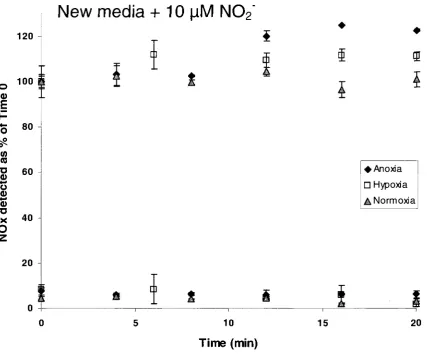
various lengths of time. As shown in Figure 2.1, when there was no nitrite added to the
cells, there was no measurable change in NOx detected by the NOA over the 20-minute
time course for any of the three conditions. There was also no significant change
exhibited by the cells exposed to normoxia in the presence of nitrite. However, an 11%
increase in NOx levels was detected under hypoxia and a 22% increase was observed
from the cells exposed to anoxia, confirming that a component of the cells was reducing
nitrite to NO in the absence of oxygen. Error bars on the data points were the calculated
120
O 1 0°
E P "S 80 W «J •o 0) o 0) © X O 60 40 20
New media + 10 |aM N0
2"
M.
•
• Anoxia D Hypoxia A Normoxia
ft 0 2
10
Time (min)
15 20
Figure 2.1 Detection of NO release from nitrite under normoxia, hypoxia and
anoxia
Endothelial cells were trypsinized, concentrated and resuspended in either the media in
which they were grown or in fresh media supplemented with 10 uM nitrite. These were
subjected to treatment with argon (anoxia), 2ppm O2 in argon (hypoxia) or incubated in
room air (normoxia) for various lengths of time. Results show that cells placed in the old
media displayed no change in NOx production over time regardless of oxygen levels.
When exposed to 10 [xM nitrite, in normal oxygen conditions (gray triangles) there was
little change in NOx detected. However, after incubation in 2ppm O2 in argon (white
squares), levels of NOx increased by 11%. Anoxic cells (black diamonds) released the
2.4.2 Effect of inhibitors on NO release by endothelial cells under anoxia
Since the endothelial cells contain several enzymes which may be responsible for
the reduction of nitrite to NO under anoxia, we treated cells with different inhibitors to
determine the correct enzyme based on their effects. L-NMMA is a non-specific NOS
inhibitor; 7-NI targets the specific isozyme nNOS and to a lesser extent iNOS; and
oxypurinol inhibits xanthine oxidase. Glass vials were prepared as described above, with
fresh media and 10 \iM nitrite. There were five categories: no cells, cells that were not
treated with any inhibitor, and cells treated with one of the three inhibitors listed. After
10-minute argon exposure, 10 ul of media was injected into the NOA in duplicate either
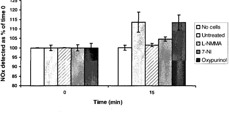
immediately or following 15 additional minutes of anoxia. Results after the 15 minutes
are shown in Figure 2.2 as a percentage of time 0, which was set to 100% for each group
of vials. There was no change in NOx detected by the NOA in the absence of cells.
Untreated cells had a similar NOx increase as that seen in Figure 2.1. Oxypurinol had no
effect on the cells' nitrite reduction; as an inhibitor specific to xanthine oxidase, it shows
that this enzyme is not involved in the process. The non-specific NOS inhibitor L-NMMA
nearly eliminated the increase in NOx while the nNOS- and iNOS-specific 7-NI reduced
it. This verifies the identification of NOS as the responsible enzyme for nitrite reduction
but does not conclusively point to eNOS alone. Data are represented as the average of two
125 •]
° 1 2 0
| 1 1 5
-1 " 0 "
m 105 •
re "g
100V 9 5
•O 9 0 -X O 8 5
-T -T X 0 T
1,
T 1 Time (min) 15 T JL• No cells • Untreated m L-NMMA • 7-NI H Oxypurinol
I
Figure 2.2 Effect of inhibitors on nitrite reduction under anoxia
Endothelial cells were trypsinized and distributed into glass vials as described above.
Media was supplemented with 10 fxM nitrite and either L-NMMA, 7-NI, oxypurinol or no
treatment. In addition, vials containing cell-free media and nitrite were included. These
were exposed to anoxia and incubated for 0 or 15 minutes and media was injected into the
NOA. As shown in the figure, in the absence of cells (white), there was no difference in
NOx detected before or after anoxia treatment. Oxypurinol-treated cells (black) exhibited
the same trend as untreated cells (light gray) indicating a lack of involvement by xanthine
oxidase. Nitrite reduction was somewhat inhibited by 7-NI (dark gray) and almost
completely inhibited by L-NMMA (gray hatched) which shows that NOS is the
2.4.3 NO formed by in vitro NOS or NOS reductase domain under anoxia
As stated in 2.4.1 above, Dr. Slama-Schwok's group showed that eNOS is
efficient at reducing nitrite to NO, and that nNOS has a limited ability. We then sought to
determine whether the entire enzyme or simply the reductase domain was necessary to
perform this reduction. Vials were prepared which contained 10 uM eNOS, eNOSred,
nNOS or nNOSred; 10 \iM nitrite or nitrate; and all of the required cofactors except
NADPH. Half of the vials did not have NADPH added at all as a negative control; these
were blown in argon and then incubated for 15 minutes. The other half had NADPH
added immediately before the argon treatment. These were then incubated for 15 minutes
as well. After the incubation period, 1 ml of air above the solution was injected into the
NOA. There were no detectable peaks generated from either eNOSred or nNOSred, and
therefore they did not produce NO under anoxia in the presence of all cofactors.
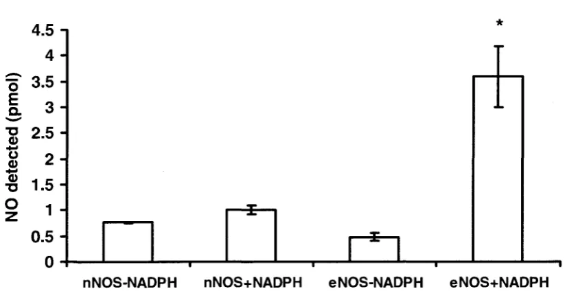
Wild-type nNOS and eNOS released a low basal level of 0.76 and 0.48 pmol of NO,
respectively, in the absence of NADPH (Figure 2.3). However, in the presence of all
cofactors including NADPH, NO levels were 1.0 ± 0.08 pmol for nNOS, a ~ 1.3-fold
increase, and 3.6 ± 0.59 pmol for eNOS which corresponds to a ~7.5-fold increase. NO
was not detected in any condition in which nitrate was present rather than nitrite. Data is
o E
O
4-1
o
0)
4.5 -I 4
- 3.53 - 2.52 - 1.51 -
0.5-0 - ' i '
1—
—sl
*
1 1
nNOS-NADPH nNOS+NADPH eNOS-NADPH eNOS+NADPH
Figure 2.3 Detection of NO released by nNOS and eNOS under argon
Vials were made containing nitrite, calmodulin, calcium, BH4, arginine and either nNOS
or eNOS in 50 mM Tris buffer. These were blown with argon either immediately or after
the addition of 100 uM NADPH and the released NO was detected in duplicate using the
NOA. nNOS in the presence of NADPH exhibited a ~ 1.3-fold increase in NO production
compared to its control, while eNOS in the presence of NADPH showed a ~7.5-fold