Outcome of Supracondylar Femur Fracture in Adults
Managed By Distal Femur Locking Compression Plate
Gyanendra Kumar Jha
1*, Deepesh Kumar Yadav
2and Pravin Timsina
31Lecturer Department of Orthopaedics, College of Medical Sciences, Nepal
2,3Department of Orthopaedics College of Medical Sciences Bharatpur Chitwan Nepal
Received: February 27, 2018; Published: March 12, 2018
*Corresponding author: Gyanendra Kumar Jha, Lecturer Department of Orthopaedics, College of Medical Sciences, Bharatpur Chitwan, Nepal,
Tel: ; Email:
ISSN: 2574-1241 Biomed J Sci & Tech Res
Introduction
Distal femoral fractures currently account for less than 1% of all fractures and about 3% to 6% of all femoral fractures; however, the incidence rate increases with increasing age [1-3]. The distal femur fractures have bimodal incidence. One group includes patients below 40 years of age (nearly 40 % of cases), predominantly males,
sustaining high-energy trauma such as Road traffic accident or a
fall from heights and the other group consists of patients more than 50 years, (nearly 60 % of cases) predominantly females, with osteoporosis, who sustain relatively low energy trauma [4-8].
However, in both instances, axial load to the leg is the most common cause. Management of distal femoral fracture remains a challenge to orthopaedic surgeons. They usually result from high-velocity injury leading to comminuted and unstable fracture pattern [9,10]. Challenges associated with these types of fractures are adequate exposure of particular surface, particularly of the medial femoral condoyle and coronal plane fractures. In the setting of medial combinations and short distal segment, there is a high incidence
of loss of fixation and varus collapse on weight bearing if improper Abstract
Introduction: Fractures of distal femur are one of the most prevalent fractures encountered in high-velocity trauma which are associated with high morbidity and mortality if not managed well. The isolated fracture can itself lead to complications such as Acute Respiratory Distress
Syndrome (ARDS) and pulmonary embolism (PE). This necessitates early stabilization of fractures. Open reduction and internal fixation
(ORIF) with locking compression plate is the treatment of choice for closed fractures of the distal femur. Distal femur anatomic contoured locking compression plate (LCP) has shown to give one of the best results regarding recovery, fracture union, return to work and the functional outcome. We present our experience of management of distal femur fracture at our centre.
Methods: 30 cases of supracondylar femur fracture were treated by ORIF with LCP between 1st October 2014 to 30th September 2016 at our tertiary level trauma centre. The patients were evaluated clinically and radiologically for outcomes. All patients were followed up for an average of 6 months. The outcome was assessed using Neer’s Score. The fracture was considered united clinically when there was no pain on palpation, no discomfort on full weight bearing. Radiologically fracture was considered united when serial roentgenograms demonstrated bone trabaculae is crossing the fracture site. The functional outcome was evaluated by Neer’s knee score criteria.
Result: In our study 30 cases of supracondylar femur fractures were treated. 18 patients were males, and 12 patients were females. The
median age was 34.97 years ranging from 18 to 62 years? All the fractures were caused by road traffic accidents. 19 patients were with fracture
on the right side and 11 on the left side. Using Neer’s scoring system excellent outcome was seen in 53.33%, good in 36.67%, fair in 6.67% and failure in 3.33%. The range of motion of the knee was excellent to very good. Gait and weight bearing after the complete union was satisfactory.
Conclusion: The method of treatment is standard with good exposure of fracture fragments, less operative time and good functional and radiological outcome. LCP is an important modality in the treatment of fractures around knee especially when the fracture is severely communited and in situations of osteoporosis.
Keywords: Supracondylar fracture; locking compression plate; Open reduction internal fixation
Abbreviations: ARDS: Acute Respiratory Distress Syndrome; PE: Pulmonary Embolism; ORIF: Open Reduction and Internal fixation; LCP:
Locking Compression Plate
and inadequate fixation technique is used for the treatment of such
fractures.
Similarly, these fractures are also associated with higher incidence of infections, joint stiffness, melanin and early onset of osteoarthritis. So these fractures have to be appropriately managed to overcome these factors [11-15]. This led to the concept of indirect
reduction and biological fixation of fractures with preservation of
vascularises and soft tissue integrity to the bone fragments [16-21]. Principle’s of management includes anatomic restoration, axial alignment and rotational stability of particular surface. The management of distal femur fracture with anatomical distal locking
compression plate (LCP) has multiple benefits. It provides multiple points of fixed plate to screw contact, generating greater stability
than is provided by a single lateral construct, which potentially reduces the tendency for varus collapse. The locking screws also
provide stronger fixation of the plate in the proximal fragment by
eliminating any potential for toggle and sequential screw loosening. These have a particular advantage in osteoporotic bone and have a possible biological advantage over conventional plates [22]. There was no local study available to assess the results of LCP for the management of this types of fractures in our part of the world having the different demographic combination. This study was carried out to assess the functional and radiological outcome of patients with supracondylar femur fracture managed with LCP.
Material and Methods
Figure 1: Pre op X-ray Anteroposterior(AP) and lateral X–Ray of thigh.
30 cases of supracondylar femur fracture were treated by
open reduction and internal fixation (ORIF) with LCP between 1st
October 2014 to 30th September 2016 at our tertiary level trauma centre. The patients were evaluated clinically and radiologically for
outcomes using various parameters. The fracture was classified according to the AO classification system preoperatively on the
Anteroposterior and lateral X-Rays (Figure 1). All the patients in
this study were treated by open reduction and internal fixation
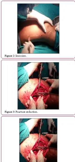
by using standard lateral approach within four days of injury (Figure 2). The fracture was adequately exposed and reduced
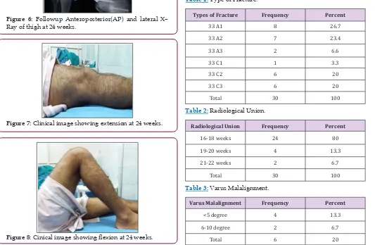
under C-arm guidance. K-wires were used to provisionally fix the
fracture (Figures 3 & 4). LCP of appropriate length was used in each case according to the type of fracture (Figure 5). All patients were
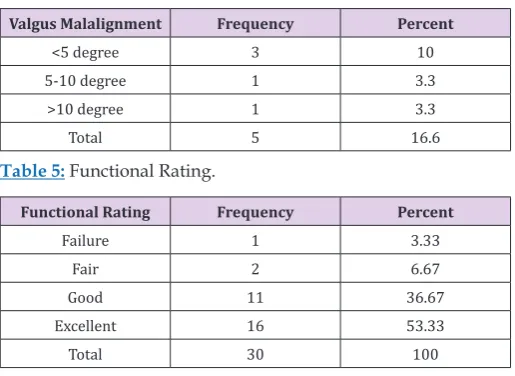
followed up for an average of 6 months. Check X-Ray was done at regular intervals to see the progress of the fracture union (Figures 6-8). The outcome was assessed using Neer’s Score [23-26]. The fracture was considered united clinically when there was no pain on palpation, no discomfort on full weight bearing. Radiologically fracture was considered united when serial roentgenograms demonstrated bone trabaculae is crossing the fracture site. The functional outcome was evaluated by Neer’s knee score criteria.
Figure 2: Incission.
Figure 3: Fracture reduction.
Figure 5: LCP with screws placed over fracture.
Figure 6: Followup Anteroposterior(AP) and lateral X– Ray of thigh at 24 weeks.
Figure 7: Clinical image showing extension at 24 weeks.
Figure 8: Cinical image showing flexion at 24 weeks.
Results
The mean age of 30 treated patients was 34.97 years (range 18-62 years), 18 were male, and 12 were female, 19 patients had a fracture in the right side and 11 on the left side. The number of patients in each fracture type is described in Table 1. All the 30 fractures were closed type fractures. Average blood loss was 85.33 ml (range 50-150ml). The average duration of surgery was 93.83
minutes (range 60-150 minutes). Average flexion achieved by
the patients in this study is 103.5º (range 60- 120º). The average duration of full weight bearing was 16.3 weeks (range 14-20 weeks). Average duration of radiological union was 17.83 weeks (range 16-22 weeks) as described in Table 2. 20 percent cases had varus misalignment out of which only 6.7 percent had more than 5 degree of varus misalignment as described in Table 3. 16.7 percent of cases had values misalignment out of which 6.6 percent had more than 5 degree of values misalignment (Table 4). 10 percent cases had knee extension lag out of which only 6.25 percent cases had more than 5 degrees of extension lag. Average Neer score was 83.467 (range 47- 96). Out of 30 cases of supracondylar fracture 16 patients had an excellent outcome, 11 patients had good outcome, two had fair and one case had failure outcome. Out of the 17 cases of AO type, a fracture 14 had excellent outcome and three cases had a good outcome. Out of the 13 cases of AO type three fractures two had excellent outcome eight had good outcome, two had fair outcome and only one had failure outcome (Table 5).
Table 1:Type of Fracture.
Types of Fracture Frequency Percent
33 A1 8 26.7
33 A2 7 23.4
33 A3 2 6.6
33 C1 1 3.3
33 C2 6 20
33 C3 6 20
Total 30 100
Table 2:Radiological Union.
Radiological Union Frequency Percent
16-18 weeks 24 80
19-20 weeks 4 13.3
21-22 weeks 2 6.7
Total 30 100
Table 3:Varus Malalignment.
Varus Malalignment Frequency Percent
<5 degree 4 13.3
6-10 degree 2 6.7
Table 4:Valgus Malalignment.
Valgus Malalignment Frequency Percent
<5 degree 3 10
5-10 degree 1 3.3
>10 degree 1 3.3
Total 5 16.6
Table 5:Functional Rating.
Functional Rating Frequency Percent
Failure 1 3.33
Fair 2 6.67
Good 11 36.67
Excellent 16 53.33
Total 30 100
Discussion
Intraarticular fractures require anatomic restoration and maintenance of the congruence of the two particular surfaces. Distal femoral alignment is one of the treatment priorities. The femoral shaft is oriented 7° of values in relation to the knee joint. Maintaining this alignment is critical to the function and durability of the limb [27-29]. Coronal plane alignment has been shown to be
the most difficult factor to control and the most crucial to the overall
outcome [30-32]. Malalignment in the axial and sagittal planes also affects knee kinematics and range of motion. When combination is present, supracondylar femoral fractures are especially prone to
varus collapse [33,34] Patients with more significant loss of fixation
tend to have a worse outcome. Open fractures are related to high-energy injury mechanism and a higher prevalence of infection. The outcome of distal femoral fractures, similar to other major injuries, not only depends on bony reconstruction but also on soft tissue management. The locking compression plate offers some
advantages in fracture fixation combining angular stability through
the use of locking screws with conventional plating and bridging plate osteosynthesis [35].
However, the system is complex, requiring careful attention to biomechanical principles and good surgical technique. The angular stability provided by LCP at the plate-screw interface,
allows extraperiosteal fixation of the plate to bone. By preserving
periosteum blood supply to the bone, it addresses the importance of the biological factors involved in fracture healing. The principles
of flexible fixation are employed where the goal is for indirect
healing with the formation of callus. Although the LCP system offers a number of advantages in fracture management, its successful use requires careful pre-operative planning, consideration of biomechanical principles, and the use of the appropriate plate and screws combined with good surgical technique. A study conducted by [36] showed that mean age group for distal femur fracture was 35 years. Similarly, in the studies conducted by [37], it was found to be 44 years [38], 40.22 years [39], 44 years [40], 40.44 years [41], 32.37 years. In our study, the mean age group is 34.97 years which
can be attributed to the high incidence of road traffic accident in
the younger age group which is very common in this part of the country due to its proximity to the east-west highway [42]. In the study conducted by [43] 64 percent of patients were male. Similarly in the study conducted by [44] 76 percent of patients were male, and in the study conducted of 69.2 percent cases were male.
It is comparable to our study in which 60 percent cases were male, and 40 percent were female. This can be attributed to the
fact that all the cases in our study were due to road traffic accident.
A study conducted by [45] showed that average surgery injury
interval was five days. Similarly, in the study conducted by [46], it
was seven days, [47] 9.9 days [48], 7.16 days [49], 6.1 days. They concluded that any delay in surgery injury interval predisposes to unsatisfactory results compared to those cases which were done earlier. This is also comparable to our study in which all the cases
were managed with five days of injury. A study conducted by [50]
showed that average duration of surgery was 85 min with a range of 40-135 min. In the studies by [51] average time was 119.2 minutes with a range of 80-180 minutes, and [52] 129.6 minutes.
They concluded that longer duration of surgery is associated with higher incidence of complications such as infections, delayed or non-union and impaired functional outcome. It is comparable to our study in which average duration of surgery was 93.83 minutes and range of 60-150 minutes. A study conducted by Panchal et al. [53,54] showed that 70% of patients started full wt bearing at 14 weeks. Similarly, Khurseed et al. [55] found it to be 14.32 weeks. In our study it was found to be 16.3 weeks. This can be attributed to the patient factor. The patients were not compliant and started delayed weight bearing for fear of losing the stability from patient side. A study by Krishna et al. [56] showed that average duration of radiological union was 15.36 weeks [56]. Similarly in the study conducted by Malik A et al. [57] showed that 96 % of patients achieved radiological union by 20 weeks and 100 % achieved union by 24 to 36 weeks of follow up, Panchal et al. [53] showed that 85% patients achieved radiological union by 18 weeks, Weight and Collinge [58] found it to be 13 weeks, Kregor et al. [59] found it to be 12 weeks, Bae et al. [60] found it to be 14.3 weeks, Khurseed et al. [55] found it to be 16.88 weeks, Kiran et al. [61] found it to be 14 weeks, Shriharsha et al. [62] found it to be 18.53 weeks, Yeap and Deepak [63] found it to be 18 weeks, Malik I et al. [64] found it to be 13.88 weeks, Malik A et al. [57] found it to be 16 weeks.
In our study mean duration of radiological union was 17.83 weeks and 93.3% of patients achieved radiological union by 20 weeks and 100% achieved union by 22 weeks. The higher union time compared to some of the study can be attributed to the fact that all of the cases in our study were managed by open reduction and
not by minimally invasive technique. Normal knee flexion is 140º.
Laubenthal [65] has demonstrated that average motion required for normal Sitting is 93º, stair climbing is 100º and squatting is 117º. In the study conducted by Krishna et al. [56] average range
of knee flexion obtained postoperatively was 111.30º. Similarly in
to be 111º, Yeap and Deepak [64] found it to be 107.7º, Malik I et
al. [65] found it to be 107.6º. In our study average knee flexion achieved was 103.5º. Slightly less average knee flexion in some of
the patients can be attributed to the fact that some of the patients had multiple injuries involving both distal femur and proximal tibia and some of the patients were not regular with their physiotherapy and knee range of motion exercises.
A study conducted by Panchal et al. [53] showed that 15% of patients had extensor lag more than 10 degrees and 60% had extensor lag less than 5 degrees [53]. Similarly in the study conducted by Scutz et al. [69] 4 percent patients had extensor lag of more than 5 degrees. In our study 3.75 percent cases had extensor lag of less than 5 degree and 6.25 percent cases had more than 5 degrees of extensor lag. A study conducted by Scutz et al. [69] showed that 20 percent cases had less than 10 degrees of varus misalignment which is comparable to our study in which 6.7 percent of cases had more than 5 degrees of varus misalignment. A study conducted by Scutz et al. [69] showed that 3.2 percent cases had more than10 degrees of values misalignment which is comparable to our study in which 3.3 percent cases had more than 10 degrees of values misalignment. A study conducted by Krishna et al. [56] showed that Mean Neer’s score was 80.20. It is comparable to our study in which Mean Neer’s score was 83.4.
A study conducted by Krishna et al. [56] showed that excellent results were seen in 15 cases, good in 11, satisfactory in 3 and 1 case of failure was seen. Similarly in the study conducted by Shriharsha et al. [69] 80% type A and 40% Type C fractures had excellent or good results. They concluded that extra articular (type A) fractures had better outcome than intra articular (type C) fractures. In the study conducted by Yeap and Deepak et al. [64] 4 patients showed excellent results, 4 good, 2 fair and 1 failure. In the study conducted by Panchal et al. [53] 15 patients had excellent, 5 had good, 4 had fair and only 1 patient had failure outcome. Similarly in the study conducted by Shriharsha et al. [69] 82 percent cases had excellent/ good outcome, and in the study conducted by Bohra et al. [70] 77.5 percent patients had excellent outcome.
These studies are comparable to our study in which 82 percent of type A fractures had excellent results and 76.9 percent of type C fractures had excellent or good outcome. Similarly excellent results were seen in 16 cases good results in 11 cases satisfactory in 2 cases and failure was seen in 1 case. A study conducted by Bohra
et al. [70] showed that only 2 cases had complications of superficial
infection [70]. In another study conducted on 2 patients developed
superficial infection and 2 patients developed implant discomfort/
tenderness. Similarly in the study conducted by Kiran et al. [62] no cases of infection were reported. Similarly in the study conducted by 2 cases of contracture, 1 case of implant failure and 1 case of residual infection was observed. In our study only 2 patients had
superficial infection which was managed with regular dressings
and oral antibiotics (cephalexin) for 7 days after sending swab for culture and sensitivity reporting [71-73]. The incidence of infection in our case can be attributed to the fact that open reduction technique was used in our study [74].
The limitations of the study includes that the study was carried out in a single institution in small number of cases, so a large scale study is required to formulate further inference. The long term follow up could not be assessed because of limitation of study period. So long term outcome of the patient could not be assessed in the long run. The need for secondary operation for plate removal has not been discussed in this study due to short follow up time.
Conclusion
Over the years, different treatment options have been proposed for supracondylar femur fracture like conservative management with skeletal traction and surgical management with dynamic condoler screw, retrograde intramedullary nailing, but recently it has been found especially in communities, Intraarticular fractures and osteoporotic people that the functional outcome in these patients is not very good. In this study 30 patients with supracondylar femur fracture were managed with distal femur locking compression plate with good to excellent outcome. The method of treatment is standard with good exposure of fracture fragments, less operative time and good functional and radiological outcome. To conclude, locking compression plate is an important modality in treatment of fractures around knee especially when fracture is severely communities and in situations of osteoporosis.
References
1. (2010) Lower Extremity Fractures and Dislocations. In: Egol KA, Koval KJ, Zuckerman JD (Eds.). Handbook of fractures. (4th edn), Philadelphia:
Wolters Kluwer/Lippincott Williams & Wilkins Health; p. 420-428.
2. Court-Brown CM, Caesar B (2006) Epidemiology of adult fractures: A review. Injury 37(8): 691-697.
3. Martinet O, Cordey J, Harder Y, Maier A, Buhler M, et al. (2000) The epidemiology of fractures of the distal femur. Injury 31(Suppl 3): C62-63.
4. Auffarth A, Bogner R, Koller H, Tauber M, Mayer M, et al. (2009) How severe are initially undetected injuries to the knee accompanying a femoral shaft fracture? J Trauma 66(5): 1398-1401.
5. Stewart MJ, David TS, Wallace SL (1966) Fractures of the distal third of the femur. J Bone Joint Surg Am 48(4): 784-807.
6. Neer CS, Grantham SA, Shelton ML (1967) Supracondylar fracture of the adult femur. A study of one hundred and ten cases. J Bone Joint Surg Am 49(4): 591-613.
7. Schatzker J, Tile M (1996) the rationale of operative fracture care (2nd
edn). Berlin, Springer, New York, USA.
8. Kregor PJ (2002) Distal femur fractures with complex particular involvement: management by particular exposure and sub muscular
fixation. Orthop Clin North Am 33(1): 153-175.
9. Hierholzer C, von Ruden C, Potzel T, Woltmann A, Buhren V (2011) Outcome analysis of retrograde nailing and less invasive stabilization system in distal femoral fractures: A retrospective analysis. Indian J Orthop 45(3): 243-250.
10. Pettine KA (1990) Supracondylar fractures of the femur: long-term
follow-up of closed versus no rigid internal fixation. Contemp Orthop
21(3): 253-261.
11. Thomas HO (1996) distal femur fractures. In: Rockwood CA, Green DP (Eds.). Rockwood and Green’s fractures in adults (4th edn).
12. Steinman F (1996) distal femur fractures. In: Rockwood CA, Green DP (Eds.). Rockwood and Green’s fractures in adults (4th edn). Philadelphia:
Lippincott-Raven pp. 1972-1993.
13. (1983) Anastomosis around knee joint. In: Grant JCB, Anderson JE (Eds.). Grant’s Atlas of anatomy. 8th (Edn.). Baltimore: Williams & Wilkinsm,
USA p. 54-60.
14. Modlin Quoted by Stewart MJ, David TS, Wallace SL (1966) Fractures of distal third of Femur-A compression Method of treatment. J Bone Joint Surg Am 48(4): 784-807.
15. Hampton Quoted by Stewart MJ, David TS, Wallace SL (1966) Fractures of the distal third of Femur-A compression Method of treatment. J Bone Joint Surg Am 48(4): 784-807.
16. (1982) Injuries of the Thigh. In: Wilson JN (Eds.). Watson-Jones Fractures and Joint Injuries. (6th Edn). Edinburgh; Churchill Livingstone, New York,
USA, pp. 1003-1070.
17. White, Russin Quoted by Stewart MJ, David TS, Wallace SL (Fractures of the distal third of Femur-A compression Method of treatment. J Bone Joint Surg Am 48(4): 784-807.
18. Charnley J (1961) the closed treatment of common fractures (3rd edn).
Edinburgh, E&S. Livingstone, USA.
19. Banks HH (1965) The Healing of Intra-articular Fractures. Clin Orthop Relat Res 40: 17-29.
20. Anderson RL (1967) Conservative treatment of fractures of the femur. J Bone Joint Surg Am 49(7): 1371-1375.
21. Mooney V, Wardlaw D, McLauchlan J, Pratt DJ, Bowker P (1981) A Biomechanical study of cast-brace treatment of femoral shaft fractures. J Bone Joint Surg Br 63B(1): 7-11.
22. Olerud S (1972) Operative treatment of supracondylar-condylar
fractures of the femur. Technique and results in fifteen cases. J Bone Joint
Surg Am 54(5): 1015-1032.
23. Zickel RE, Fietti VG, Lawsing JF, Cochran GV (1977) A new intramedullary
fixation device for the distal third of the femur. Clin Orthop Relat Res
(125): 185-191.
24. Austin Brown JDA (1970) internal fixation for supracondylar fracture
femur in elderly patients. J Bone Joint Surg Am 53-B: 420-424.
25. Enneking W, Horowitz M (1972) The intra-articular effects of immobilization on the human knee. J Bone Joint Surg Am 54(5): 973-985.
26. Pavel A, Smith RL, Ballard A, Larsen AJ (1974) Prophylactic Antibiotics in Clean Orthopaedic Surgery. J Bone Joint Surg Am 56(4): 777-782.
27. Schatzker J, Home G, Waddell J (1974) The Toronto experience with the supracondylar fracture of the femur, 1966-72. Injury 6(2): 113-128.
28. Gustilo RB, Anderson JT (1976) Prevention of infection in the treatment
of one thousand and twenty-five open fractures of long bones:
retrospective and prospective analyses. J Bone Joint Surg Am 58(4): 453-458.
29. Zimmerman AJ (1979) Intra-articular fractures of the distal femur. Orthop Clin North Am 10(1): 75-80.
30. Mize RD, Bucholz R, Grogan D (1982) Surgical treatment of displaced, comminuted fractures of the distal end of the femur. J Bone Joint Surg Am 64(6): 871-879.
31. Giles JB, DeLee JC, Heckman JD, Keever JE (1982) Supracondylar-intercondylar fractures of the femur treated with a supracondylar plate and lag screw. J Bone Joint Surg Am 64(6): 864-870.
32. Yang RS, Liu HC, Liu TK (1990) Supracondylar fractures of the femur. J Trauma 30(3): 315-319.
33. Sanders R, Swiontkowski M, Rosen H, Helfet D (1991) Double-plating of comminuted, unstable fractures of the distal part of the femur. J Bone
34. Shewring DJ, Meggitt BF (1992) Fractures of the distal femur treated with the AO dynamic condylar screw. J Bone Joint Surg Br 74(1): 122-125.
35. Iannacone WM, Bennett FS, DeLong WG,Born CT, Dalsey RM (1994) Initial experience with the treatment of supracondylar femoral fractures using the supracondylar intramedullary nail: a preliminary report. J Orthop Trauma 8(4): 322-327.
36. Butt MS, Krikler SJ, Ali MS (1996) Displaced fractures of the distal femur in elderly patients. Operative versus non-operative treatment. J Bone Joint Surg Br 78(1): 110-114.
37. Schuetz M, Müller M, Krettek C, Höntzsch D, Regazzoni P, et al. (2001) Minimally invasive fracture stabilization of distal femoral fractures with the LISS: A prospective multicenter study results of a clinical study with
special emphasis on difficult cases. Injury 32(supply 3): 48-54.
38. Armstrong R, Milliren A, Schrantz W, Zeliger K (2003) Retrograde interlocked intramedullary nailing of supracondylar distal femur fractures in an average 76-year-old patient population. Orthopedics 26(6): 627-629.
39. Zlowodzki M, Williamson S, Cole PA, Zardiackas LD, Kregor PJ (2004) Biomechanical evaluation of the less invasive stabilization system, angled blade plate, and retrograde intramedullary nail for the internal
fixation of distal femur fractures. J Orthop Trauma 18(8): 494-502.
40. Goesling T, Frenk A, Appenzeller A, Garapati R, Marti A, et al. (2003) LISS PLT: design, mechanical and biomechanical characteristics. Injury 3(Suppl 1): A11-A15.
41. Egol KA, Kubiak EN, Fulkerson E, Kummer FJ, Koval KJ (2004) Biomechanics of locked plates and screws. J Orthop Trauma 18(8): 488-493.
42. (2006) Ahmad M, Nanda R, Bajwa A, Candal-Couto J, Green S, et al. (2006) Biomechanical testing of locking compression plates: is distance
between bone and implant significant? Orthopaedic Proceedings.
43. Yeap E, Deepak A (2007) Distal femoral locking compression plate
fixation in distal femoral fractures: early results. Malays Orthop J 1(1):
12-17.
44. Ricci W, Tornetta III P, Zheng Y, Jones B, Cartner J (2007) Does locked plating provide improved fatigue properties over non-locked plating and does bone quality matter. Orthopaedic Trauma Association.
45. Isapure WA , Sunil patil (2013) Outcomes of Some Surgical Fixation Techniques for Supracondylar Femoral Fractures: A Comparative Study. IOSR Journal of Dental and Medical Sciences 10(3): 44-47.
46. Singh AK, Rastogi A, Singh V (2013) Biomechanical comparison of
dynamic condylar screw and locking compression plate fixation in
unstable distal femoral fractures: An in vitro study. Indian J Orthop 47(6): 615-620.
47. Dasaraiah C, Rao AS, Sahini SC (2016) Study of Surgical Management of Supracondylar Femoral Fracture by Locking compression Plate. IOSR Journal of Dental and Medical Sciences 1(15): 23-33.
48. O’ Brien PJ MR, Blachut PA, Broekhuyse HM (2006) Fractures of distal femur. In: Rockwood CA, Green DP, Bucholz RW, Eds. Rockwood and Green’s fractures in adults. 6th ed. Philadelphia: Lippincott Williams & Wilkins.
49. Wiss D WJ, Johnson EE (1996) Fractures of the knee. In: Rockwood CA, Green DP (Eds.), Rockwood and Green’s fractures in adults (4th edn).
Philadelphia: Lippincott-Raven, USA.
50. Schatzker J, Lambert DC (1979) Supracondylar fractures of the femur. Clin Orthop Relat Res 138: 77-83.
51. (2013) Fractures and Dislocations in Adults. In: Campbell WC, Canale ST, Beaty JH (Eds,). Campbell’s operative orthopaedics (12th Edn).
Philadelphia, PA: Elsevier/Mosby p. 2690-26701.
53. Panchal P, Patel C, Poptani A (2016) Treatment of distal end of fracture femur by locking compression plate. Int J of Med Sci Public Health 5(9): 1754-1758.
54. Schütz M, Müller M, Kääb M, Haas N (2002) Less invasive stabilization system (LISS) in the treatment of distal femoral fractures. Acta Chir Orthop Traumatol Cech 70(2): 74-82.
55. Khursheed O, Wani M, Rashid S, Lone A, Manaan Q, et al. (2017) Results of treatment of distal extra: particular femur fractures with locking plates using minimally invasive approach-experience with 25 consecutive geriatric patients. Musculoskelet Surg 99(2): 139-1347.
56. Krishna KR, Nayak B, Amrit G (2015) Study of surgical management of distal femoral fractures by distal femoral locking compression plate osteosynthesis. Indian Journal of Orthopaedics Surgery 1(1): 22-26.
57. Malik AL, Siddque M, Niazi NS (2015) Outcome of Locking Compression Plate in Supracondylar Fracture of Distal Femur by Minimally Invasive Plate Osteosynthesis. Pakistan Journal of Medical and Health Sciences 9(1): 31-33.
58. Weight M, Collinge C (2004) Early results of the less invasive stabilization system for mechanically unstable fractures of the distal femur (AO/OTA types A2, A3, C2, and C3). J Orthop Trauma. 18(8): 503-508.
59. Kregor PJ, Stannard JA, Zlowodzki M, Cole PA (2004) Treatment of distal femur fractures using the less invasive stabilization system: surgical experience and early clinical results in 103 fractures. J Orthop Trauma 18(8): 509-20.
60. Bae SH, Cha SH, Suh JT (2010) Treatment of Femur Supracondylar Fracture with Locking Compression Plate. Journal of the Korean Fracture Society 23(3): 282-288.
61. Kiran Kumar G, Sharma G, Farooque K, Sharma V, Ratan R, et al. (2014) Locking Compression Plate in Distal Femoral Intra-Articular Fractures: Our Experience. Int Sch Res Notices.
62. Shriharsha R, Sapna M (2015) Utility and outcomes of locking compression plates in distal femoral fractures. International Journal of Research in Orthopaedics 1(1): 15-21.
63. Yeap E, Deepak A (2007) Distal femoral locking compression plate
fixation in distal femoral fractures: early results. Malays Orthop J 1(1):
12-17.
64. Malik I, Khan R, Sharma S, Ravi khurana (2015) Comparative study of management of distal femoral fractures managed by dynamic condylar screw and distal femoral locking compression plate. P. 1-14.
65. Laubenthal KN, Smidt GL, Kettelkamp D (1972) A quantitative analysis of knee motion during activities of daily living. Phys Ther 52(1): 34-43.
66. Siliski JM, Mahring M, Hofer HP (1989) Supracondylar-intercondylar
fractures of the femur treatment by internal fixation. J Bone Joint Surg
Am 71(1): 95-104.
67. Schütz M, Müller M, Kääb M, Haas N (2002) Less invasive stabilization system (LISS) in the treatment of distal femoral fractures. Acta Chir Orthop Traumatol Cech 70(2): 74-82.
68. Kregor PJ, Stannard JA, Zlowodzki M, Cole PA (2004) Treatment of distal femur fractures using the less invasive stabilization system: surgical experience and early clinical results in 103 fractures. J Orthop Trauma 18(8): 509-20.
69. Shriharsha R, Sapna M (2015) Utility and outcomes of locking compression plates in distal femoral fractures. International Journal of Research in Orthopaedics 1(1): 15-21.
70. Bae SH, Cha SH, Suh JT (2010) Treatment of Femur Supracondylar Fracture with Locking Compression Plate. Journal of the Korean Fracture Society 23(3): 282-288.
71. (2008) Fractures and Dislocations. In: Campbell WC, Canale ST, Beaty JH (Eds.). Campbell’s operative orthopaedics (11th edn). Philadelphia, PA:
Elsevier/Mosby, USA.
72. Rigoins RS, Garrick JG, Lipscomb PR (1972) Supracondylar Fractures of the Femur A Survey of Treatment. Clin Orthop Relat Res 82: 32-36.
73. Müller ME, Allgöwer M, Perren S (1991) Manual of internal fixation:
techniques recommended by the AO-ASIF group: Springer Science & Business Media.
74. (2002) Thompson J Thigh/hip. Netter’s Concise Atlas of Orthopedic Anatomy pp. 167-198.
Submission Link: https://biomedres.us/submit-manuscript.php Assets of Publishing with us
• Global archiving of articles
• Immediate, unrestricted online access • Rigorous Peer Review Process • Authors Retain Copyrights • Unique DOI for all articles
https://biomedres.us/