ABSTRACT
CURTIS, EMILY MARIE. A Novel, Systematic Multiscale Modeling Method to Calculate Coarse-Grained Parameters for the Simulation of Biomolecules. (Under the direction of Carol K. Hall.)
We developed new intermediate resolution implicit solvent models for lipids, “LIME” and DNA molecules, “DIME,” designed for use with discontinuous molecular dynamics (DMD) simulations. A multi-scale modeling approach was used to extract both the LIME and DIME parameters from explicit solvent atomistic simulations. We applied LIME to study the spontaneous formation of lipid bilayers, the behavior of mixed lipid systems at different pH values and the interaction between membranes and nanoparticles. DIME was used to investigate the structural properties of DNA and the process by which two DNA strands hybridize in solution.
In LIME, 14 coarse-grained sites that are classified as 1 of 6 types represent DPPC. DMD simulations performed on a random solution of DPPC lipids resulted in the
spontaneous formation of a defect free bilayer in less than 4 hours. The speed at which the formation of the bilayer was observed is close to an order of magnitude faster than the fastest reported speed for a coarse-grained, implicit solvent model. The bilayer formed
quantitatively reproduces the main structural properties (e.g. area per lipid, bilayer thickness, bond order parameters) that are observed experimentally. In addition, the bilayer transitions from a liquid-crystalline phase to a tilted gel phase when the temperature is reduced.
Our initial LIME model was extended to include the description of the geometry and energetics of DPPC, distearoyl-sn-glycero-3-phospho-L-serine (DSPS) and
1,2-dihenarachidoyl-sn-glycero-3-phosphocholine (21PC) at both neutral and low pH at 310K. In the model, 14 coarse-grained sites represent DPPC, 17 coarse-grained sites represent DSPS and 18 coarse-grained sites represent 21PC. Each of these coarse-grained sites is classified as 1 of 10 types. LIME/DMD simulations performed on bilayers containing different compositions of DPPC/DSPS and 21PC/DSPS showed similar heterogeneous domain formation at both a neutral and low pH.
In DIME, three coarse-grained sites are used to represent each nucleotide (one for each sugar, phosphate and base molecule). Each of these coarse-grained sites is classified as 1 of 6 types for sugar, phosphate, cytosine, guanine, adenine and thymine. DMD simulations performed on an initial random configuration of two single-stranded Dickerson-Drew
dodecamer chains resulted in the formation of a double-helical structure within
approximately 0.17 CPU hours. An alternative procedure for calculating the square-well width for each pair of interaction sites, which involves the second virial coefficient, was also investigated. Simulations run using this second set of parameters did not result in the
A Novel, Systematic Multiscale Modeling Method to Calculate Coarse-Grained Parameters for the Simulation of Biomolecules
by
Emily Marie Curtis
A dissertation submitted to the Graduate Faculty of North Carolina State University
in partial fulfillment of the requirements for the degree of
Doctor of Philosophy
Chemical Engineering
Raleigh, North Carolina 2013
APPROVED BY:
_______________________________ ______________________________
Carol K. Hall Erik Santiso
Committee Chair
________________________________ ________________________________
BIOGRAPHY
ACKNOWLEDGMENTS
I would like to express my sincere gratitude to all of the individuals who made it possible for me to complete this dissertation.
First, I would like to express my sincere gratitude to Professor Carol K. Hall for her wisdom, guidance and encouragement. Her work ethic and dedication to research has been an inspiration to me and has helped motivate me whenever I encountered roadblocks. Professor Hall has been an incredible mentor, providing support and advice, both personal and professional, throughout the pursuit of my doctorate. I greatly appreciate the research environment she established for me in which I felt as though my work had a clearly established focus and goal, and yet, I had the freedom to pursue my own ideas.
I would like to thank all of the Hall research group members that I currently work with and that I have worked with in the past. Their friendship and enthusiasm about our work has made our lab environment an extremely enjoyable place to work. I acknowledge them for all of their illuminating discussions and for their assistance with debugging code and improving algorithms. I would like to especially thank the following group members: Dr. Victoria Wagoner, for teaching me the basics about discontinuous molecular dynamics simulations and writing computer code; Lauren Ridge for her invaluable help debugging EMBLEM, the code that was used to run all of the discontinuous molecular dynamics simulations presented in this work; Dr. Amir Bahrami for his collaboration on the
I would also like to thank all of the system administrators, Lauren Ridge, David Latshaw, David Rutkowski and Gary Gatling, for the work that they have done to provide the Hall Lab with one of the most up to date and reliable computer clusters.
I would like to thank my parents and my sister for their love and encouragement throughout my life. My parents raised me to love math and science and have cheered me on at all stages of my career.
TABLE OF CONTENTS
LIST OF TABLES ... viii
LIST OF FIGURES ... x
CHAPTER 1 ... 1
Introduction ... 1
1.1 Motivation ... 1
1.2 Overview ... 3
1.3 References ... 6
CHAPTER 2 ... 7
Molecular Dynamics Simulations of DPPC Bilayers Using “LIME,” a New Coarse-grained Model ... 7
2.1 Introduction ... 7
2.2 Theoretical Methods ... 14
2.3 Results and Discussion ... 17
2.4 Conclusion ... 32
2.5 References ... 35
2.6 List of Tables ... 39
CHAPTER 3 ... 49
The Extension of LIME to Model the Phase Separation Behavior of Mixed Lipid Systems at Neutral and Low pH ... 49
3.1 Introduction ... 49
3.2 Methods and Model ... 56
3.3 Results ... 67
3.5 References ... 96
CHAPTER 4 ... 100
Modeling the Interaction Between Hydrophilic and Hydrophobic Nanoparticles with Bilayer Membranes Using LIME, an Intermediate-Implicit Solvent Model Designed for Use with Discontinuous Molecular Dynamics ... 100
4.1 Introduction ... 100
4.2 Methods and Model ... 107
4.3 Results and Discussion ... 115
4.4 Conclusion ... 126
4.5 References ... 128
CHAPTER 5 ... 131
Discontinuous Molecular Dynamics Simulations of DNA Hybridization using “DIME,” a New Coarse-Grained Implicit-Solvent Model ... 131
5.1 Introduction ... 131
5.2 Model and Methods ... 135
5.3 Results and Discussion ... 138
5.4 Conclusion ... 157
5.5 References ... 158
CHAPTER 6 ... 161
Future Work ... 161
6.1 Investigation of the Change in Orientation Observed for DSPS Lipids in 21PC/DSPS Bilayers at Low pH ... 161
6.2 Expanding LIME to Include Parameters for Cholesterol ... 162
6.3 Expanding LIME to Include Coarse-Grained Parameters for Doxorubicin ... 162
LIST OF TABLES
CHAPTER 2 ... 7 Molecular Dynamics Simulations of DPPC Bilayers Using “LIME,” a New Coarse-grained Model ... 7
Table 2.1: The type, number of atoms, and mass for all of the coarse-grained sites in the LIME representation. ... 39 Table 2.2: The hard sphere diameters, square well widths and interaction energy for each pair of coarse-grained type. ... 40 CHAPTER 3 ... 49
The Extension of LIME to Model the Phase Separation Behavior of Mixed Lipid Systems at Neutral and Low pH ... 49
Table 3.1: The type, number of atoms, and mass for all of the coarse-grained sites in the LIME representation. ... 61 3.3 Results ... 67 Table 3.2: The type of lipids, molar ratio of lipids, pH and bilayer plane box lengths for each set of simulation parameters. ... 68 CHAPTER 4 ... 100 Modeling the Interaction Between Hydrophilic and Hydrophobic Nanoparticles with Bilayer Membranes Using LIME, an Intermediate-Implicit Solvent Model Designed for Use with Discontinuous Molecular Dynamics ... 100
Discontinuous Molecular Dynamics Simulations of DNA Hybridization using
“DIME,” a New Coarse-Grained Implicit-Solvent Model ... 131 Table 5.1: The type, number of atoms and mass for all of the coarse-grained
LIST OF FIGURES
CHAPTER 2 ... 7 Molecular Dynamics Simulations of DPPC Bilayers Using “LIME,” a New Coarse-grained Model ... 7
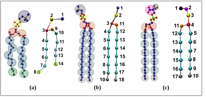
Figure 2.1: (a) United atom and (b) coarse-grained representation of DPPC. The color scheme is; purple (choline entity – type I for site 1); yellow (phosphate group – type II for site 2); red (ester group – type III for site 3); orange (ester group – type IV for site 9); cyan (alkyl tail groups – type V for sites 4-7 & 10-13); green (terminal tail groups - type VI for sites 8&14). The coarse-grained site size does not represent the actual size of each site. ... 41 Figure 2.2: A schematic of the approach used to calculate the LIME interaction energies for two coarse-grained types: (a) the radial distribution function is calculated (b) the one-step Boltzmann inversion scheme is used to calculate the potential of mean force by inverting the RDF; ε is chosen as the minimum U(r) value (blue line); the depth of the square well potential or the interaction energy is assigned the ε value (red line) ... 42 Figure 2.3: A radial distribution function for sites 1 & 1 obtained during a LIME simulation. The first non-zero value is located at the hard sphere diameter (4.75 Å) and the small discontinuity is located at the square-well width (12.55 Å). The shape of the radial distribution function differs significantly from the shape of a distribution function associated with a more traditional Lennard Jones potential. ... 42 Figure 2.4: (a) The intermolecular radial distribution functions, hard-sphere
collisions (d). An additional 100 million collisions are required for the aggregate to adopt the conformation of a defect-free bilayer (h). ... 45 Figure 2.8: Comparison of the orientational bond order parameter SBOND for intra-molecular bonds in LIME/DMD (green line) and GROMACS simulations (blue line) versus the bond number: the latter is defined in the inset. ... 46 Figure 2.9: Snapshots of a lipid bilayer in DMD/LIME as the system temperature is cooled from (a) a liquid-crystalline phase at T* = 0.77, (b) a tilted gel phase at T* = 0.30 and (c) a cross-tilted gel phase which is only observed in some simulations. .... 46 Figure 2.10: Snapshots of a lipid (spherical representation) as it flips from the
bottom leaflet of a bilayer to the top leaflet. The tail beads of the lipid that flips are highlighted in yellow and the head beads are highlighted in lime. (a) – (c) = 866, 883, 885 million collisions, respectively. ... 47 Figure 2.11: Mass density distribution of coarse-grained sites in DMD/LIME
simulations (dotted lines) and GROMACS simulations (solid lines) versus the
distance from the bilayer center (z = 0 Å). ... 48 CHAPTER 3 ... 49
The Extension of LIME to Model the Phase Separation Behavior of Mixed Lipid Systems at Neutral and Low pH ... 49
Figure 3.4: The number of DSPS lipids with a DSPS nearest neighbor versus the collision time for Systems 1 through 4. The data displayed is time averaged in 50 million collision increments from 0 to 1000 million collisions. In addition, the data was averaged for the 3 replicates run for each system. ... 75 Figure 3.5: Snapshots of the side profile of replicate 3 from System 4 at (a) 750 million collisions and (b) 1 billion collisions. The color scheme is: DSPS lipids (black) and 21PC lipids (cyan). ... 76 Figure 3.6: Snapshots (aerial images) of the bilayer formed at 1 billion collisions in simulations of Systems 2 (a and b), 4 (c and d), 9 (e and f) and 10(g and h). The color scheme is: black (DSPS lipids), lime (DPPC lipids), blue (21PC lipids). ... 78 Figure 3.7: The number of DSPS lipids with a DSPS nearest neighbor verses the collision time for Systems 2, 4, 9 and 10. The values for the y-axis were averaged for each of the 3 replicates run for each system number. ... 83 Figure 3.8: Snapshots (aerial images) of bilayers at 1 billion collisions for Systems 2, 4, 5, 6, 7, and 8. The color scheme is: black (DSPS lipids), lime (DPPC lipids), blue (21PC lipids). ... 85 Figure 3.9: The number of DSPS lipids with a DSPS nearest neighbor verses the collision time for Systems 2, 4, 5, 6, 7 and 8. The values for the y-axis were averaged for each of the 3 replicates run for each system number. ... 89 Figure 3.10: Snapshots of the DPPC/DSPS/doxorubicin liposome after (a) 1 million collisions and (b) 1.5 billion collisions. Snapshots of the 21PC/DSPS/doxorubicin liposome after (c) 1 million collisions and (d) 1.5 billion collisions. The color scheme is: black (DSPS lipids), lime (DPPC lipids), blue (21PC lipids), purple (doxorubicin) ... 91 CHAPTER 4 ... 100 Modeling the Interaction Between Hydrophilic and Hydrophobic Nanoparticles with Bilayer Membranes Using LIME, an Intermediate-Implicit Solvent Model Designed for Use with Discontinuous Molecular Dynamics ... 100
Figure 4.1: (a) Coarse-grained representation of DPPC (b) Coarse-grained
Figure 4.2: Snapshots of simulations run on the interaction between hydrophilic nanoparticles of different sizes and mass/volume with a DPPC bilayer membrane. (a) run #1, (b) run #2, (c) run #3, (d) run #4, (e) run #5, (f) run #6, (g) run #7, (h) run #8. The color scheme is: purple (DPPC choline entity), orange (DPPC phosphate group), red (DPPC ester groups), cyan (DPPC alkyl tail groups), yellow (nanoparticles). .. 119 Figure 4.3: Snapshots from run #6 in which a hydrophilic nanoparticle with diameter 40Å is wrapped by a bilayer membrane. The nanoparticle (a) reaches the surface of the bilayer at 25 million collisions, the wrapping process at (b) 625 million collisions, (c) 1250 million collisions and (d) 3250 million collisions. ... 122 Figure 4.4: Snapshots from run #10 of a simulation of a hydrophobic nanoparticle with a diameter of 20Å and a DPPC bilayer composed of 1500 lipids. The
nanoparticle (a) reaches the surface of the bilayer after 25 million collisions, (b) is embedding itself within the membrane after 50 million collisions ,and (c) is fully embedded within the inner hydrophobic core of the membrane after 275 million collisions. ... 123 Figure 4.5: Snapshots from run #12 of a simulation of a hydrophobic nanoparticle with a diameter of 40Å and a DPPC bilayer composed of 1500 lipids. The
nanoparticle (a) reaches the surface of the bilayer after 50 million collisions, (b) is embedding itself within the membrane after 75 million collisions and (c) is fully embedded within the inner hydrophobic core of the membrane after 200 million collisions. ... 125 Figure 4.6: Snapshots from simulations of hydrophobic nanoparticles with a mass of 43.6 amu (a) and 0.82 amu (b). ... 126 CHAPTER 5 ... 131
Discontinuous Molecular Dynamics Simulations of DNA Hybridization using
“DIME,” a New Coarse-Grained Implicit-Solvent Model ... 131 Figure 5.1: (a) Atomistic and (b) coarse-grained representation of a Dickerson-Drew dodecamer duplex. The color scheme for the coarse-grained representation is; cyan (sugar group – type S); red (phosphate group – type P); white (cytosine and guanine bases – types C&G); blue (adenine and thymine bases – types A&T). The coarse-grained size does not represent the actual size of each site. ... 136 Figure 5.2: Snapshots from a simulation of the spontaneous formation of a
Dickerson-Drew dodecamer duplex formation. The following color scheme is used to represent each of the coarse-grained types: S (blue), P (green), C (yellow), G
CHAPTER 1 Introduction
1.1 Motivation
The technological advances that have been made to date in the many fields of research aimed at exploiting the unique properties of biomolecules and nanoparticles are astonishing. For example, lipid bilayers and liposomes are now being used to create novel devices for the targeted delivery of proteins, nucleic acids and drugs to treat a wide variety of diseases.[1,2,3] In addition, DNA microarrays are used to measure gene expression levels [4,5,6,7] and the potential of DNA to replace silicon transistors as the next generation of storage technology [8] was recently demonstrated. Drugs that function by targeting DNA molecules have also been developed.[9,10,11] The interaction between nanoparticles and biomolecules has also become a popular area of study due to the increasing applications of nanoparticles in drug delivery and the concerns associated with nanoparticle toxicity. In order to fully realize the potential of the numerous emerging biomolecular technologies, a tool that would provide a better understanding of biomolecules and nanoparticles on a molecular level is needed. One approach that is commonly used to gain insight into the behavior of such biomolecular systems on a molecular level is computer simulation.
Molecular dynamics computer simulations can be divided roughly into two
One drawback associated with atomistic models is that the detail that makes these models so realistic and appealing also makes them extremely computational intensive and prevents them from examining large conformation changers or long time scales. In contrast to high-resolution models, low-high-resolution models, which are also known as coarse-grained models, are based on a simplified representation of molecular geometry and energetics. In a coarse-grained model a single interaction site is used to represent the behavior of a group of several atoms. This reduces the total number of sites whose trajectories must be calculated, thereby increasing the speed of the simulation.
This thesis describes our work to develop new intermediate resolution implicit solvent models for lipids, “LIME,” and DNA molecules ,“DIME,.” designed for use with discontinuous molecular dynamics computer simulations We provide a detailed explanation regarding the multi-scale modeling approach that was used to extract both the LIME and DIME parameters from explicit solvent atomistic simulations. We applied LIME to study the spontaneous formation of lipid bilayers, the behavior of mixed lipid systems at different pH values and the interaction between membranes and nanoparticles. DIME was used to investigate the structural properties of DNA and the process by which two DNA strands hybridize in solution. The long term goal of our work is to develop a systematic approach that can be used to gather the geometric and energetic parameters required to run implicit-solvent, coarse-grained simulations with discontinuous molecular dynamics of any system of biomolecules. In addition, we would like to use these models to provide researchers with molecular level insights in order to facilitate the exploration and design of novel
1.2 Overview
In this section, we summarize Chapters 2 – 6 of this thesis. Each chapter contains a literature review and a bibliography.
Chapter 2 describes in detail the development of “LIME,” which is a new
intermediate resolution implicit model for lipid molecules. LIME was designed for use with discontinuous molecular dynamics (DMD) simulations. The model was developed using a multi-scale modeling approach in which the geometric and energetic parameters are obtained by collecting data from atomistic simulations of a system composed of 1,2-dipalmitoyl-sn-glycero-3-phosphocholine (DPPC) molecules and explicit water. In the model, 14 coarse-grained sites are used to represent DPPC and each of these sites is classified as 1 of 6 types. DMD/LIME simulations performed on a random solution of DPPC resulted in the formation of a defect free bilayer in less than 4 CPU hours. The bilayer formed quantitatively
reproduces the main structural properties (e.g. area per lipid, bilayer thickness, bond order parameters) that are observed experimentally. In addition, the bilayer transitions from a liquid-crystalline phase to a tilted gel phase when the temperature is reduced. Transbilayer movement of a lipid from the bottom leaflet to the top leaflet is observed when the
temperature is increased.
In Chapter 3 we discuss the expansion of LIME to describe the geometry and energetics of DPPC, distearoyl-sn-glycero-3-phospho-L-serine (DSPS) and
the model, 14, 17 and 21 coarse-grained sites are used to represent DPPC, DSPS and 21PC, respectively. Each of these coarse-grained sites is classified as 1 of 10 types. LIME/DMD simulations of equimolar bilayers show the following: 21PC/DSPS bilayers separate slightly faster at low pH than at neutral pH, but DPPC/DSPS bilayers separate at approximately the same rate at neutral and low pH, 21PC/DSPS bilayers separate slightly more than
DPPC/DSPS bilayers. Our results also show that at low pH equimolar DPPC/DSPS bilayers without surface area restrictions separate faster than those with restrictions but surface area restrictions on equimolar low pH 21PC/DSPS bilayers did not affect the separation rate. Simulations of DPPC/DSPS and 21PC/DSPS bilayers with different molar ratios of PC:PS lipids showed that the higher the concentration of PS lipids, the faster the separation rate. Simulations of DPPC/DSPS and 21PC/DSPS liposomes containing doxorubicin showed domain formation in both types of liposomes. However, no drug molecules escaped from either type of liposome after 1.5 billion collisions.
these nanoparticles become embedded on the surface of the lipid bilayers among the hydrophilic head groups of the lipid molecules. Finally, our simulations showed that the mass per volume of a nanoparticle did not significantly affect its interaction with a lipid bilayer.
Chapter 5 provides a description of initial work performed to develop a new
intermediate-resolution implicit-solvent model for DNA molecules, which we call “DIME” for DNA Intermediate Resolution Model. The same multiscale modeling approach used to develop LIME was followed to calculate parameters for DIME. The parameters for this model are obtained by collecting data from an atomistic simulation of a Dickerson dodecamer duplex with explicit solvent and counterions. A single coarse-grained site is used to represent each sugar, phosphate and base molecule in this model. Each of these coarse-grained sites is classified as 1 of 6 types for sugar, phosphate, cytosine, guanine, adenine and thymine. Similar to LIME, DIME was designed for use with discontinuous molecular dynamics simulations. We are able to use this new model to show the spontaneous hybridization that occurs when two Dickerson-Drew dodecamer strands are placed in an initial random configuration. In addition, we discuss the results of alternative method used to calculate the square-well width between coarse-grained sites that involves the second virial coefficient. Simulations run using this second set of parameters did not result in the
spontaneous formation of a double helix even though the helix remained stable at low temperatures.
Chapter 2 is adapted from the following publication:
Curtis, E.; Hall, C. J. Phys. Chem. B., 2013, 117, 5019-5030.
1.3 References
1. Karve, S.; Bandekar, A.; Ali, M.; Sofou, S. Biomaterials. 2010, 31, 4409 – 4416.
2. Schroeder, A.; Levins, C.; Cortez, C.; Langer, R.; Anderson, D. Journal of Internal Medicine. 2009, 267, 9 – 21.
3. Almeida, A.; Souto, E. Adv. Drug Delivery Rev. 2007, 59, 478-‐479.
4. Schena, M.; Shalon, D.; Davis, R.; Brown, P. Science, 1995, 270, 467 – 470.
5. DeRisi, J.; Penland, L.; Brown, P.; Bittner, M.; Meltzer, P.; Ray, M.; Chen, Y.; Su, Y.; Trent, J. Nature Genetics, 1996, 14, 457 – 460.
6. Lockhart, D.; Winzeler, E. Nature, 2000, 405, 827 – 836.
7. Chon, H.; Lancaster, J. Cancer Control, 2011, 18, 8 – 15.
8. Goldman, N.; Bertone, P.; Chen, S.; Dessimoz, C.; LeProust, E.; Sipos, B.; Birney, E. Nature, 2013, 494, 77-‐80.
9. Hurley, L. Nature Reviews Cancer, 2002, 2, 188 – 200.
10. Palchaudhuri, R.; Hergenrother, P. Current Opinion in Biotechnology, 2007, 18, 497 – 503.
CHAPTER 2
Molecular Dynamics Simulations of DPPC Bilayers Using “LIME,” a New Coarse-grained Model
2.1 Introduction
The lipid bilayer, the primary constituent of cellular and intracellular membranes in
all living organisms, plays a central role in many biological processes including cell signaling
and protein function.[(1),(2),(3)] In addition to its physiological significance, lipid bilayers
are now being used to create devices for targeted delivery of proteins, nucleic acids and drugs
in the treatment of a wide variety of diseases.[(4),(5),(6)] Significant progress has been
made by scientists working to use these structures to develop therapeutic
agents.[(7),(8),(9),(10),(11),(12)] A tool that would allow researchers to visualize the
structure and function of the lipid bilayer on a molecular level could help enhance the rate of
advancement in these areas. For example, we are now using the model discussed in this
manuscript to study the release of drug molecules from liposomes composed of lipid
mixtures as a result of a change in pH. [(13),(14),(15)]
In this paper we take a multiscale modeling approach to develop a new
implicit-solvent intermediate-resolution lipid model, “LIME” for Lipid Intermediate Resolution
Model, that enables molecular dynamics simulation of the self-assembly of a lipid bilayer.
The model system chosen for study is the lipid 1,2-dipalmitoyl-sn-glycero-3-phosphocholine
(DPPC) in water. We show that discontinuous molecular dynamics (DMD) simulations
including the area per lipid, bilayer thickness, bond order and mass density profiles. This
model is the culmination of a systematic program of research aimed at developing simulation
tools based on coarse-grained lipid models that are fast enough to simulate self assembly of
large structures yet have accuracy that is comparable to that found in atomistic simulations.
By using multiscale modeling, which translates the atomistic details from well established
force fields, GROMOS96 53a6 in this case, into coarse grained simulations, we avoid the
pitfalls associated with fitting many molecular parameters to the limited data on lipid
systems.
Molecular dynamics studies of phospholipid bilayers can be divided roughly into two
categories: high-resolution models and low-resolution models. High-resolution or atomistic
models are based on a realistic representation of membrane geometry and energetics and
typically account for the motion of every atom on the membrane and every solvent atom.
Atomistic simulations of lipid bilayers have been performed to study the permeation of small
molecules through a lipid bilayer [6], the interaction between lipid bilayers and substrates
[7], the behavior of charged and neutral bilayers [8], and a large variety of additional bilayer
properties and behaviors.[(19),20,(21)] In a recent united-atom study, Kukol demonstrated
that the GROMOS96 53a6 forcefield [(22)] and the Kukol DPPC3 topology could be used
with GROMACS [(23),(24)] to reproduce the experimental area per lipid of a preformed
DPPC bilayer for a united-atom system composed of 128 lipids and 3655 water molecules
with 3% accuracy without assuming constant surface area or including surface
pressure.[(25)] , one drawback associated with atomistic models is that the detail that makes
and prevents them from examining large conformational changes or long time scales. For
example, the 90 ns atomistic simulation of 1017 DPPC lipids and 106,563 water molecules
by de Vries et al. in 2004, which shows the spontaneous formation of a DPPC vesicle, was
run on four or eight processors at a rate of only 1 ps per processor CPU hour (1.7 GHz Intel
Pentium IV processors).[(26)]
Coarse-grained models of lipids have been developed in order to reduce simulation
time so as to access longer time scales than are achievable in atomistic simulations. In these
models clusters of atoms are grouped together into single sites to reduce the number of
events that require calculation.[(27] A popular explicit-solvent coarse-grained model for
lipids is MARTINI which was developed by Marrink et. al. and has been used to simulate the
spontaneous aggregation of a DPPC bilayer.[(28)] In this model, an average of four atoms
are represented by a single interaction site, 10 different types of interaction sites are defined,
and the interaction strengths between any two sites are assigned one of five values.[(28)]
The Marrink predictions of the DPPC area per head group, bending modulus, area
compressibility, lipid lateral diffusion coefficient and water permeation rate closely matched
the experimentally measured quantities.[(28)] Marrink et. al improved the MARTINI force
field (creating version 2.0) by increasing the number of types of possible interaction sites
from 10 to 18 and increasing the number of interaction strength levels from 5 to 10.[(29)]
MARTINI (version 2.0) was applied to model molecular raft formation in model
membranes.[(29),(30)] Although we have looked, we have been unable to locate any
information about the computational speed of the MARTINI model for lipid systems. Orsi
represents the 118 atoms of DMPC by 10 coarse-grained sites; the predicted structure,
elasticity, electrostatics and dynamics of a DMPC bilayer quantitatively matched
experimental data.[(27)] In this model, the spherical units representing the headgroup
choline and phosphate groups interact via the Lennard Jones potential and the glycerol and
hydrocarbon groups of the lipids are modeled as soft uniaxial ellipsoids through the
Gay-Berne potential.[(27)] The Orsi model was extended to dioleylphosphatidylcholine (DOPC)
by adopting a 12 site coarse-grained representation.[(31)] Their simulations in 2010 of the
formation by 128 DOPC lipids of a defect-free bilayer required approximately 2.5 days;
those of the formation by 128 DMPC lipids of a defect-free bilayer with an embedded water
pore required approximately 25 days (Intel 2.8 GHz processors in serial).[(31)]
Subsequently, Orsi and Essex developed the electrostatics-based “ELBA” 1.0 force field for
coarse-grained models of lipid membranes which explicitly represents charges and
dipoles.[(32)] In this new model the Gay-Berne components are replaced with Lennard
Jones potentials. DOPC, DOPE and DSPC are each represented by 15 spherical CG sites and
water is treated explicitly. The ELBA 1.0 force-field was found to accurately reproduce
several of the experimentally observed physical properties of single-species lipid bilayers
composed of DOPC, DOPE or DSPC in the liquid crystal phase and DSPC in the gel phase.
ELBA 1.0 was later refined to become ELBA 1.1 to correctly reproduce the hexagonal
inverse phase for DOPE-water systems.[(33)] ELBA 1.1 was also used to simulate mixed
DOPC-DOPE bilayers at various compositions and to calculate the first reported values for
the lateral pressure and electrical potential profiles for mixed DOPC-DOPE bilayers.[(33)]
model for phospholipids for use with hybrid particle field molecular dynamics
simulations.[(34)] In this work, the MARTINI coarse-grained mapping scheme is used to
represent DPPC and the model parameters are optimized so that the coarse-grained model
reproduces the structural properties of the reference particle-particle simulations.[(34)]
Some coarse-grained models ignore the motion of solvent atoms to further enhance
the computational efficiency associated with coarse-graining. Instead, the effect of solvent
atoms is included implicitly through the use of effective potentials, or potentials of mean
force. Recently, Wang and Deserno presented an implicit solvent, coarse-grained model for
POPC bilayers derived using a multiscale modeling approach based on structure-matching
methodology.[(35)] In this model the 134 atoms in each POPC lipid molecule are
represented by 16 coarse-grained sites of 8 different types. The coarse-grained potentials
were optimized iteratively to reproduce radial distribution functions and the area per lipid of
the bilayer obtained from all-atom simulations performed with the molecular dynamics
program NAMD [(36)] and the fully atomistic CHARM27 [(37)] parameters. In order to
promote lipid aggregation in this model, it was necessary to introduce additional cohesive
interaction potentials between the alkyl tails and between the interfacial head group sites.
The strength of the cohesive interaction potentials were chosen to promote bilayer stability,
to match RDFs for the coarse-grained and atomistic simulations, and to optimize the lateral
stress profile; without it, the bilayer falls apart. This model was used to simulate the
self-assembly of 288 POPC lipids into a bilayer from a random lipid dispersion that quantitatively
matched experimental bilayer properties. A defect-free bilayer formed in approximately 32
similar approach was taken by Lyubartsev who constructed a coarse-grained, implicit-solvent
lipid model containing 10 coarse grained sites to represent the 118 atoms of DMPC; the
parameters were optimized to reproduce the radial distribution functions from all-atom
molecular dynamics simulations performed with the MDynaMix [(38)] package and the
all-atomic CHARMM27 [(20),(37)] force field.[(39)] Simulations of DMPC lipids using this
model show the formation of bicelles and vesicles starting from a disordered system of
lipids.[(39)] Izvekov et. al. used a multiscale coarse-graining approach to develop a model in
which 11 different coarse-grained types are used to represent DMPC and cholesterol
molecules.[(40)] The Izvekov model accurately reproduced the structural and elastic
properties of a DMPC lipid bilayer and was used to simulate pre-formed DMPC/cholesterol
liposomes. However, simulations of a DMPC/cholesterol system starting from a random
dispersion did not form a bilayer. Instead, this system assembled into aggregates composed
of DMPC and cholesterol and aggregates composed primarily of cholesterol.[(40)]
In this paper, we describe the development of an implicit-solvent, coarse-grained
model, LIME, derived using a multi-scale modeling approach that enables simulations of
large numbers of phospholipids in aqueous solution. The number of coarse-grained sites per
lipid, 14, is very similar to the coarse-grained phospholipid representation of DPPC in other
models,[(28),(31),(35),(39)] but the interactions are not. Instead the interactions between
coarse-grained sites are represented by hard sphere and square well potentials as opposed to
Lennard Jones potentials, thereby allowing us to use discontinuous molecular dynamics, a
fast alternative to traditional molecular dynamics. The multiscale modeling procedure used to
ns GROMACS simulation using the GROMOS96 53a6 united-atom forcefield for a system
containing 30 DPPC phospholipids is coarse-grained into 14 sites. United-atom simulations
are essentially the same as atomistic simulations with the exception that hydrogen atoms
bonded to carbon atoms are represented as a single site. Radial distribution functions (RDF)
between all bonded and non-bonded pairs of coarse-grained sites are calculated and used to
determine LIME geometrical and energetic parameters. The hard sphere diameter between
non-bonded coarse-grained sites is estimated to be the smallest distance at which the RDF
takes a non-zero value. The RDFs were also used to estimate the square well width, and the
minimum and maximum bond lengths between bonded pairs. The relative stiffness of each
lipid is maintained by imposing pseudobonds, which limit the bond length fluctuations to the values observed in the GROMACS simulations. Interaction energies between non-bonded coarse-grained sites are determined by calculating the potential of mean force using a
one-step Boltzmann inversion scheme.[(41),(42)] In the model each coarse-grained site has its
own realistic mass.
Highlights of our results include the following; The model successfully simulates the spontaneous assembly of a DPPC bilayer composed of 256 lipids in less than 4 CPU hours starting from a random initial configuration, which is approximately an order of magnitude faster than the fastest reported coarse-grained implicit solvent model. The area per lipid of
our bilayer is within 2% of the value calculated from our GROMACS simulation data and the
literature value. The thickness of our bilayer is within 5-7% of the literature value and
within 4-8% of the thickness of the bilayer in our GROMACS simulations. The orientational
calculated from the GROMACS model. The mass density profiles of the LIME and
GROMACS models closely match each other. Finally, LIME is able to simulate transbilayer
flip-flop in which a lipid flips from the bottom leaflet of a membrane to the top leaflet.
2.2 Theoretical Methods
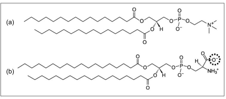
In LIME six different coarse-grained types (I – VI) are used to represent the 130
atoms that make up a DPPC molecule. DPPC is composed of a polar head group that
includes a choline, phosphate and two ester linkages, and two nonpolar hydrophobic acyl
tails. Figure 2.1 illustrates the coarse-graining of a DPPC molecule from 50 united-atom
(Figure 2.1a) to the 14 coarse-grained sites in the LIME representation. This figure and all
other figures depicting lipid molecules throughout the paper were generated with Visual
Molecular Dynamics (VMD).[(43) Each coarse-grained site that represents a unique set of
atoms is assigned a different coarse-grained type. Each coarse-grained type is represented by
a different color in Figure 2.1b. Table 2.1 lists the atoms included in each coarse-grained
site, the “type” assigned to each coarse-grained site and the mass of each coarse-grained site.
Types I and II represent the choline entity and the phosphate group, respectively. Ester
coarse-grained sites 3 and 9 are assigned types III and IV, respectively. The coarse-grained
sites in the hydrocarbon tails (excluding the terminal sites) are assigned type V. Finally, the
terminal tail coarse-grained sites are classified as type VI. We considered treating the
terminal tail beads (sites 8 & 14) as the same type as the non-‐terminal tail beads (sites
4-‐7 & 10-‐13) because type V differs from type VI only by a single hydrogen atom.
different than the parameters for type VI&VI pairs (ε = -‐0.070 eV). This is probably
because of the differences in connectivity between the two types. Therefore, we felt
that it was important that the different groups have their own unique types.
In addition to coarse-graining and treating solvent implicitly, we employ
discontinuous molecular dynamics (DMD) simulation to further increase the speed of our
code. DMD [(44)] is a very fast alternative to traditional molecular dynamics simulation that
is applicable to systems of molecules interacting via discontinuous potentials, e.g.,
hard-sphere and square-well potentials. For this reason, all of the inter- and intra- molecular
interactions in our lipid model are represented by a combination of hard-sphere and square
well potentials, as opposed to the Lennard Jones, Coulombic and harmonic potentials found
in traditional molecular dynamics simulations. Unlike continuous potentials, such as the
Lennard-Jones potential, discontinuous potentials exert forces only when particles collide.
This enables the exact (as opposed to the numerical) solution of the collision dynamics. This
imparts great speed to the algorithm and allows sampling of much wider regions of
conformational space, longer time scales and larger systems than in traditional molecular
dynamics. Molecules are given an initial random configuration that satisfies both excluded
volume and angular constraints. Initial velocities are chosen randomly from a
Maxwell-Boltzmann distribution about the desired temperature. Particle trajectories are followed by
calculating the time between each collision and advancing the simulation to the next event.
Types of events include a collision between two spheres, a bond event when the distance
between two bonded spheres reaches a minimum or maximum limit, and a square well event
leave (dissociation) a square-well attraction.[(44),(45),(46),(47)] The simulations are
performed with the number of particles, the temperature and the volume held constant. The
temperature is maintained constant using the Andersen thermostat, which uses ghost
collisions with randomly selected particles in the system to maintain the Maxwell-Boltzmann
velocity distribution about the desired temperature.[(48)] The simulations were run at
constant volume as this is the most straightforward ensemble for use in DMD. In the
Conclusion section we discuss the possibility of simulating lipids in the NPT ensemble using
a combination of a hybrid Monte Carlo/DMD approach.
Data used to calculate the coarse-grained model parameters was obtained by running
explicit-solvent NPT ensemble united-atom simulations on a system containing 30 DPPC
phospholipids and 8655 water molecules. The GROMACS simulation package [(23),4],
version 4.5.4, was used with the GROMOS96 53a6 forcefield [(22)] and the Kukol DPPC3
topology, which has been shown previously to accurately reproduce the experimental area
per lipid, lateral self diffusion constant and deuterium order parameters for the acyl chains in
DPPC bilayers in solution.[(25)] The initial configuration of this system was random in a box
with equal sides of length 100.0 Å. The Berendsen thermostat [(49)] was used to keep the
temperature constant at 325K throughout the simulation with a time constant of 0.1ps. The
simulation was run for 20 ns with a time step of 0.002 ps for approximately 48 CPU hours.
Periodic boundary conditions were applied and the pressure was maintained at 1.0 bar.
Throughout the GROMACS simulation the coordinates of each atom were written to an
output trajectory file every 1 ps. These output files were used to calculate the centers of mass
We chose to run atomistic simulations of a small system to gather data for use in
calculating coarse-grained parameters because we did not want to restrict the movement of
the lipids during the atomistic simulation. It is not uncommon to gather data for a
coarse-grained model from a small system. For example, Lyubartsev performed atomistic
simulations of only 16 DMPC lipids to obtain data for use in calculating coarse-grained
parameters.[(39)] To ensure that our atomistic simulation of 30 DPPC lipids was not too
small or at too low a density, we compared the radial distributions we calculated from this
simulation with those calculated from a GROMACS simulation of 128 DPPC lipids in a box
with dimensions of 64Å x 64Å x 90Å. The sigma and lamda values calculated from each
GROMACS simulation were nearly identical. Furthermore, the epsilon values calculated
from each simulation differed by minimal amounts.
2.3 Results and Discussion
The LIME interaction energies were determined using a one-step Boltzmann
inversion procedure inspired by the iterative Boltzmann inversion scheme, which is a popular
strategy used to systematically compute potentials for coarse-grained
simulations.[(35),(41),(42)] We begin by reminding the reader of the iterative Boltzmann
inversion scheme, an approach based on the idea that the effective potential, or potential of
mean force, U(r), between two molecules in a sea of molecules can be obtained from the
radial distribution g(r) using:
where kB is the Boltzmann constant and T is the temperature of the system. The iterative
Boltzmann inversion scheme involves the following steps: (1) Data from an atomistic
simulation is used to calculate the intermolecular radial distribution function g(r) between
coarse-grained sites. (2) An initial guess for the potential of mean force, U(r), between the
coarse-grained sites is determined using Equation 1. (3) A coarse-grained simulation using
the initial guess for the potential of mean force is run and a new g(r) between coarse-grained
sites is calculated. (4) The difference between the coarse-grained and the atomistic potentials
of mean force is used to generate a correction to the coarse-grained potential of mean force.
(5) This process is repeated iteratively until the coarse-grained and atomistic potentials of
mean force match each other within a prescribed tolerance.[(41),(42)]
Instead of using the iterative Boltzmann inversion procedure described above, we use
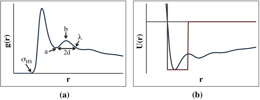
a simplified, one-step Boltzmann inversion to obtain the LIME interaction energies. Figure
2.2 outlines this approach, the procedure is the following: (1) the average radial distribution
function between two intermolecular coarse-grained sites is determined, (2) the potential of
mean force is calculated using Equation 1, and (3) the minimum value of the potential of
mean force between the coarse-grained sites, ε, is chosen to be the depth of the square well
potential. Mathematically this can be expressed as:
ε = -kBTln[g(r)MAX] Equation 2
where g(r)MAX is the maximum value of g(r) in the radial distribution function and T is the
temperature of the system. If the ε between two coarse-grained sites is greater than -0.005
The iterative Boltzmann inversion approach was not used to obtain the LIME
parameters because the shape of the radial distribution function for a discontinuous potential
obtained by coarse-graining the GROMACS simulation is dramatically different from the
shape of the radial distribution function associated with a square-well potential. See for
example the radial distribution function for sites 1 & 1 obtained during a LIME simulation
shown in Figure 2.3. As is expected for square-well systems, there are no oscillations and
there are discontinuities in g(r) at distances that correspond to the discontinuity in the
potential. Thus it does not make sense to use an iterative procedure to try to match the
GROMACS and coarse-grained potentials. By using the one-step Boltzmann inversion
scheme we are able to develop a model that very accurately matches experimental
observations for a lipid bilayer. Since we were able to obtain very good agreement between
the physical properties of our LIME bilayer with experimental values we did not attempt to
go beyond a one-step scheme.
The LIME hard sphere (σHS) diameters and square-well (λ) widths were determined
from the radial distribution functions between pairs of non-bonded coarse-grained sites in the
GROMACS simulations which were run on 30 DPPC and 8655 water molecules. As shown
in Figure 2.2a the hard sphere diameter (σHS) for each pair of interaction sites was
determined by locating the smallest non-zero separation between the two sites. The square
well sphere diameter (λ) for each pair of interaction sites was determined by examining the
radial distribution function between those sites. First, the local maximum (labeled b in
minimum (labeled a in Figure 2.2a) preceding this local max was identified. Finaly, λ was
calculated using:
λ = a + 2d Equation 3
where d = (b-a). If a λ value greater than 15 Å was calculated, the procedure for calculating
λ was repeated using a local maximum closer to the origin. Sample radial distributions for
intermolecular coarse-grained types 1&1, 1&2 and 5&5 are provided in Figure 2.4. The hard
sphere diameters (σHS) determined for coarse-grained types 1&1, 1&2 and 5&5 are 4.35 Å,
3.85 Å and 3.75 Å, respectively. The values of λ for coarse-grained types 1&1, 1&2 and
5&5 were found to be 12.65 Å, 9.85 Å and 11.56 Å, respectively. A complete list of all hard
sphere diameters, square well widths and interaction energies is provided in Table 2.2.
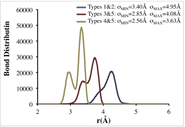
The minimum and maximum bond lengths were determined by plotting the radial
distribution functions for bonded coarse-grained sites. The minimum bond length (σMIN) was
chosen as the smallest possible distance between two bonded coarse-grained sites. The
maximum bond length (σMAX) was chosen as the largest distance for which a non-zero g(r)
was observed. Sample distributions for intramolecular coarse-grained types 1&2, 3&5, and
5&6 used to determine σMIN and σMAX for bonds and the resulting values of σMIN and σMAX are
provided in Figure 2.5.
The relative stiffness of the lipid molecule is maintained by imposing pseudobonds,
which limit the fluctuation of coarse-grained sites to the angles and torsional angles observed
during the GROMACS simulations. Bond angles were maintained by imposing pseudobonds
with pseudobonds between next next nearest neighboring sites along the chain. Bond
distributions for intramolecular sites calculated from the GROMACS simulations were used
to determine the minimum and maximum values for the pseudobond lengths in LIME. The
minimum pseudobond length was determined by finding the smallest distance at which the
intramolecular bond distribution function exceeds 30% of its maximum value. The
maximum pseudobond length was determined by finding the smallest distance, larger than
the distance at which the bond distribution function maximum occurs, where the
intramolecular bond distribution function falls below 30% of its maximum value.
Pseudobonds were also added between intramolecular coarse-grained sites 5&11, 6&12,
7&13 and 8&14 on the same chain to restrict the separation between the tails to the distances
observed during the GROMACS simulations. This was done to prevent the tails from
adopting conformations that were not frequently observed during the GROMACS
simulations. The minimum pseudobond lengths between intramolecular sites 5&11, 6&12,
7&13, and 8&14 were determined by locating the smallest distance at which the
intramolecular bond distribution function exceeds 30% of its maximum value. The
maximum pseudobond lengths between intramolecular sites 5&11, 6&12, 7&13 and 8&14
were determined by finding the smallest distance, larger than the distance at which the
intramolecular bond distribution function maximum occurs, where the bond distribution
function falls below 30% of its maximum value.
In all the simulations run using the LIME force field, periodic boundary conditions
were implemented to eliminate any artifacts that might be caused by the box walls. In
particles, the temperature and the volume are held constant. Simulation temperature in LIME
is expressed in terms of the reduced temperature, T*:
T*= kBT/ε* Equation 4
where kB is Boltzmann’s constant, T is the temperature, and ε* is the reference interaction
strength.[(50)] The reference interaction strength, ε*, was calculated using:
ε
*
=
n
ijε
ij ij∑
n
ij ij∑
Equation 5where nij is the number of coarse-grained sites with a type i and type j interaction and εij is
the interaction energy between coarse-grained types i and j. The εij values were obtained
from the GROMACS simulations at T=325K. The resulting value for ε* calculated from
Equation 4 is 0.0363. Thus when T* = kBT/ε* = (8.6173x10
-5
eV/K)*(325K)/(0.363eV) =
0.77 in our DMD/LIME simulations, the lipid molecules will behave as they would at a real
temperature of 325K. The Andersen thermostat is used to hold the temperature constant. In
this method randomly selected particles collide infrequently with ghost particles, effectively
reassigning the particle’s velocity randomly so as to maintain a Maxwell-Boltzmann
distribution centered at the simulation temperature.[(48)] All LIME/DMD simulations were
run with a DMD software program developed in the Hall research lab called EMBLEM.
This program is written in C++. The Intel compiler was used to compile this code and all
serial. DMD can be run in parallel and in the future it is likely that we will parallelize our
code.[(51)]
Five independent DMD simulations starting from different random configurations
were run using the LIME force field to determine whether or not a bilayer could be formed
starting from a random configuration of 256 DPPC phospholipids at a T* = 0.77. The
lengths of the sides of the simulation cell were set to 90 Å. A bilayer was formed in all five
simulations. Simulations of the 256-lipid system were run with several different box sizes to
ascertain which box size should be used to evaluate bilayer properties. First, we simulated a
system of 256 lipids in a box with dimensions of 100Å x 100Å x 100Å. This system formed
a bilayer with a large hole in it as shown in Figure 2.6a. The area per lipid of this bilayer,
counting only portions external to the hole, was 63.3 Å2
±0.1 Å2
. (If it had spanned the box,
the area per lipid would have been 78.1 Å2
). Although at first glance the hole in our bilayer
may appear to be a hole of vacuum, this is not the case. In the implicit-solvent approach that
we are using all of the empty space is meant to represent a structureless solvent. This is a
consequence of using the McMillan-Mayer solution approach in which a two-component
system is mapped onto to a one-component system by integrating out the degrees of freedom
of solvent and hence increasing the speed of our simulations.[(52)] Since our lipid
parameters are calculated using a method that accounts for the effect of water, they are
expected to behave as they would in an aqueous solution, not a vacuum. See the
Conclusions section for more discussion of this issue. Next, we ran a simulation of 256
DPPC lipids in a box with dimensions of 80Å x 80Å x 80Å. The bilayer formed in this
sides of the simulation box. Thus there were obviously too few lipids in the first box size
(100Å3
) and too many lipids in the second box size (80 Å3
). Since the bilayer formed in the
first simulation adjusted its size naturally, unencumbered by the constraints imposed by
periodic boundary conditions, we surmised that its area per lipid (63.3 Å2
) was likely to be
characteristic of an equilibrium structure. Hence we decided to perform all of our 256-lipid
production simulations in boxes of size of 90Å x 90Å x 90Å, since this gave the best chance
of having an area per lipid of 63.3 Å2
. Figure 2.6b shows an aerial image of a bilayer
formed during one of those simulations.
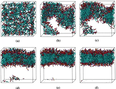
Snapshots of one system at different time points throughout the simulation are
provided in Figure 2.7. Each lipid is represented according to the following color scheme:
purple (choline entity – type I); orange (phosphate group – type II); red (ester groups – type
III and type IV); cyan (alkyl tail groups – type V and type VI). The initial random
configuration of the system is shown in Figure 2.7(a). The lipids begin aggregating at
around 15 million collisions (Figure 2.7(b)) and form a single disordered aggregate (Figure
2.7(c)) at 50 million collisions. The snapshots in Figures 2.7(d – f) show the aggregate as it
rearranges to form a defect free bilayer. The 150 million collisions required to form the
defect free bilayer shown in Figure 2.7f took approximately 3.8 CPU hours. The time scale
for aggregate formation, 50 million collisions, is rather quick compared to the time it takes,
150 million collisions, to organize into a defect free bilayer.
It is of interest to compare the structural properties of the DPPC bilayer formed using
LIME/DMD simulations with the structural properties obtained from a GROMACS
composed of 128 DPPC lipids was performed using the GROMOS96 53a6 forcefield in the
NPT ensemble. The initial coordinates for the bilayer were obtained from the supporting
information provided by Kukol.[(25)] The Berendsen thermostat was employed to maintain
the temperature at 325K; the pressure was held constant at 1.0 bar.[(49)] The simulation was
run for 20 ns with a time step of 0.002 ps. The bilayer remained stable throughout the
simulation.
The bilayer thickness and the area per lipid of the bilayers formed during the LIME
simulations closely match both experimental values and the values calculated from the
GROMACS simulation described above. The LIME values for the bilayer thickness and the
area per lipid were calculated by averaging the data from the five independent DMD
simulations, which were all started from different initial configurations. After a bilayer
formed, the simulation was continued for an additional billion collisions. Data from the
billion collisions following the formation of a defect free bilayer was used to calculate the
physical properties of that bilayer, including the bilayer thickness, area per lipid and bond
order parameters. The bilayer thickness is defined as the distance along the direction
perpendicular to the bilayer normal between the average location of phosphate groups in the
top and bottom leaflets of the bilayer. The LIME/DMD value for the thickness of the DPPC
bilayer is 35.7 Å + 0.3 Å at a reduced temperature of 0.77. The experimental value for
the DPPC bilayer thickness, which is measured as the distance between phosphate
groups in the upper and lower leaflets of the bilayer in the electron density profile is
approximately 38.0 Å at 50oC.[(53),(54)] The bilayer thickness measured between two
+ 0.6 Å at 52oC. Thus, the LIME bilayer thickness is within 5-‐7% the experimental value
and 4-‐8% the GROMACS bilayer thickness. The area per lipid in our LIME/DMD and
GROMACS simulations was calculated by multiplying the length and width of the bilayer
and dividing by half of the number of the lipids in the system (to approximate the
number of lipids in each leaflet). The area per lipid for the bilayer formed by DPPC in our
DMD/LIME simulations is 63.3 Å2
, which is very close to the experimental area of 63.0 + 1.0
Å2 at T = 323K reported by Kucerka et al.[(54)] The area per lipid calculated for the DPPC
bilayer in our GROMACS simulations at a T = 325K is 64.6 + 0.1 Å2.
To further evaluate the structural properties of our coarse-grained model the
orientational order parameter, sbond, for different bonds along the chain was calculated; it is
defined to be:
Sbond =
1 2 3cos
2
θ−1 Equation 2
where θ is the angle between the vector along a coarse-grained bond and the bilayer
normal.[(28),(35)] Sbond values of 1, -1/2 and 0 correlate to bonds with a parallel, an
alignment perpendicular to the bilayer normal and completely random alignment with the
bilayer normal.[(28),(35)] To obtain the orientational bond order parameters from the
GROMACS simulation of the DPPC bilayer, the data was coarse-grained and the bond order
parameters for different site types along the chain were calculated. The GROMACS
simulation was run at a temperature of 325K and the LIME/DMD simulation was run at the
equivalent reduced temperature of 0.77. Figure 2.8 shows the values of the orientational
GROMACS simulation (blue line) versus the bond numbers, which are defined in the figure
insert. The orientational bond order parameters from the DMD model and the GROMACS
simulations are in very close agreement. The bond order parameters range from
approximately 0.26 to 0.52 in Figure 2.8, indicating that the bonds in both the GROMACS
and the DMD simulations are not very well ordered and have between a parallel and a
completely-random alignment with the bilayer normal. These bond order parameters are
very similar to those calculated by Marrink and co-workers for a DPPC bilayer at a
temperature of 323K; their values ranged from approximately 0.3 to 0.5.[(28)] A direct
comparison cannot be made between the LIME forcefield and the model developed by
Marrink and co-workers because in LIME each alkyl tail of DPPC is represented by 5
grained sites and in the Marrink model each alkyl tail of DPPC is represented by 4
coarse-grained sites.
As the temperature is cooled in our DMD system, the bilayer undergoes a phase
transition from a liquid-crystalline phase to a tilted gel phase. Experimentally, as a DPPC
bilayer is cooled it also undergoes a phase transition from a liquid-crystalline phase to a tilted
gel phase in which the lipid tails are tilted with respect to the bilayer normal.[(55),(56)] The
tilted gel phase generally has a smaller area per lipid than in the liquid-crystalline phase and
the lipid tails are straighter.[(56)] The temperature at which a DPPC bilayer transitions from
the liquid-crystalline phase to the gel phase is reported experimentally as 314.4K.[(57)]
Three LIME/DMD simulations, each starting from a different configuration of a preformed
DPPC bilayer composed of 128 lipids at T* = 0.77 and at constant volume in a box with
procedure was implemented: T* was decreased from 0.77 to 0.30 at a rate of 0.01 T*/million
collisions and then maintained at 0.30 for 550 million collisions. All properties were
calculated using the last 100 million collisions that a bilayer was at T*=0.30. Figure 2.9
provides snapshots of a DPPC bilayer in a DMD/LIME simulation at (a) a reduced
temperature of 0.77 where a liquid crystalline phase is observed and (b) a reduced
temperature of 0.30 where a tilted gel phase is observed. Figure 2.9c shows a cross-tilted gel
phase, which we observe in some of our simulations but not in others. We are currently
investigating the conditions that lead to the tilted gel phase and the cross-tilted gel phase.
We believe that the cooling rate may determine whether the tilted or cross-tilted gel phase is
formed. As the temperature of the DPPC bilayer is decreased the tails become more straight
and rigid causing the bilayer thickness to increase from 35.7 Å + 0.3 Å at T* = 0.77 to 40.9
Å + 1.0 Å at T* = 0.30. Consistent with our predictions, experimental observations also
show that the bilayer thickness increases with decreasing temperature. For example, the
experimental bilayer thicknesses at T=323K and T=293K are reported as 38.3 Å and
44.2 Å, respectively.[(58)] We find that as the DPPC bilayer transitions to the gel phase,
the area per lipid decreases; e.g. the area per lipid at T* = 0.77 and T* = 0.30 was 63.3 Å2
and 49.6 Å2 + 1.4 Å2
, respectively. The decrease in area per lipid with decrease in
temperature agrees with experimental observations. The experimental value of the area per
lipid at T=323K and T=293K are 64.0 Å2 and 47.9 Å2
, respectively.[(58)] These simulations
were run at constant volume with box dimensions of 64Å x 64Å x 90Å. At a T*=0.77 the
bilayer spans the entire x-y plane. As the bilayer is cooled it stops spanning the entire x-y
shows that the bilayer is present in only a small portion of the x-y plane. The volume of the
simulation box was constant with dimensions of 64Å x 64Å x 90Å as the bilayer was cooled
to T*=0.30. In our constant volume production run simulations, we adjusted the box
volume so that our bilayer would span the entire x-‐y plane throughout the simulation.
We did not adjust the box volume during the cooling simulations for the bilayer in the
interest of computational efficiency because this would have required us to constantly
change the box volume. In the future we plan to adjust the box volume during cooling
simulations. The tilted gel phase, which we observe in our LIME/DMD simulations is not
usually observed in coarse-grained simulations. For example, coarse-grained simulations
performed by Marrink and co-workers and by Wang and Deserno also show the formation of
untilted gel phase lipid bilayers.[(56),(59)] However, atomistic simulations performed by
Leekumjorn and Sum did show the formation of a tilted gel phase of DPPC.[(60)] The
question naturally arises as to why we had to go to such low reduced temperatures to observe
the gel phase when it is typically reached experimentally at 314.4K [(57)] when the liquid
crystalline phase is simulated at a temperature of 325K. Our explanation is that the LIME
epsilon values were only calculated at a temperature of 325K, which is equivalent to a
LIME/DMD reduced temperature of 0.77 and that they were then assumed to be independent
of temperature. In the future, a multiscale modeling procedure may be used to determine the
temperature dependence of the epsilons. This would require data from atomistic simulations
at various temperatures. We speculate that once the temperature dependence of the epsilon
values is incorporated into our DMD/LIME simulations, it will be unnecessary to reduce T*
The transbilayer movement of phospholipids from one leaflet to another, known as
“translocation” or “flip-flop,” was measured during the DMD/LIME simulations.
Translocation is thought to play an important role in numerous cellular processes including
cell apoptosis and drug function.[(61)] Unless protein-mediated, this lipid migration is
thought to occur very slowly, with half-lives on the order of hours.[(61),(62),(63)] We did
not observe any flip flops in any of our simulations of lipid systems at T*=0.77. However,
when we performed a simulation of DPPC bilayer composed of 128 lipids at a slightly higher
temperature, T*=0.85, we did see one flip flop. Figure 2.10 shows snapshots of a lipid (in
yellow) that flips from the bottom leaflet of the membrane to the top leaflet of the membrane
during one of these simulations. Over the course of 1 billion collisions only one lipid
successfully flipped from one leaflet to another. It required approximately 19 million
collisions for the lipid to complete the flip-flop.
A comparison of the mass density profile of different coarse-‐grained types along
the bilayer normal between the LIME and GROMACS simulations is presented in Figure
2.11. The mass density profile is the mass per unit volume at a distance (z) from the
bilayer normal. Since the GROMACS simulation from which the mass density profile
was obtained used a bilayer consisting of 128 DPPC molecules, we ran a DMD/LIME
simulation on a bilayer consisting of 128 lipids. The LIME/DMD simulation was started
from a random initial configuration of 128 lipids and formed a defect free bilayer in 1.1
CPU hours. The mass density distributions from the DMD/LIME simulation were taken
from conformations once the lipids had formed a defect free bilayer (after 41 million