Molecular Characterization of
Pax6
2NeuThrough
Pax6
10Neu: An Extension of the
Pax6
Allelic Series and the Identification of Two Possible Hypomorph
Alleles in the Mouse
Mus musculus
Jack Favor,* Heiko Peters,*
,1Thomas Hermann,
†Wolfgang Schmahl,
‡Bimal Chatterjee,*
Angelika Neuha¨user-Klaus* and Rodica Sandulache*
*Institute of Mammalian Genetics, GSF-Research Center for Environment and Health, Neuherberg D-85764, Germany,
†Memorial Sloan-Kettering Cancer Center, New York, New York 10021 and‡Lehrstuhl fu
¨r Allgemeine Pathologie und Neuropathologie, Tiera¨rztliche Fakulta¨t, Ludwig-Maximilians-Universita¨t, Mu¨nchen D-80539, Germany
Manuscript received April 27, 2001 Accepted for publication September 28, 2001
ABSTRACT
Phenotype-based mutagenesis experiments will increase the mouse mutant resource, generating muta-tions at previously unmarked loci as well as extending the allelic series at known loci. Mapping, molecular characterization, and phenotypic analysis of nine independentPax6 mutations of the mouse recovered in mutagenesis experiments is presented. Seven mutations result in premature termination of translation and all express phenotypes characteristic of null alleles, suggesting that Pax6 function requires all domains to be intact. Of major interest is the identification of two possible hypomorph mutations: Heterozygotes express less severe phenotypes and homozygotes develop rudimentary eyes and nasal processes and survive up to 36 hr after birth. Pax64Neu results in an amino acid substitution within the third helix of the homeodomain. Three-dimensional modeling indicates that the amino acid substitution interrupts the homeodomain recognition␣-helix, which is critical for DNA binding. Whereas cooperative dimer binding of the mutant homeodomain to a paired-class DNA target sequence was eliminated, weak monomer binding was observed. Thus, a residual function of the mutated homeodomain may explain the hypomorphic nature of thePax64Neuallele.Pax67Neuis a base pair substitution in the Kozak sequence and results in a reduced level of Pax6 translation product. The Pax64Neu andPax67Neualleles may be very useful for gene-dosage studies.
I
N the mouse a number of large-scale mutagenesis ment ofPax6 in eye development suggested thatPax6is the candidate gene affected in this large group of screens have been undertaken to systematically
re-cover mutations with relevant phenotypes to comple- mutations.
The mousePax6gene belongs to the family of paired-ment the forthcoming genome sequence data and to
provide the genetic variants to initiate functional ge- box-containing genes, which function as transcription factors regulating developmental processes. Pax6 en-netic studies (Hrabe´ de Angeliset al.2000;Nolanet
al. 2000). We have conducted an extensive series of codes a protein with both a paired domain and homeo-domain, separated by a linker segment, and followed by mutagenesis experiments to recover dominant
muta-tions that affect eye morphology (Favor and Neu- a C-terminal proline-, serine-, and threonine-rich region (Walther and Gruss 1991). The highly conserved
ha¨user-Klaus2000). A total of 192 independent
muta-tions are available for genetic and molecular analyses, paired domain and homeodomain are DNA-binding do-mains while the proline-, serine-, and threonine-rich which represents one of the largest collections of
muta-tions affecting the development and function of a de- domain functions in transcriptional activation (Glaser
et al.1994). Mutant analyses have shown thatPax6plays fined organ. We are systematically mapping all
recov-ered mutations to identify the genes/loci responsible a role in the development of the eye (Theiler et al.
1978;Hoganet al. 1988), nasal derivatives (Hogan et
for eye development. Our results to date indicate that
the most mutable locus is at chromosome 2, centi- al.1988;Heinzmannet al.1991;Grindleyet al.1995;
Quinnet al.1996), additional craniofacial traits (
Kauf-morgan 58 in the vicinity of the Pax6 gene, which is
known to function in eye development. This combina- man et al. 1995), the central nervous system (CNS;
Schmahlet al. 1993;Grindleyet al. 1997;Stoykova
tion of chromosomal location and the known
involve-et al.1996, 1997, 2000;Go¨ tzet al.1998), the pancreas (St-Ongeet al.1997), and the pituitary gland (Bentley
Corresponding author:Jack Favor, Institute of Mammalian Genetics, et al.1999;Kioussi et al. 1999). Heterozygous carriers GSF-Research Center for Environment and Health, Ingolsta¨dter
ofPax6null mutations express phenotypic effects, which
Landstr. 1, D-85764, Neuherberg, Germany. E-mail: [email protected]
suggests that a defined concentration of Pax6 protein
1Present address: University of Newcastle upon Tyne, Institute of
Human Genetics, Newcastle upon Tyne, NE1 7RU United Kingdom. activity is necessary for normal development (Hill et
mapped mutation (ENU-65 or ENU-5011) to heterozygous
al. 1991). This suggestion is supported by studies in
Pax6Sey-Neu/⫹mice. Pregnant females were dissected at stages
which the wild-typePax6 allele was overexpressed and
E15 to E17 and embryos were classified for the typical
homozy-resulted in abnormal eye development (Schedl et al. gote mutantPax6phenotype indicative of
noncomplementa-1996). tion (anophthalmia and craniofacial abnormalities).
RT-PCR and sequence analysis:Total RNA was isolated from
Human patients heterozygous forPAX6mutant alleles
heads of E15 embryos expressing the typical wild-type or
ho-express eye abnormalities similar to those observed in
mozygous mutantPax6phenotype recovered ininter secrosses
the mouse (Brownet al.1998;Prosserandvan
Hey-of heterozygotes using the RNeasy kit (QIAGEN, Chatsworth, ningen1998). Most describedPAX6mutant alleles re- CA). RNA was reverse transcribed and the entire coding region sult in premature termination of translation. The under- was PCR amplified with a Titan one Tube RT-PCR system
(Roche Diagnostics, Mannheim, Germany) as recommended
representation of missense mutations may be due to the
by the supplier. Two overlapping primer sets, A, 5⬘-CAGAA
fact that hypomorph alleles likely result in a less severe
GACTTTAACCAAGGGC [nucleotide (nt) 33–53] and 5⬘-AT
phenotype and patients who carry hypomorph alleles
CCTTAGTTTATCATACATGCCG (nt 647–623), and B,
5⬘-might not have been included in a mutation screen. AACAGAGTTCTTCGCAACCTGG (nt 573–595) and 5⬘-GCT The identification of Pax6 hypomorph alleles in a GTGTCCACATAGTCATTGGC (nt 1545–1522), were used for
the amplification of thePax6cDNA. The PCR products were
laboratory animal would be useful to experimentally
HPLC purified and sequenced with a Taq Dye-Deoxy
termina-address the question of Pax6 protein activity dosage
tor cycle sequencing kit by SequiServe (Vaterstetten,
Ger-and phenotypic defects. Here we report nine newPax6
many). Additional primers were used for the complete
se-mutant alleles in the mouse. Seven mutations result quencing of the coding region as well as for the confirmation in a truncated gene product. Of major interest is the of the mutation employing genomic DNA as a template.
Numbering of the Pax6 nucleotides and codons/amino
identification of two possible hypomorph alleles. One
acids follows that ofWaltherandGruss(1991).
is an amino acid substitution in the homeodomain. The
Assay for nasal development:To assay the extent of nasal
second is a base pair substitution in the Kozak sequence.
development inPax6mutant mice we utilized thePax9lacZ re-porter gene, which is expressed in nasal mesenchyme (Peters et al. 1998). Double heterozygotes, consisting of Pax9lacZ/⫹ MATERIALS AND METHODS withPax6Sey-Neu/⫹ orPax64Neu/⫹, were constructed and bred to heterozygous carriers of the respectivePax6mutant allele.
Animals:All inbred strain (C3H, C57BL/6, 102), F1hybrid
Embryos (E12) were identified as homozygous wild type and (102⫻C3H) and Tester-stock animals were obtained from
heterozygous or homozygous mutant at the Pax6 locus by breeding colonies maintained by the GSF-Department of
Ani-phenotype. HeterozygousPax9lacZcarriers were identified by mal Resources at Neuherberg.
a PCR assay as previously described (Peterset al.1998). Whole
Mutation recovery:Mutants were recovered in the offspring
mountPax9lacZ/⫹embryos were stained with X-Gal for 8–12 of (102⫻C3H)F1hybrid or C3H males that were mutagenically
hr according to established protocols (GosslerandZachgo treated and mated to untreated Tester-stock or C3H females,
1993). To completely visualize Pax9lacZexpression, embryos respectively. Prior to the initiation of the mutagenesis
experi-were dehydrated in methanol and cleared with benzyl benzo-ments, all parental animals were ophthalmologically examined
ate/benzyl alcohol. and only those that did not express an eye morphological
Assay for eye phenotype:Offspring from the cross-mutant abnormality were employed in order to eliminate any
preex-heterozygote with inbred strain C3H/J were ophthalmologi-isting eye mutations.
cally examined at 21 days of age and categorized for the degree
Ophthalmological examination:Mice were examined
biomi-of lens/corneal opacity. At 30 days biomi-of age the biomi-offspring were croscopically for eye abnormalities at weaning. Pupils were
sacrificed by cervical dislocation, and eyes were enucleated, dilated with a 1% atropin solution applied to the eyes at least
washed once in PBS, rinsed in H2O, blotted dry on filter paper, 10 min prior to examination. Both eyes of the mice were
and weighed. Eye weight data were statistically analyzed by examined by slit lamp biomicroscopy (Zeiss SLM30) at⫻48
Factorial ANOVA, General Linear Models Procedure, em-magnification with a narrow beam slit lamp illumination at a
ploying SAS Software release 6.12 (Cary, NC). 25⬚–30⬚angle from the direction of observation.
Histology:Pregnant females fromPax64Neu/⫹orPax67Neu/⫹
Genetic confirmation crosses: The recovered presumed
inter secrosses were sacrificed on day 16 postconception and dominant eye mutants were outcrossed to wild-type strain 102,
embryos assigned a genotype on the basis of phenotype (het-C3H, or (102⫻C3H)F1partners and at least 20 offspring were
erozygotes have a slightly smaller eye with distorted shape of ophthalmologically examined for transmission of the specific
the pupil; homozygous mutants are anophthalmic). Embryos eye abnormality. The offspring were carefully examined to
were fixed in 10% buffered formalin, and heads were embed-arrive at a more complete description of the phenotype
ex-ded in paraffin and serially sectioned (coronal) at 5m. Sec-pressed by heterozygous carriers. A detailed consideration of
tions were stained with hematoxylin and eosin. the ophthalmological examination procedures and genetic
Three-dimensional modeling:A three-dimensional model
confirmation test has been published (Favor1983). All
con-of the Pax6 homeodomain (Leu1 through Glu60) was con-firmed mutations were maintained by outcrossing
heterozy-structed by homology modeling using the atomic coordinates gous carriers to homozygous wild-type partners. Before
initiat-of the homeodomains initiat-of the genespaired(Wilsonet al.1995), ing the mapping experiments each mutation was backcrossed
Antennapedia(Qianet al.1994b),VND/NK-2(Tsaoet al.1995), at least 10 generations to strain C3H.
engrailed(Kissingeret al.1990), andFushi Tarazu(Qianet al.
Mapping and allelism tests:Complete details of our
map-1994a). Protein sequences were aligned with FASTA (Pearson ping procedures have been published recently (Favoret al.
1990). Three-dimensional modeling was performed with the 1997). Segregation data were analyzed with Map Manager,
InsightII software (Molecular Simulations, San Diego) follow-Version 2.6.5 (Manly1993), and the gene order was
deter-ing a previously described procedure (Kru¨ ger et al.1998). mined by minimizing the number of multiple crossovers.
recognition site, guided by the atomic coordinates of the ment). The original mutant recovered for each of these paired/DNA complex (Wilsonet al.1995). independent mutations expressed bilateral anterior
po-Expression of Pax6 homeodomains of the wild-type and
lar cataract, corneal adhesions, and, depending on the
Pax64Neualleles in bacteria:The appropriate fragments were
mutant allele, slight to more severe microphthalmia.
amplified from cDNA templates and inserted between the
PstI andHindIII sites of the pQE-41 vector (QIAGEN). The Genetic confirmation tests indicated that the original
expression constructs result in the in-frame fusion of the mutants were heterozygous for an autosomal dominant desired sequence (the Pax6 amino acids 218 to 288, which
allele. Two mutations (ENU-65 and nPMS-9) were
recov-includes the homeodomain and six amino acid residues
up-ered in offspring derived from fertilizations in which
stream and four amino acid residues downstream) to the
the participating parental male germ cells were
muta-C-terminal end of the mouse dihydrofolate reductase protein.
The pQE expression constructs were transformed intoEsche- genically exposed at the spermatocyte stage. The
fertil-richia colistrain M15 (pREP4), which carries thelacIqrepressor
ization producing the mutant ENU-65 occurred 24 days
gene. Cultures of exponentially growing bacteria were induced
after treatment. The fertilization producing the mutant
with 0.1 mmisopropyl thiogalactoside for 2 hr at 30⬚. Bacterial
nPMS-9 occurred 20 days after treatment. The original
pellets were lysed in 50 mmNaH2PO4, 300 mmNaCl, pH 8.0,
0.5 mg/ml lysozyme, 5g/ml DNaseI and 1 mmphenylmethyl- mutant ENU-65 expressed unilaterally anterior polar
sulfonyl fluoride. Crude extracts were electrophoresed in 10% opacity, corneal adhesions, and microphthalmia. Ge-SDS-PAGE and proteins were visualized by staining with
Coo-netic confirmation and subsequent breeding results
sug-massie brilliant blue.
gested that the original mutant was a gonosomic mosaic
Western blotting: Crude extracts were fractionated on a
for an autosomal dominant mutation (Favor 1983;
10% SDS-PAGE and then electrophoretically transferred to
fluorotrans membranes (Pall Europe Ltd, Portsmouth, UK) Favoret al.1990;FavorandNeuha¨user-Klaus1994).
and probed with rabbit anti-Pax6-homeodomain antiserum The original mutant nPMS-9 expressed bilateral eye de-(1:200; serum 13, gift from Dr. S. Saule, Institut Pasteur de
fects and genetic confirmation crosses were consistent
Lille), which was detected with anti-rabbit IgG alkaline
phos-with the assumption that the mutant was heterozygous
phatase conjugate (1:1000; Promega, Madison, WI).
Electrophoretic mobility shift analysis:Crude extracts from for an autosomal dominant mutation.
the transformed bacteria were incubated with 15 fmol of the The expressivity of all mutations was variable with target oligonucleotide, which was 3⬘end-labeled with digoxy- mutant carriers expressing a range of phenotypes from genin-11-ddUTP as recommended by the supplier (Roche
Di-small anterior polar cataracts to the more extreme
phe-agnostics, Mannheim, Germany). The target oligonucleotide
notype of anterior polar opacity, corneal adhesions, iris
contained the Pax6 homeodomain binding sequence P3
(CzernyandBusslinger1995), in which the two halves of abnormalities, and microphthalmia. Furthermore, the the palindromic sequence were separated by TGA as in the degree of phenotype expressed between the eyes of an rhodopsin promoter (Sheng et al. 1997). The sequence of
individual mutation carrier was variable.
the double-strand oligonucleotide (with P3 underlined)is as
Examination of the eyes of animals not treated with
follows: 5⬘-TCGAGGGCATCAGGATGCTAATTGAATTAGCA
TCCGATCGGG-3⬘ and 3⬘-CCCGTAGTCCTACGATTAACTT atropin revealed that heterozygous carriers for all
mu-AATCGTAGGCTAGCCCAGCT-5⬘. tant alleles expressed greatly reduced or absent iris.
Protein-DNA complexes were separated on a native 10% Breeding results are given in Table 2. Segregation polyacrylamide gel (in 0.25⫻Tris-borate-EDTA), blotted onto
analyses for the outcross of heterozygous carriers to wild
BM nylon membranes, and detected by autoradiography
fol-type indicated a 1:1 ratio of wild fol-type to mutant carrier
lowing an enzyme immunoassay with antidigoxygenin
(Fab)-alkaline phosphatase and the chemiluminiscent substrate offspring consistent with an autosomal dominant
muta-CSPD (Roche). tion without viability or penetrance effects for six
muta-The sequence data presented in this article have been
sub-tions (Pax62Neu,Pax63Neu,Pax64Neu,Pax65Neu,Pax66Neu, and mitted to the EMBL/GenBank Data Libraries under the
acces-Pax610Neu). The segregation analyses of three mutations sion numbers: Pax62Neu, Y19193; Pax63Neu, Y19195; Pax64Neu,
Y19196;Pax65Neu, Y19197; Pax66Neu, Y19198; Pax67Neu, Y19199; (Pax67Neu, Pax68Neu, and Pax69Neu) deviated significantly Pax69Neu, AJ292077; andPax610Neu, AJ307468.
from the expected 1:1 ratio. For Pax68Neu andPax69Neu
there was an excess of wild-type offspring relative to mutants. The deviations in segregation observed for
RESULTS these two mutations may be due to a conservative
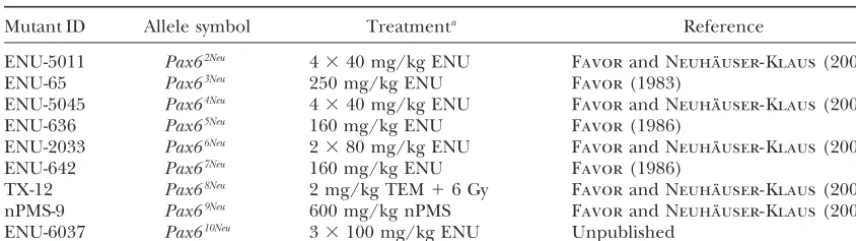
classi-fication of offspring in the initial generations of breed-Mutations:Table 1 gives the original mutant
identifi-ing or to genetic background effects since, as will be cation, the proposed allele symbol (based on the
molec-seen below, (a)Pax68Neu(with an observed deviation in
ular characterization results below), the mutagenic
treat-segregation) is an exact repeat mutation of that carried ment of the parental male mice, and reference to the
byPax66Neu (with no deviation in segregation) and (b)
mutagenesis experiments in which the original mutants
once the spectrum of phenotypes associated with the were recovered. Seven mutations (5011,
ENU-mutations became known we were able to classify the 5045, 636, 2033, 642, TX-12, and
ENU-genotypes on the basis of phenotype with no deviation 6037) were recovered in offspring derived from
fertiliza-in the segregation ratio fertiliza-in the mappfertiliza-ing studies. The tions in which the participating parental male germ cells
segregation results forPax67Neushowed an excess of
mu-were mutagenically treated at the stem cell
TABLE 1
Origin and proposed allele symbols of the mousePax6mutations characterized in this study
Mutant ID Allele symbol Treatmenta Reference
ENU-5011 Pax62Neu 4⫻40 mg/kg ENU FavorandNeuha¨user-Klaus(2000)
ENU-65 Pax63Neu 250 mg/kg ENU Favor(1983)
ENU-5045 Pax64Neu 4⫻40 mg/kg ENU FavorandNeuha¨user-Klaus(2000)
ENU-636 Pax65Neu 160 mg/kg ENU Favor(1986)
ENU-2033 Pax66Neu 2⫻80 mg/kg ENU FavorandNeuha¨user-Klaus(2000)
ENU-642 Pax67Neu 160 mg/kg ENU Favor(1986)
TX-12 Pax68Neu 2 mg/kg TEM⫹6 Gy FavorandNeuha¨user-Klaus(2000)
nPMS-9 Pax69Neu 600 mg/kg nPMS FavorandNeuha¨user-Klaus(2000)
ENU-6037 Pax610Neu 3⫻100 mg/kg ENU Unpublished
aENU, ethylnitrosourea; TEM, triethylenemelamine; nPMS,n-propyl methanesulfonate.
segregation ratio in the mapping analyses and we con- shown to be alleles ofPax6on the basis of noncomple-clude the deviation to be spurious. mentation with Pax6Sey-Neu. Phenotypic analyses of the
Examination of embryos from inter se crosses of mutationPax610Neusuggested thatPax6was mutated. The
heterozygous carriers of seven mutations (Pax62Neu,Pax- remaining six mutations were localized relative to the
63Neu,Pax65Neu,Pax66Neu,Pax68Neu,Pax69Neu, andPax610Neu)
chromosome 2 markersD2Mit249andAgouti(Table 3). revealed that the resultant phenotypes were identical All mutations mapped to the same region, expressed a similar phenotype, and, therefore, were assumed to be to those previously described (Hoganet al.1988):
Het-mutant alleles of the same gene. Combined mapping erozygotes express obvious microphthalmia with a
trian-results yielded the following gene order:D2Mit249-(1.4 gular-shaped pupil and homozygotes express
anoph-cM)-Pax6-(25.3 cM)-Agouti. thalmia with severe craniofacial defects (Figure 2A).
Molecular characterization: Sequence analyses indi-Embryos from two mutations deviated from this general
cated that the Pax6 gene was affected in all nine mu-phenotypic classification.Pax64NeuandPax67Neu
heterozy-tations (Table 4). Seven muheterozy-tations (Pax62Neu,
Pax64Neu,
gotes expressed abnormal pupil shape but eye size
reduc-Pax65Neu,Pax66Neu,Pax67Neu,Pax68Neu, andPax610Neu) were
tion was only slight; homozygous mutants were
anoph-base pair substitutions, one mutation (Pax63Neu) was a
thalmic but some were observed to have a distinct optic
base pair insertion, and one mutation (Pax69Neu) was a
pit with underlying pigmentation (Figure 2, B and C).
7-bp deletion. Mapping:Two mutations (Pax62NeuandPax63Neu) were
TABLE 2 TABLE 3
Breeding results for heterozygotePax6mutant Mapping results of six independent dominant
outcrosses orinter secrosses eye mutations of the mouse in the cross
(C3H-Pax6#Neu⫻C57BL/6)⫻C57BL/6 Offspring resulting from the cross
Markers Haplotypesa
M/⫹ ⫻ ⫹/⫹ M/⫹ ⫻M/⫹
D2Mit249 䊏 䊐 䊏 䊐 䊏 䊐 䊏 䊐
Mutation ⫹/⫹ M/⫹ ⫹/⫹ M/⫹ M/M
Pax6 䊏 䊐 䊐 䊏 䊏 䊐 䊐 䊏
Agouti 䊏 䊐 䊐 䊏 䊐 䊏 䊏 䊐
Pax62Neu 83 67 13 32 12
Pax63Neu 93 93 24 30 18 Pax64Neu 37 40 0 1 22 14 0 0
Pax65Neu 39 42 0 0 9 4 0 0
Pax64Neu 64 61 5 6 4a
Pax65Neu 67 63 5 13 7 Pax66Neu 39 40 0 0 11 16 0 0
Pax67Neu 41 30 1 1 14 15 1 2
Pax66Neu 132 127 9 13 11
Pax67Neu 54 91 8 9 6a Pax68Neu 42 44 0 0 16 9 0 0
Pax69Neu 43 30 0 0 14 9 1 2
Pax68Neu 221 39 4 10 4
Pax69Neu 77 57 3 14 4 Total 241 226 1 2 86 67 2 4
Pax610Neu 91 90 3 5 1
For each independent mutation, the number of offspring carrying the haplotypes inherited from the C3H-Pax6#Neu⫻ Offspring from outcrosses were classified at weaning by slit
C57BL/6 outcross parent are given. lamp biomicroscopy. Embryos frominter secrosses were
classi-aThe solid boxes designate the alleles carried by the con-fied at E15–E17.
genic C3H-Pax6#Neustrains; the open boxes designate the al-aTwo embryos observed with pronounced optic pits and
TABLE 4
Molecular characterization ofPax6mutations of the mouse
Allele Mutation Position Codon Domain
Pax62Neu T to C Intron 9, 2 5⬘splice site After 269 Homeobox
Pax63Neu A insert Exon 7, after 598 After 145 Linker
Pax64Neu T to C Exon 10, 979 Ser273 to Pro Homeobox
Pax65Neu A to T Exon 6, 517 Arg119 to Stop Paired box
Pax66Neu C to A Exon 10, 1092 Tyr310 to Stop P/S/T
Pax67Neu A to T Exon 4,⫺3 Kozak sequence
Pax68Neu C to A Exon 10, 1092 Tyr310 to Stop P/S/T
Pax69Neu TCACAGC deletion Exon 5, 261–267 After 33 Paired box
Pax610Neu C to T Exon 6, 469 Gln103 to Stop Paired box
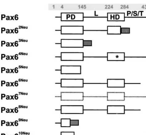
The predicted Pax6 protein products are given in normal amino acid sequence through codon 145 of exon 7 followed by 12 aberrant amino acids and a pre-Figure 1. Seven mutations (Pax62Neu,Pax63Neu,Pax65Neu,
Pax66Neu, Pax68Neu, Pax69Neu, and Pax610Neu) result in a mature stop. The gene product contains a normal
paired domain but the linker region, the homeodo-truncated Pax6 protein product.Pax62Neuis a base pair
substitution at nucleotide position 2 of the intron 9 main, and the P/S/T domain are all deleted. The base pair substitution in thePax65Neumutation creates a
pre-donor splice site. The isolated cDNA was 87 bp longer
than wild type due to the destruction of the splice site mature stop site in the C terminus of the paired box, resulting in a predicted gene product that is truncated and the use of a cryptic splice site. Translation of the
aberrant mRNA is predicted to result in a normal amino for the last 27 amino acids of the paired domain as well as the linker region, the homeodomain, and the P/S/T acid sequence up to codon 269 of exon 9 followed
by 23 amino acids encoded by the included intron 9 domain.Pax66Neu is also a base pair substitution, which
creates a premature stop codon and results in a trun-sequence and a premature stop. The mutation results
in the truncation of the last 15 amino acids of the C cated gene product from the N-terminal end of the P/S/T domain. Pax68Neu is a repeat mutation such as
terminus of the homeodomain and the entire P/S/T
domain. ThePax63Neu frameshift mutation results in a that observed inPax66Neu. The 7-bp deletion associated
withPax69Neuis predicted to result in an extremely
trun-cated gene product consisting of the first 32 amino acids followed by 17 aberrant amino acids and a premature stop. Finally, thePax610Neubase pair substitution creates
a stop codon in the C terminus of the paired box. The predicted mutant gene product consists of the first 102 amino acids and a truncation of the last 43 amino acids of the paired domain, as well as the linker region, the homeodomain, and the P/S/T region.
One missense mutation has been identified. The
Pax64Neumutation results in an amino acid substitution,
Ser273 to Pro, in the third helix of the homeodomain. The predicted gene product contains a single amino acid substitution at a site known to be critical for DNA binding activity. Finally, thePax67Neumutation is a base
pair substitution in the Kozak sequence in the 5⬘ -untranslated region of exon 4 and is predicted to affect translation.
We detected two differences between our wild-type
Pax6sequence and the published mousePax6sequence (WaltherandGruss1991) at position 354 (T instead
Figure1.—Predicted protein product of wild-type and mu- of C) and at position 1263 (G instead of A). The
differ-tant Pax6 alleles. PD, paired domain; L, linker region; HD, ences do not affect the amino acids specified. Our se-homeodomain; P/S/T, proline-/serine-/threonine-rich re- quence data are for strain C3H, were based upon analy-gion (the codon positions at the domain borders of thePax6
ses of cDNA, and were confirmed by analysis of genomic
wild type are shaded numbers); shaded boxes, aberrant amino
DNA. All mutant sequences have been submitted to
acid sequences; *, missense mutation; dashed lines, reduced
TABLE 6 TABLE 5
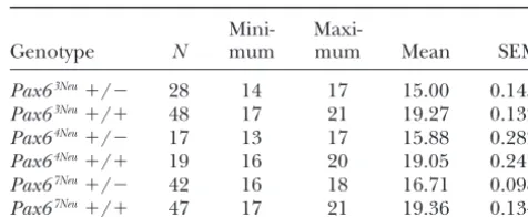
Degree of lens/corneal opacity inPax6mutation Eye weight (mg) inPax6mutation heterozygotes
and wild-type litter mates heterozygotes and wild-type litter mates
Phenotypic class Mini-
Maxi-Genotype N mum mum Mean SEM
Genotype 0% 25% 50% 75% 100%
Pax63Neu⫹/⫺ 28 14 17 15.00 0.1455
Pax63Neu⫹/⫺ 2 4 9 17
Pax63Neu⫹/⫹ 48 17 21 19.27 0.1322
Pax63Neu⫹/⫹ 48
Pax64Neu⫹/⫺ 17 13 17 15.88 0.2828
Pax64Neu⫹/⫺ 6 11 5 2
Pax64Neu⫹/⫹ 19 16 20 19.05 0.2474
Pax64Neu⫹/⫹ 32
Pax67Neu⫹/⫺ 42 16 18 16.71 0.0980
Pax67Neu⫹/⫺ 9 25 6 2
Pax67Neu⫹/⫹ 47 17 21 19.36 0.1340
Pax67Neu⫹/⫹ 48
processes were present but of reduced size in homozy-accession numbers: Pax62Neu, Y19193; Pax63Neu, Y19195; gotes for the Pax64Neu allele (Figure 2C). Histological
Pax64Neu, Y19196; Pax65Neu, Y19197; Pax66Neu, Y19198; examination indicated that, in comparison to
homozy-Pax67Neu, Y19199; Pax69Neu, AJ292077; and Pax610Neu, gous wild type (Figure 3A), there was hyperplasia of the
AJ307468. cartilaginous parts of the nasal anlage and a concomi-Heterozygote and homozygote phenotypes of the hy- tant reduction in size of the nasal cavities and their pomorph Pax64Neu and Pax67Neu mutant alleles:The de- associated nasal epithelia in homozygous Pax64Neu
mu-gree of lens/corneal opacity in mutant heterozygotes tant embryos (Figure 3B).
and wild-type littermates is given in Table 5. Heterozy- The extent of eye development in homozygous mu-gous carriers of thePax63Neutruncation mutation express
tantPax64Neu and Pax67Neu embryos is most interesting.
a severe lens/corneal opacity, the median and modal Whereas in homozygous wild type (Figure 3A) eye devel-values being 100% opaque. In contrast, carriers of the opment is complete, with a well-developed retina and two presumed hypomorph allelesPax64NeuandPax67Neu
lens, eye development in homozygous mutants termi-express a less severe phenotype. For both mutations nated prematurely (Figure 3, B–E). The optic stalk is the median and modal values were 25% opaque. As not in contact with the surface ectoderm. However, the a measure of eye size, individual eyes were weighed. tip of the optic stalk is enlarged and invaginated to form Data were first analyzed for the effects of mutant allele a “pseudo-optic cup.” In homozygous mutant Pax64Neu
(Pax63Neu,Pax64Neu, orPax67Neu), genotype (heterozygous embryos, the enlarged and invaginated tip of the optic
or wild type), and sex of animal. The effects of both stalk is surrounded by cartilage. Invaginations from the mutant allele (F2, 196⫽ 6.00, P ⬍0.002) and genotype surface ectoderm are evident but remain a considerable
(F1, 196 ⫽ 591.07, P ⬍ 0.0001) were highly significant. distance from the tip of the optic stalk. Lens did not
The sex of the animal had no effect on eye weight develop. Homozygotes for Pax6 null mutations die (F1, 196⫽ 0.12,P ⵑ0.729). Data were pooled for sex of shortly after birth. In contrast, homozygousPax64Neuand
animal and the range, mean, and standard error of the Pax67Neu offspring survive up to 24–36 hr after birth.
mean eye weights for heterozygous carriers and wild- Together, the above observations suggest Pax64Neu and
type littermates are given in Table 6. Eye weights of Pax67Neuare hypomorph alleles.
Pax63Neucarriers were 78% that of wild-type littermates.
Three-dimensional model and DNA-binding activity The corresponding values for thePax64NeuandPax67Neu
of the Pax64Neu mutant gene product: The site of the
alleles were less severe (83 and 86%, respectively). Ex- Pax64Neu
missense mutation, Ser273, is located in the third amination of embryos from inter se crosses of Pax64Neu helix of the homeodomain (position 50 in the
homeo-heterozygotes (Figure 2B) revealed that the resultant domain, position 9 in the third helix). The substitution phenotypes were less severe than that observed for carri- of Pro for Ser is predicted to interrupt the structure of ers of aPax6null allele (Hoganet al.1988). We further the third helix (Figure 4), which, in three-dimensional characterized the extent of nasal development, since structures of homologous homeodomain proteins
com-Pax6is essential for normal development of this organ plexed with DNA, inserts into the major groove of the (Hoganet al.1988;Grindleyet al.1995;Quinnet al. DNA target. We compared the binding activity of the wild 1996). Previous studies have shown thatPax9expression type and Pax64Neu mutant homeodomains to the
palin-marks the mesenchymal domains of the medial and dromic P3 target oligonucleotide, since the Pax6 homeo-lateral nasal processes after E10.5 of mouse develop- domain binds specifically to the P3 sequence (Czerny
ment (Peterset al. 1998). Using thePax9lacZ
allele as a andBusslinger1995). Comparable concentrations of reporter, we found that the lateral and medial nasal wild-type and mutant homeodomain proteins were as-processes were completely absent in homozygotes for sayed (Figure 5A). The binding activity of thePax64Neu
be greatly reduced in comparison to the binding activity results in loss of the homeodomain against which the anti-Pax6 antiserum reacts.
of the wild-type homeodomain (Figure 5B). Only very faint traces of monomer protein-DNA complex were observed. The mutant protein competes weakly with the
DISCUSSION
wild-type protein, reducing the level of dimer
protein-DNA complex formation. Here we have extended thePax6allelic series in the mouse. We have shown by mapping, molecular charac-Levels of Pax6 expression in homozygousPax64Neuand
Pax67Neumutant mice: The levels of Pax6 expression in terization, and phenotype analyses that seven alleles
result in truncated gene products and express pheno-homozygous mutant embryos are given in Figure 6. In
comparison to homozygous wild-type embryos, normal typic defects characteristic for null mutations. Most im-portantly, we have identified two potential hypomorph levels of Pax6 expression were observed in homozygous
Pax64Neuembryos. For homozygous
Pax67Neumutant em- alleles.
Truncation mutations:SixPax6mutations have been bryos, the level of Pax6 expression was greatly reduced.
The lack of signal associated with homozygousPax63Neu previously described. The
Pax6LacZ(
St-Ongeet al.1997) was constructed and the initiation start site as well as the embryos can be taken only as a negative control for
the Western blot since thePax63Neutruncation mutation entire paired box were replaced by the-galactosidase
reporter gene and the neo-selector gene. Pax6Sey-Dey is
due to an intergenic deletion including thePax6 and
Wt1 genes (Glaser et al. 1990). Pax6Sey is a base pair
substitution creating a premature stop codon in the linker region (Hillet al.1991).Pax6Sey-Neuis a splice site
mutation resulting in a predicted gene product trun-cated from the N-terminal end of the P/S/T region (Hill et al. 1991). Pax6Coop is a base pair substitution
that creates a premature stop codon between the ho-meodomain and the P/S/T region (Lyonet al.2000).
Pax6Sey-H
is a large deletion and most likely involves addi-tional linked loci (Hillet al.1991). Together with the presently described mutations, an allelic series exists for truncation mutations with the order from the longest gene product to total gene ablation as follows: Pax6Sey-Neu,
Pax66Neu, Pax6Coop, Pax62Neu, Pax6Sey, Pax63Neu, Pax65Neu,
Pax610Neu,Pax69Neu, andPax6LacZ.Lyonet al.(2000) have
observed very small vestiges of unpigmented eyes in homozygousPax6Coopembryos, raising the possibility that
this mutation may be a hypomorph allele. Although a direct comparison of the phenotypic effects of the different Pax6 alleles in one laboratory has not been
carried out, some appropriate data are available. In ho-mozygousPax6SeyandPax6Sey-Neumutants, the optic stalk
develops distally, there is a condensation of mesenchy-mal-like cells underneath the surface ectoderm, and the overlying surface ectoderm appears to form a pit, but neither a lens nor an eye develops (Grindley et al.
1995;Schedlet al.1996). We observed pits in the pre-sumptive eye region of homozygous embryos for all mu-tations reported here, including the shortest truncation allele in which none of the Pax6 domains remain intact. Thus, the development of such pits may be a component of the phenotype associated with null alleles. The gross morphology of heterozygous and homozygous carriers of all truncation alleles reported here is similar, which suggests thatPax6 gene function requires all domains to be intact. Destruction of the paired domain or the homeodomain should lead to loss of gene function since each participates directly in DNA binding. The amino acid composition of the P/S/T region of the Pax6 pro-tein resembles the activation domains of the transcrip-tion factors Oct-1 and Oct-2 (TanakaandHerr1990). Such activation domains are important for the interac-tion of DNA-bound transcripinterac-tion factors with other pro-teins required for transcription.
Potential hypomorph alleles: In homozygous Pax6
null mutants the optic vesicle makes initial contact with the surface ectoderm but their contact is subsequently
Figure4.—Three-dimensional model of thePax6 homeo-domain (green) docked to the DNA recognition sequence (cyan). The protein is represented as a backbone ribbon. The position of the mutated residue 9 is marked in red. (A) Wild-type homeodomain with the recognition␣-helix inserted into the DNA major groove. (B) In the mutantPax64Neu homeo-domain the substitution of serine to proline leads to disruption of the recognition␣-helix such that the DNA major groove cannot be recognized.
lost. The tip of the optic vesicle is broader but does not Figure5.—DNA-binding assay of the wild-type and mutant Pax6 homeodomain. (A) SDS-PAGE and Western blot analysis
invaginate in the early steps of optic cup formation and
of the homeodomain ofPax6wild-type and mutant Pax64Neu
the surface ectoderm does not thicken or invaginate,
alleles expressed inE. colias fusion proteins. The efficiency
a process that normally marks the early steps of lens
of expression was similar for wild-type and mutant alleles as
formation (Hogan et al. 1986; Schmahl et al. 1993; observed by SDS-PAGE (lanes 1–4) and Western blotting
Grindleyet al. 1995). Studies utilizing chimeric mice (lanes 5–7). To determine the amounts of protein, Coomassie staining intensities of 3l of 1:10 and 1:50 crude lysate were
have demonstrated that Pax6 activity is essential for
sur-compared with known amounts of marker proteins. For the
face ectoderm cells to participate in lens formation
Western blots a Pax6-homeodomain-specific antiserum was
(Quinn et al. 1996). Our preliminary description of
used (gift from Dr. S. Saule, Institut Pasteur de Lille). Lane
homozygousPax64NeuandPax67Neumutants indicates that
1, marker; lane 2, vector; lane 3, wild type; lane 4,Pax64Neu;
eye development proceeds further than that observed lane 5, wild type; lane 6,Pax64Neu; lane 7, marker. (B) DNA-binding properties of the homeodomain ofPax6wild-type and
for homozygous null mutations. Both the tip of the optic
mutantPax64Neu alleles analyzed by electrophoretic mobility
vesicle and the surface ectoderm invaginate. However,
shift assay. The target oligonucleotide was P3. Whole cell
ex-contact is not maintained and neither a lens nor a true tracts from the transformed E. coli strain M15pREP4 were optic cup develops. incubated with 15 fmol of the digoxygenin-labeled P3 oligonu-The most conserved segment of the 300 homeodo- cleotide. Binding specificity was demonstrated by competition of the labeled olignucleotide with an excess of unlabeled
oligo-mains known to date is helix III, which makes extensive
nucleotide and by a supershift assay with Pax6
homeodomain-DNA contacts in the major groove of the homeodomain-DNA target
specific antibody. Lane 1, oligonucleotide alone; lanes 2–4,
sequence (Gehringet al.1994). In allPax6homologs, oligonucleotide with the wild-typePax6 homeodomain (25, residue 9 of the third helix is serine. Mutagenesis studies 50, or 100 ng, respectively); lane 5, as lane 4 with 1.5 pmol
unlabeled oligonucleotide; lanes 6–7, oligonucleotide with the
in vitrohave confirmed that the amino acid at position
mutantPax64Neuhomeodomain (50 or 100 ng, respectively);
9 of the third helix is a major determinant for the
DNA-lane 8, oligonucleotide with 100 ng wild type and 100 ng
binding specificity of the homeodomain (Hanes and
Pax64Neuhomeodomain; lane 9, as lane 4 with 2l of thePax6 -Brent1989;Treismanet al.1989). The interaction of homeodomain-specific antiserum. The positions of the free the N-terminal end of the Pax6 homeodomain and the oligonucleotide (O), the monomeric (M) and dimeric (D) DNA-protein complexes, and the antibody DNA-protein
com-minor groove of the P3 target sequence has been
ob-plex shifted band (S) are marked.
served in structural studies (Gehringet al.1994;Qian
et al.1994b). Cooperative dimerization is a two-step pro-cess that cannot take place without an efficient
mono-site, CC(A/G)CCaugG emerged as the consensus se-mer binding event (Wilsonet al.1993). Thus our
obser-quence (Kozak1987a). Within this consensus motif the vation of faint traces of Pax64Neu mutant monomer
purine at position⫺3 is the most highly conserved in protein-DNA complex may be due to the weak
interac-all eukaryotic mRNAs and mutations at this position tion of the intact N-terminal end of the homeodomain
affect translation more profoundly than point mutations with the minor groove of the target DNA.
at other positions (Kozak 1986). In the absence of a ThePax67Neuallele is an A-to-T transversion in the 5⬘
-purine at position⫺3, however, a G residue following untranslated mRNA region, at position⫺3 of the Kozak
the AUG codon (position⫹4) is essential for efficient sequence that surrounds the translation start codon.
(Grønskov et al. 1999). Only one missense mutation in the homeobox was detected, Q255H (Gln to His), at a highly conserved site in the recognition helix. The mutation is likely a hypomorph allele since it is associ-ated with sporadic aniridia (Chaoet al.2000). The value of an extensive allelic series of mousePax6 mutations
Figure6.—Levels of Pax6 expression in wild-type and ho- is that it provides animal models with which to study in mozygous mutant embryos. Protein extracts from heads of E15
detail gene function in development. The hypomorph
embryos were analyzed by Western blot for Pax6 expression.
mutant alleles may be of special interest for studying
Estimation of protein concentration, electrophoresis
proce-the effect of a Pax6 gene product dose on proce-the resulting
dures, and Pax6-homeodomain-specific antiserum were as
given in Figure 5. Fourteen micrograms of total protein were phenotypes that develop. Our continued efforts to char-electrophoresed per lane. Lane 1,Pax6⫹/⫹; lane 2,Pax67Neu
acterize dominant eye mutations recovered in
mutagen-⫺/⫺; lane 3,Pax64Neu⫺/⫺; lane 4,Pax63Neu⫺/⫺.
esis experiments in the mouse should lead to the identi-fication of additionalPax6 mutant alleles.
We thank Bianca Hildebrand and Irmgard Zaus for their expert technical assistance; Utz Linzner, Institute of Pathology/BIODV
Work-The sequence surrounding the AUG translation start
group, for the synthesis of the oligonucleotide primers; and Professor
site, CCAGCaugC, (Walther andGruss1991) in the S. Saule for the gift of the rabbit anti-mouse-Pax6-homeodomain. The wild-typePax6gene agrees well with the Kozak consen- Tskgel DNA-NPR-S ion exchange column for the HPLC purification sus sequence. The transversion of A to T at position⫺3 of PCR products was kindly provided by TosoHaas, Stuttgart, Germany. Research was supported in part by contract no. CHRX-CT93-0181
of the Kozak sequence in Pax67Neu combined with C at
from the Commission of the European Communities and National
position⫹4 suggests that the efficiency of translation is
Institutes of Health grant R01EY10321.
reduced. The reduced levels of Pax6 product in the West-ern blot analysis confirmed this prediction. Our observa-tion of homozygous mutant embryos with rudimentary
LITERATURE CITED
eyes suggests that the mutationPax67Neuretains some Pax6
rest activity and may represent a hypomorph allele. Azuma, N., S. Nishina, H. Yanagisawa, T. OkuyamaandM. Yamada, 1996 PAX6missense mutation in isolated foveal hypoplasia. Nat.
HumanPAX6mutation database:Human patients
ex-Genet.13:141–142.
pressing aniridia, Peters’ anomaly, congenital cataract,
Bentley, C. A., M. P. Zidehsarai, J. C. Grindley, A. F. Parlow, S.
keratitis, or foveal hypoplasia have been shown to be Barth-Hallet al., 1999 Pax6 is implicated in murine pituitary endocrine function. Endocrine10:171–177.
heterozygous carriers ofPAX6mutations (Glaseret al.
Brown, A., M. McKie, V. van HeyningenandJ. Prosser, 1998 The
1992, 1994; Jordan et al. 1992; Epstein et al. 1994; humanPAX6mutation database. Nucleic Acids Res.26:259–264.
Hansonet al.1994;Mirzayanset al.1995;Azumaet al. Chao, L. Y., V. Huff, L. C. StrongandG. F. Saunders, 2000 Muta-tion in the PAX6 gene in twenty patients with aniridia. Hum.
1996). In addition, one patient expressing
anophthal-Mutat.15:332–339.
mia and CNS defects has been shown to be a compound Czerny, T., andM. Busslinger, 1995 DNA-binding and transactiva-heterozygote forPAX6mutations (Glaseret al.1994). tion properties of Pax-6: three amino acids in the paired domain are responsible for the different sequence recognition of Pax-6
A database of humanPAX6mutations has been created
and BSAP (Pax-5). Mol. Cell. Biol.15:2858–2871.
(Brown et al. 1998) and recently reviewed (http:// Epstein, J. A., T.Glaser, J.Cai, L.Jepeal,D. S.Waltonet al., 1994 www.hgu.mrc.ac.uk/Softdata/PAX6). A total of 201 mu- Two independent and interactive DNA-binding subdomains of the Pax6 paired domain are regulated by alternative splicing.
tations were considered, of which 143 were base pair
Genes Dev.8:2022–2034.
substitutions, 56 were small deletions, insertions, or de- Favor, J., 1983 A comparison of the dominant cataract and recessive letion/insertions, one was a large deletion, and one was specific-locus mutation rates induced by treatment of male mice
with ethylnitrosourea. Mutat. Res.110:367–382.
a CA repeat in an intron. The majority of mutations
Favor, J., 1986 The frequency of dominant cataract and recessive
were predicted to result in premature termination of specific-locus mutations in mice derived from 80 and 160 mg translation and 38 missense mutations were recovered. ethylnitrosourea per kg body weight treated spermatogonia.
Mu-tat. Res.162:69–80.
Most of the recovered missense mutations expressed
Favor, J., and A. Neuha¨user-Klaus, 1994 Genetic mosaicism in
total aniridia similar to truncation alleles. However, the house mouse. Annu. Rev. Genet.28:27–47.
some missense mutations expressed an intermediate Favor, J., andA. Neuha¨user-Klaus, 2000 Saturation mutagenesis for dominant eye morphological defects in the mouseMus
muscu-phenotype. The mutation R26G (Arg to Gly) was
de-lus. Mamm. Genome11:520–525.
scribed as a hypomorph allele (Tang et al. 1997) and Favor, J., A. Neuha¨user-KlausandU. H. Ehling, 1990 The fre-the mutation A33P (Ala to Pro) was associated with a quency of dominant cataract and recessive specific-locus muta-tions and mutation mosaics in F1mice derived from
post-sper-partial aniridia with significant iris remnants (Hanson
matogonial treatment with ethylnitrosourea. Mutat. Res. 229:
et al. 1999). Both mutations are in the N terminus of 105–114.
the paired domain, which is involved in the binding to Favor, J., P. Grimes, A. Neuha¨user-Klaus, W. PretschandD. Stam-bolian, 1997 The mouseCat4locus maps to Chromosome 8
the paired domain target DNA. The missense mutations
and mutants express lens-corneal adhesion. Mamm. Genome8:
A79E (Ala to Glu) in the paired domain and R208Q (Arg 403–406.
Gehring, W. J., Y. Q. Qian, M. Billeter, K. Furukubo-Tokunaga,
A. F. Schieret al., 1994 Homeodomain-DNA recognition. Cell Kozak, M., 1987a An analysis of 5⬘-noncoding sequences from 699 vertebrate messenger RNAs. Nucleic Acids Res.15:8125–8148.
78:211–223.
Kozak, M., 1987b At least six nucleotides preceding the AUG
initia-Glaser, T., J. LaneandD. Housman, 1990 A mouse model of the
tor codon enhance translation in mammalian cells. J. Mol. Biol. aniridia-Wilms tumor deletion syndrome. Science250:823–827.
196:947–950.
Glaser, T., D. S. WaltonandR. L. Maas, 1992 Genomic structure,
Kru¨ ger, K., T. Hermann, V. ArmbrusterandF. Pfeifer, 1998 The evolutionary conservation and aniridia mutations in the human
transcriptional activator GvpE for the halobacterial gas vesicle
PAX6gene. Nat. Genet.2:232–239.
genes resembles a basic region leucine-zipper regulatory protein.
Glaser, T., L. Jepeal, J. G. Edwards, S. R. Young, J. Favoret al.,
J. Mol. Biol.279:761–771. 1994 PAX6gene dosage effect in a family with congenital
cata-Lyon, M. F., D. Bogani, Y. Boyd, P. GuillotandJ. Favor, 2000 racts, aniridia, anophthalmia and central nervous system defects.
Further genetic analysis of two autosomal dominant mouse eye Nat. Genet.7:463–471.
defects,CcwandPax6coop. Mol. Vis.6:199–203.
Gossler, A., andJ. Zachgo, 1993 Gene and enhancer trap screens in
Manly, K. F., 1993 A Macintosh program for storage and analysis of ES cell chimeras, pp. 181–227 inGene Targeting: A Practical Approach,
experimental genetic mapping data.Mamm. Genome4:303–313. edited by A. L.Joyner. Oxford University Press, New York.
Mirzayans, F., W. G. Pearce, I. M. MacDonaldandM. A. Walter,
Go¨ tz, M., A. StoykovaandP. Gruss, 1998 Pax6 controls radial
1995 Mutation of the PAX6 gene in patients with autosomal glia differentiation in the cerebral cortex. Neuron21:1031–1044.
dominant keratitis. Am. J. Hum. Genet.57:539–548.
Grindley, J. C., D. R. DavidsonandR. E. Hill, 1995 The role of
Nolan, P. M., J. Peters, M. Strivens, D. Rogers, J. Haganet al.,
Pax-6 in eye and nasal development. Development121:1433–
2000 A systematic, genome-wide, phenotype-driven mutagene-1442.
sis programme for gene function studies in the mouse. Nat.
Grindley, J. C., L. K. Hargett, R. E. Hill, A. RossandB. L. Hogan,
Genet.25:440–443. 1997 Disruption of Pax6 function in mice homozygous for the
Pearson, W. R., 1990 Rapid and sensitive sequence comparison with
Pax6Sey-1Neumutation produces abnormalities in the early
develop-FASTP and FASTA, pp. 63–98 inMethods in Enzymology, Vol. 183, ment and regionalization of the diencephalon. Mech. Dev.64:
edited by R. F.Doolittle. Academic Press, San Diego. 111–126.
Peters, H., A. Neubu¨ ser, K.Kratochwiland R. Balling, 1998
Grønskov, K., T. Rosenberg, A.Sandand K.Brøndum-Nielsen,
Pax9-deficient mice lack pharyngeal pouch derivatives and teeth 1999 Mutational analysis of PAX6: 16 novel mutations including
and exhibit craniofacial and limb abnormalities. Genes Dev.12:
5 missense mutations with a mild aniridia phenotype. Eur. J.
2735–2747. Hum. Genet.7:274–286.
Prosser, J., andV. van Heyningen, 1998 PAX6mutations reviewed.
Hanes, S. D., and R. Brent, 1989 DNA specificity of the Bicoid
Hum. Mutat.11:93–108. activator protein is determined by homeodomain recognition
Qian, Y. Q., K. Furukubo-Tokunaga, D. Resendez-Perez, M.
helix residue 9. Cell57:1275–1283.
Mu¨ ller, W. J. Gehringet al., 1994a Nuclear magnetic
reso-Hanson, I. M., J. M. Fletcher, T. Jordan, A. Brown, D. Tayloret al.,
nance solution structure of the fushi tarazu homeodomain from 1994 Mutations at thePAX6locus are found in heterogeneous
Drosophila and comparison with the Antennapedia homeodo-anterior segment malformations including Peters’ anomaly. Nat.
main. J. Mol. Biol.238:333–345. Genet.6:168–173.
Qian, Y. Q., D. Resendez-Perez, W. J. GehringandK. Wu¨ thrich,
Hanson, I., A. Churchill, J. Love, R. Axton, T. Mooreet al., 1999
1994b The des(1–6)Antennapedia homeodomain: comparison Missense mutations in the most ancient residues of the PAX6
of the NMR solution structure and the DNA-binding affinity with paired domain underlie a spectrum of human congenital eye
the intact Antennapedia homeodomain. Proc. Natl. Acad. Sci. malformations. Hum. Mol. Genet.8:165–172.
USA91:4091–4095.
Heinzmann, U., J. Favor, J.Plendl and G.Grevers, 1991
Ent-Quinn, J. C., J. D.Westand R. E.Hill, 1996 Multiple functions wicklungssto¨rung des olfaktorischen Organs. Ein Beitrag zur kau- forPax6in mouse eye and nasal development. Genes Dev.10: salen Genese bei einer Mausmutante. Verh. Anat. Ges.85(Anat.
435–446. Anz. Suppl. 170): 511–512.
Schedl, A., A. Ross, M. Lee, D. Engelkamp, P. Rashbasset al., 1996
Hill, R. E., J. Favor, B. L. M. Hogan, C. C. T. Ton, G. F. Saunders
Influence ofPAX6gene dosage on development: overexpression
et al., 1991 MouseSmall eyeresults from mutations in a
paired-causes severe eye abnormalities. Cell86:71–82. like homeobox-containing gene. Nature354:522–525.
Schmahl, W., M. Knoedlseder, J. FavorandD. Davidson, 1993
Hogan, B. L. M., G. Horsburgh, J. Cohen, C. M. Hetherington, Defects of neuronal migration and the pathogenesis of cortical G. Fisheret al., 1986 Small eyes(Sey): a homozygous lethal muta- malformations are associated with
Small eye (Sey)in the mouse, a tion on chromosome 2 which affects the differentiation of both point mutation at the Pax-6-locus. Acta Neuropathol.86:126–135. lens and nasal placodes in the mouse. J. Embryol. Exp. Morphol.
Sheng, G., E. Thouvenot, D. Schmucker, D. S. Wilsonand C.
97:95–110. Desplan, 1997 Direct regulation ofrhodopsin 1byPax-6/eyeless
Hogan, B. L. M., E. M. A. Hirst, G. HorsburghandC. M. Hether- inDrosophila: evidence for a conserved function in photorecep-ington, 1988 Small eye (Sey): a mouse model for the genetic tors. Genes Dev.11:1122–1131.
analysis of craniofacial abnormalities. Development103(Suppl.): St-Onge, L., B. Sosa-Pineda, K. Chowdhury, A. MansouriandP.
115–119. Gruss, 1997 Pax6is required for the differentiation of
gluca-Hrabe´ de Angelis, M., H. Flaswinkel, H. Fuchs, B. Rathkolb, D. gon-producing␣-cells in mouse pancreas. Nature387:406–409. Soewartoet al., 2000 Genome-wide, large-scale production of Stoykova, A., R. Fritsch, C. WaltherandP. Gruss, 1996 Fore-mutant mice by ENU mutagenesis. Nat. Genet.25:444–447. brain patterning defects in Small eye mutant mice. Development
Jordan, T., I. Hanson, D. Zaletayev, S. Hodgson, J. Prosseret al., 122:3453–3465.
1992 The human PAX6 gene is mutated in two patients with Stoykova, A., M. Go¨ tz, P. GrussandJ. Price, 1997 Pax6 -depen-aniridia.Nat. Genet.1:328–332. dent regulation of adhesive patterning,R-cadherinexpression and
Kaufman, M. H., H.-H. ChangandJ. P. Shaw, 1995 Craniofacial boundary formation in developing forebrain. Development124: abnormalities in homozygous Small eye (Sey/Sey) embryos and 3765–3777.
newborn mice. J. Anat.186:607–617. Stoykova, A., D. Treichel, M. HallonetandP. Gruss, 2000 Pax6
Kioussi, C., S. O’Connell, L. St-Onge, M. Treier, A. S. Gleiberman modulates the dorsoventral patterning of the mammalian telen-et al., 1999 Pax6 is essential for establishing ventral-dorsal cell cephalon. J. Neurosci.20:8042–8050.
boundaries in pituitary gland development. Proc. Natl. Acad. Sci. Tanaka, M., andW. Herr, 1990 Differential transcriptional activa-USA96:14378–14382. tion by Oct-1 and Oct-2: interdependent activation domains
in-Kissinger, C. R., B. Liu, E. Martin-Blanco, T. B. Kornbergand duce Oct-2 phosphorylation. Cell60:375–386.
C. O. Pabo, 1990 Crystal structure of an engrailed homeodo- Tang, H. K., L. Y. Chaoand G. F. Saunders, 1997 Functional main-DNA complex at 2.8 A˚ resolution: a framework for under- analysis of paired box missense mutations in the PAX6 gene. standing homeodomain-DNA interactions. Cell63:579–590. Hum. Mol. Genet.6:381–386.
Kozak, M., 1986 Point mutations define a sequence flanking the Theiler, K., D. S. VarnumandL. C. Stevens, 1978 Development AUG initiator codon that modulates translation by eukaryotic of Dickie’s small eye, a mutation in the house mouse. Anat.
Treisman, J., P. Go¨ nczy, M. Vashishtha, E. HarrisandC. Desplan, Wilson, D., G.Sheng, T. Lecuit, N.Dostatniand C. Desplan, 1989 A single amino acid can determine the DNA binding speci- 1993 Cooperative dimerization of paired class homeo domains ficity of homeodomain proteins. Cell59:553–562. on DNA. Genes Dev.7:2120–2134.
Tsao, D. H., J. M. Gruschus, L. H. Wang, M. NirenbergandJ. A. Wilson, D. S., B. Guenther, C. DesplanandJ. Kuriyan, 1995 High
Ferretti, 1995 The three-dimensional solution structure of the resolution crystal structure of a paired (Pax) class cooperative NK-2 homeodomain from Drosophila. J. Mol. Biol.251:297–307. homeodomain dimer on DNA. Cell82:709–719.
Walther, C., andP. Gruss, 1991 Pax-6, a murine paired box gene, is