Expression of complement components coincides with early
patterning and organogenesis in Xenopus laevis
VALÉRIE A. McLIN
1,*, CHENG-HUI HU
1, RINA SHAH
2and MILAN JAMRICH
2,3 1Department of Pediatrics, 2Department of Cellular and Molecular Biology, and3Department of Molecular and Human Genetics, Baylor College of Medicine (BCM), Houston, Texas USA
ABSTRACT The complement system is the central component of innate immunity and an impor-tant player in the adaptive immunity of vertebrates. We analyzed the expression patterns of several key members of the complement cascade during Xenopus development. We found extensive expression of these molecules already during gastrula/early neurula stage. Remarkably, several genes also showed an organ-specific expression pattern during early organogenesis. Early expres-sion is notable for two different expresexpres-sion patterns in the neuroectoderm. In one group, there is early strong neural plate and neural precursor expression. This is the case of properdin, C1qA, C3 and C9. The second pattern, seen with C1qR and C6, is noteworthy for its expression at the periphery of the neural plate, in the presumptive neural crest. Two genes stand out for their predominantly mesodermal expression. C3aR, the message for the cognate receptor for C3 in the complement cascade, is expressed at the same time as C3, but in a complementary, reciprocal pattern in the mesoderm. C1qA expression also predominates in somites, pronephros, visceral mesoderm and ventral blood islands. Finally, several genes are characterized by later expression in developing organs. C1qR displays a reticular pattern consistent with expression in the developing vasculature. The late expression of C1qA and C3bC4b is strongest in the pronephros. Finally, the expression of properdin in the hindbrain and in the developing lens are novel findings. The expression patterns of these molecules suggest that these components of the complement system may have in Xenopus a so far undefined developmental role.
KEY WORDS:
complement, organogenesis, patterning, Xenopus
Introduction
The complement system is the central component of innate immunity and an important player in the adaptive immunity of vertebrates. It is an ancestral system of soluble factors, cell-bound receptors, and numerous soluble and cell-cell-bound regula-tors, including several proteases. It functions largely as a zy-mogen cascade whereby each protein serves as an enzyme precursor for the next step of the cascade. In host defense, the initial activation is understood to occur in one of three ways: by contact with immunoglobulins bound to a pathogen (classical pathway), by binding to bacteria with mannose-containing sur-face polysaccharides (lectin pathway), or by autologous activa-tion (alternative pathway). C3 is the convergence point of all three pathways, and is upstream of the lytic pathway which is the downstream cascade leading to lysis of the offending agent or cell. In addition to interactions with other complement proteins, C3
BIOLOGY
www.intjdevbiol.com*Address correspondence to: Valérie A. McLin. Baylor College of Medicine, Texas Children’s Liver Center, 1102 Bates St MC3-3391, Houston TX 77030, USA. Fax: +1-713-798-3017. e-mail: [email protected] - web: http://www.bcm.edu/mcb/faculty/jamrich.html
Accepted: 7 March 2008; Published online: 10 September 2008.
0214-6282/2008/$35.00
© UBC Press Printed in Spain
Abbreviations used in this paper: C1qA, complement component 1, subcomponent q, alpha polypeptide; C1qR, complement component 1, subcomponent q, receptor; C3, complement component 3; C3aR, complement component 3, anaphylatoxin receptor; C9, complement component 9; MAC, membrane attack complex.
and other members of the complement system interact with extracellular matrix proteins such as fibronectin and integrins (Hautanen and Keski-Oja, 1983, Lambris, 1993, Leivo and Engvall, 1986).
there is evidence suggesting that the function of these mol-ecules is not strictly limited to immunity (Mastellos and Lambris, 2002) (Mastellos et al., 2005). For example, in urodeles, C3 is expressed in myocytes of the regenerating limb (Del Rio-Tsonis et al., 1998). In addition, C3a, C3b, C3aR, C5a and C5aR all participate in liver regeneration in mammals (DeAngelis et al., 2006, Markiewski et al., 2004, Mastellos et al., 2001, Strey et al., 2003). The C3aR receptor has also been shown to partici-pate in the homing of hematopoietic progenitor cells in mouse (Reca et al., 2003). Furthermore, homologues of the comple-ment cascade in invertebrates are known to participate in developmental processes. For example, the C2/B-like protease gastrulation defective, is involved in early dorso-ventral pat-terning of the Drosophila embryo (DeLotto, 2001). These find-ings are of interest for two reasons. First, regeneration is commonly accepted to recapitulate developmental paradigms. Second, they illustrate that complement components are ex-pressed by cells not commonly thought to be part of the immune system.
In spite of compelling functional data in regeneration models and developmental data from invertebrates, little is known of the role and expression of complement in the developing vertebrate embryo. In mammals, it is generally accepted that complement components are largely synthesized by the liver,
our results, we have chosen to report our findings in the order of the known cascade.
C1qA is expressed in multiple mesodermal derivatives
C1qA encodes for one of the three domains comprising the soluble C1q. In the complement cascade, C1q is an upstream component of the classical pathway of complement activation. Because of similarities in gene structure and function, C1q pro-teins are considered part of the tumor necrosis factor family of signaling molecules (Kishore and Reid, 2000). They also share structural similarities with mannan-binding lectins known both for their role in the lectin-pathway of complement activation and for their conserved role in molecule recognition (Petersen et al., 2001). Two major functions have been ascribed to C1q. First, it plays a key role in the recognition of immune complexes. Second, it is a potent chemoattractant for inflammatory cells (Vegh et al., 2006). There are no functional or descriptive studies to date examining the expression or function of C1q in lower vertebrates or invertebrates.
C1qA is largely expressed in mesodermal tissues of the developing Xenopus embryo. In Xenopus, the mesoderm forms during gastrulation. It gives rise to the axial mesoderm and the lateral plate mesoderm. The axial mesoderm gives rise to noto-chord and somites. The lateral plate mesoderm, in turn, gives rise
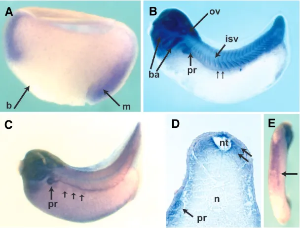
Fig. 1. Developmental expression of C1qA.(A)Bissected gastrula. Dorsal is to the right. Expression is restricted to the marginal zone mesoderm indicated by the arrow; (b) blastopore, (m) mesoderm. (B) Stage 28 embryo. Anterior is to the left and dorsal to the top. There is strong expression in the cephalic structures, especially in the branchial arches (ba) and in the otic vesicle (ov). Expression in the intersomitic veins is visible (isv). Pronephric expression has begun (pr). The small arrows indicate expression in the pronephric duct. (C)Stage 35 embryo shows detailed expression in pronephros (pr) and duct. At this stage, expression in the lateral muscle presursors (small arrows) is visible ventral to the duct.(D) Transverse section through an age-matched embryo to (C). The section is at the level of the pronephros. Dorsal is to the top. Somitic expression is most pronounced in the cells adjacent to the neural tube (nt) as indicated by the small arrows, and in the pronephros (pr). There is no staining in the notochord (n). (E)Ventral view of a stage 35 embryo reveals staining consistent with ventral blood island expression (arrow).
B
C
D
E
A
white blood cells and endothelial cells. There is limited evidence from studies in fish that comple-ment is expressed during embryonic developcomple-ment, although most of the published reports examined protein expression or gene expression in the whole embryo, with little focus on timing and organ-speci-ficity. There are a few reports of complement com-ponents isolated from Xenopus laevis and tropicalis screens showing expression in tailbud and early larval embryo (Changkyun Park et al., 2007, Costa et al., 2003, Pollet et al., 2005), but we are not aware of any systematic analysis of gene expres-sion of complement components during early verte-brate development. Based on our finding that in
Xenopus C3 mRNA expression was conserved from
the neurula stage endoderm to the adult liver, we aimed to examine the developmental expression of other complement genes. We hypothesized that expression analysis of this evolutionarily conserved cascade may be suggestive of a previously unrec-ognized developmental role. Here, we report that in Xenopus laevis, several of the complement genes are expressed during early patterning, largely in the neural precursors and mesoderm, and later during organogenesis in such organs as the kidney, intes-tine, brain and lens.
Results
to the visceral mesoderm, the kidney and blood precursors. The pronephros gives rise to the kidney, a highly vascularized organ which is also active in blood formation in the adult (Brandli, 1999). Expression of C1qA begins at gastrula stage in the mesoderm (Fig. 1A). By neurula stage, expression is most noticeable in the anterior neural plate (not shown). As development proceeds there is continued strong expression in cephalic structures, which we observed through the late tadpole stages (Fig. 1B). Additionally, as the embryo begins to elongate expression appears in the somites around stage 22-25, which is best appreciated in the tailbud embryo (Fig. 1B) and then begins to fade by tadpole stage (Fig. 1C). Expression in the developing kidney is also first notice-able in the tailbud embryo. Pronephric expression is best seen a few hours later in the stage 35-37 embryo in which the pronephros is clearly visible, as is the duct (Fig. 1C). In tandem with the pronephric expression, a subset of cells, grouped in discrete islands just ventral to the duct express C1qA. This pattern mimicks the expression of the transcription factor xFoxK1 in lateral muscle precursors as reported by Pohl and Knochel (Pohl and Knochel, 2004) (Fig. 1C). A section through an age-matched embryo reveals residual staining in the somites and tubular expression in the pronephros (Fig. 1D). Concurrently, strong expression in the ventral blood islands is noticeable (Fig. 1E). Finally, there is expression in the mesoderm of the larval gut, resembling expression of the mesodermal transcription factor FoxF1 (Tseng et al., 2004). In this tissue, expression proceeds in a cranio-caudal fashion, temporally beginning in the mesoderm of the foregut around stage 42, and then progressing throughout the mesoderm of the primitive intestine by stage 43-44 (not shown).
C1qR is expressed in the developing vasculature
In mammals, C1qR is a membrane-associated receptor which is expressed by many different cell-types including peripheral white blood cells, fibroblasts and endothelial cells. C1qR interacts with the first component of complement C1 in part by binding to its
collagen-like domains (Ghebrehiwet, 1989, Peerschke et al., 1993). C1qR has diverse functions in different adult cell types. These include leucocyte chemotaxis and calcium release (Bordin et al., 1990, Eggleton et al., 1994, Fusi et al., 1991, Ghebrehiwet et al., 1990, Ghebrehiwet et al., 1992, Peerschke and Ghebrehiwet, 1990, Peerschke and Ghebrehiwet, 1990, Peerschke et al., 1993, Vegh et al., 2006). Although C1qR was recently identified in a screen for mesenchymal genes in the developing mouse intestine (Li et al., 2007), there are no functional or expression studies to date investigating C1qR in lower vertebrates or invertebrates.
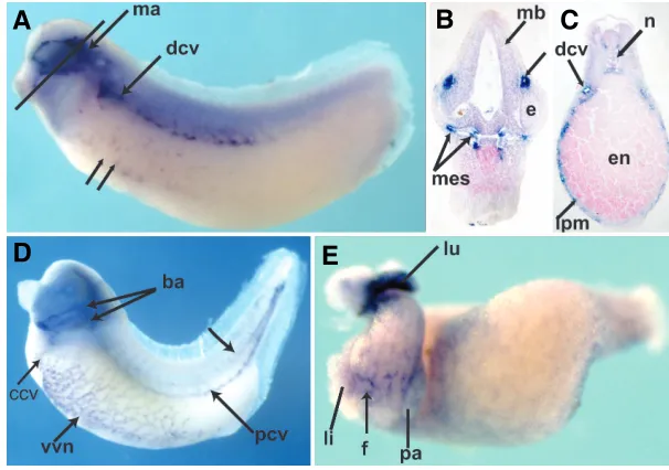
In the Xenopus embryo, the expression of C1qR is most remarkable for its expression in the developing vasculature (Fig. 2). Briefly, there are two phases of vascular development in Xenopus. The first, called vasculogenesis, consists in the forma-tion of vessels from vascular precursors. The next phase, called angiogenesis, consists in the outgrowth of vessels from these founder vessels (Cox et al., 2006). C1qR expression is first noted during the early development of the dorsal cardinal vein of the tailbud embryo (Fig. 2A). Punctate staining in the ventral portion of the embryo suggests early expression in ventral vascular precursors (Fig. 2A). Later this expression evolves into a reticular pattern observed in the ventral region of the tadpole (Fig. 2D), mimicking the expression of other vascular markers such as X-msr, Xl-fli, Dab2, Apelin, Ami and Xl Erg (Baltzinger et al., 1999, Cheong et al., 2006, Cox et al., 2006, Devic et al., 1996, Inui and Asashima, 2006, Meyer et al., 1995, Meyer et al., 1993). Expres-sion of C1qR is noted in the visceral mesoderm starting at stage 35 (Fig. 2C). The expression appears to localize to cells surround-ing small lumina, consistent with a vascular expression (Fig. 2C). In the isolated larval gut, the reticular expression is also notice-able in the intestinal precursors and in the organ buds of the liver and pancreas (Fig. 2E). Additionally, strong expression is noted in the lung buds (Fig. 2E). Interestingly, the recently-isolated Ami which has a very similar expression pattern, is homologous to the human soluble complement inhibitor Factor D (Inui and Asashima,
B
C
D
E
A
Fig. 2. Developmental expression of C1qR. Anterior is to the left and dorsal to the top unless otherwise specified. (A)
Tailbud embryo. Marked expression in the mandibular seg-ment of the neural crest is indicated (ma). Double arrows indicate punctate ventral staining consistent with early vascular precursor expression. Expression in the dorsal cardinal vein is indicated (dcv). (B) Transverse section through the head of embryo in (A) (solid black line). Dorsal is to the top. The developing mesencephalon or mid-brain is labeled (mb). The small arrow to the right of the figure points to mandibular segment expression, dorsal to the developing eye (e). Expression in the head mesenchyme is shown, consistent with neural crest expression. (C)Section through the trunk of a tailbud embryo (shown in D). Dorsal is to the top. The dorsal cardinal vein is indicated (dcv). Small struc-tures with a lumen in the lateral plate mesoderm (lpm) are consistent with expression in the developing vessels of vitelline network (vvn); (en) endoderm, (n) notochord. (D)
Tailbud embryo showing expression throughout the devel-oping vasculature. In addition to the ventral vitelline net-work, both the common (ccv) and the posterior (pcv) cardinal veins show strong expression. There is some expression in
2006). However, sequence analysis did not reveal any significant similarity between C1qR and Ami (AB238233).
In addition, C1qR expression is also noticeable in the develop-ing neural crest. The neural crest is a uniquely vertebrate cell type that arises from the peripheral regions of the neural plate. Neural crest cells are characterized by their ability to migrate long distances and contribute to many organs: cranial structures, somites, adrenal medulla and pronephros, pigment cells, fins, peripheral nervous system and enteric ganglia. At tailbud stage, expression is remarkably confined to the mandibular segment of the neural crest, outlining the developing eye and otic vesicle (Fig. 2A). A section through the head at this stage shows focal expres-sion in the head mesenchyme, similar to the expresexpres-sion of established migratory neural crest markers such twist, slug, FoxD3 and Inca (Fig. 2B) (Dirksen et al., 1993, Hopwood and Gurdon, 1991, Luo et al., 2007, Mayor et al., 1995). By tadpole stage, mandibular segment expression has ceased; in turn,
expression is noted in the branchial arches corresponding to the hyoid and branchial segments of the neural crest (Fig. 2D). Expression of vascular markers in the branchial arches has been shown previously for several genes involved in vasculogenesis (Baltzinger et al., 1999, Cheong et al., 2006, Cox et al., 2006, Devic et al., 1996, Inui and Asashima, 2006, Meyer et al., 1995).
Complement factor C3
One of the central components of the complement system is C3. It is a large protein, composed of multiple functional domains and binding sites, including motifs that recognize fibronectin and integrins. Its function is regulated by proteases, which induce conformational changes. It exerts its many functions mostly through its two active fragments, C3a anaphylatoxin, a potent chemoattractant, and C3b, which contains many binding sites for interacting with other complement and cell surface proteins (Hautanen and Keski-Oja, 1983, Janssen et al., 2005). In
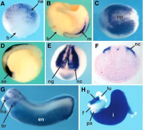
hu-Fig. 3. Developmentalexpression of C3 from gastrula to organ bud stage.(A)
Dorsal view of stage 10 embryo. Anterior is to the top. There is early, punctate expression of C3 in the neuroectoderm (ne); (b) blastopore.(B)Midline view of a bisected gastrula; dorsal is to the right. C3 is expressed in dorsal ectoderm and dorsal mesoderm (m). (C) Dorsal view of stage 13 embryo shows complex expression pattern in and around the neural plate (np). Anterior is to the left. (D)
Sagital view of early neurula embryo shows expression in anterior endoderm (ae) and continued expression in neuroectoderm at the top. Anterior is to the left and dorsal to the top. (E) Anterior view of stage 17 embryo showing defined expression in both neural crest (nc) and neural groove (ng). Dorsal is to the top.
(F)Frontal section through the embryo in (E) shows gene expression in neural crest (nc). Dorsal is to the top. (G) Stage 27 embryo shows expression in the mandibular and branchial segments of anterior neural crest. Hyoid segment is visible between the mandibular (ma) and branchial segments (br). At this stage there is strong endodermal expression (en). (H) Isolated gut tube from stage 42 embryo shows strong expression in liver and presumptive intestine, with sparing of foregut and pancreas. (f) foregut, (i) presumptive intestine, (li) liver, (lu) lung, (pa) pancreas.
mans, C3 is synthesized in the liver and macrophages. In teleost fish, C3 mRNA has been found in developing neural tissue and gastrointestinal tract (Lange et al., 2004, Lange et al., 2004).
We initially isolated a 5 kb long C3 cDNA clone in a screen designed to isolate endoderm-specific genes. The sequence showed 100% identity to the previously isolated C3 Xenopus gilli (U19253) and 97% identity to the 900bp partial coding sequence Unigene Xl.55075, LOC398666.
In situ hybridization analysis reveals that C3 has an intricate pattern of expression that shows a distinct temporo-spatial regulation. The expression pattern is remarkable for its early expression in the neural plate, followed by an intense neural crest and endodermal expression in the gastrointestinal precursor cells, con-firming the findings of others (Costa et al., 2003, Pollet et al., 2005).
By in situ hybridization, we can detect expression of C3 in the dorsal region of early gastrulae, in a wide, crescent shaped area (Fig. 3A). While most of the expres-sion is in the superficial layer of the neural plate, there is also expression in the dorsal mesoderm (Fig. 3B). At this stage, a few cells on the ventral side of the blastopore selectively express C3 (not shown). By stage 13, expres-sion in the neuroectoderm is remarkably complex. A population of cells organized in the characteristic cres-cent shape of the neural folds display solid C3 expres-sion. Medial to the crescent shaped area, in the central region of the neural plate, there is a mosaic expression of C3 (Fig. 3C). At this stage, in cells anterior to the neural plate, an area destined to form the cement gland and the placodal structures, cease expressing C3. However, a cross section of the embryo reveals an intense transcrip-tion of C3 in the prospective pharyngeal endoderm (Fig. 3D). During neurulation, C3 expression in the dorsal region of the embryos is progressively restricted to the neural crest cells (Fig. 3E, F). At the same time, C3 expression appears in the endoderm, in the presumptive intestinal cells (Fig. 3D,E), as has been previously re-ported (Costa et al., 2003). By stage 22, the neural crest expression pattern mimicked that of the known migratory
G
B
C
D
E
F
neural crest markers (Devic et al., 1996, Dirksen et al., 1993, Hopwood and Gurdon, 1991, Luo et al., 2007, Mayor et al., 1995). Following neural tube closure, in the tailbud stages, we observed strong expression of C3 in the anterior migrating neural crest - in the mandibular, hyoid, and branchial segments (Fig. 3G). Although Costa et al. had shown late neural crest expression, the early pattern in the neural plate and neural crest had not previously been analyzed in detail. Indeed, the man-dibular, hyoid, and branchial segments of the cranial migratory neural crest show high levels of C3 expres-sion. In addition, expression in the neural groove and later in the dorsal fin suggests that trunk neural crest cells also express C3.
In addition, as expected, intense C3 expression is observed in the bulk of the endoderm (Fig. 3G) (Costa et al., 2003, Pollet et al., 2005). Late expression in the larval gut is remarkable for its strong expression in the endoderm of intestinal precursors and in the early liver diverticulum, but not in the remainder of the foregut (Fig. 3H).
Expression pattern of C3aR
In the complement cascade as it is understood in immunity, one of the ways C3 signals is through the binding of C3a to the G-protein coupled transmembrane receptor C3aR. In mammals, C3aR is expressed in myeloid, non-myeloid, and endothelial cells (Morikis, 2005). It has also been identified on glial cells (Nataf et al., 1999). C3aR has been implicated in multiple cellular processes, among them leucocyte chemotaxis, vascular adhesion and smooth muscle contraction (Morikis, 2005). In light of the expression pattern of C3, we sought to examine the developmental expression of its cognate receptor. C3aR expression is markedly different from the expression of its ligand C3.
First, at gastrula stage, we observe a strong expres-sion in the mesoderm (Fig. 4A, B). During neurulation, additional diffuse expression of C3aR appears in the neural plate (not shown). By early tailbud stage, C3aR
et al., 1986). Other regions of the molecule show similarities to both epidermal growth factor and alphafeotoprotein (Catterall et al., 1987). Factor I has been isolated from Xenopus liver, and its immune function in Xenopus serum analyzed. The main difference between human and Xenopus Factor I is the presence of an additional 87 base pair domain in the amphibian (Kunnath-Muglia et al., 1993). To date, its gene expression pattern in the vertebrate embryo is unknown.
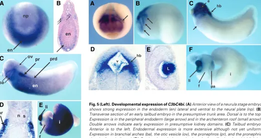
Embryonic expression begins in the neurula. Its expression is crescent-shaped lateral and ventral to the neural plate (Fig. 5A) and in the closing blastopore (not shown). In situ on a sectioned, early tailbud embryo reveals that C3bC4b is expressed at the periphery the developing endoderm, including in the cells of the archenteron roof (Fig. 5B). In the tailbud embryo, endodermal expression expands caudally before localizing to the liver and presumptive intestine in the larval gut tube (Fig. 5C). During the development of the endoderm into a gut, expression progresses radially, from the outside in, to finally express throughout the endoderm of the developing intestine (not shown). Similar to other
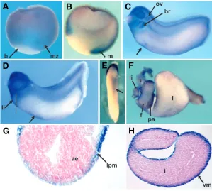
Fig. 4.Developmental expression of C3aR from gastrula to organ bud stage.(A)
Whole mount in situ hybridization on a stage 10 gastrula showing expression predominantly in the marginal zone (mz); (b) blastopore. (B) Bissecting the gastrula revealed that the expression is mesodermal (m). In both (A,B) dorsal is to the right.
(C) Stage 25 embryo showing expression in both the branchial segment of the neural crest (br) and in the otic vesicle (ov). Anterior is to the left, dorsal to the top. The arrow indicates early ventral blood island expression (VBI).(D)Stage 32 embryo. In addition to strong expression in cephalic structures, the presumptive liver area (li) is sur-rounded by cells expressing C3aR message. Arrow shows VBI expression. (E)Ventral view of same stage 32 embryo showing detail of VBI expression (arrow); head is to the top. (F)Isolated gut tube showing C3aR expression throughout the foregut (f) and presumptive intestine (i), (pa) pancreas. Anterior is to the left. (G) A section through the presumptive liver area (indicated in D) shows expression in lateral plate meso-derm (lpm) surrounding the anterior endomeso-derm (ae). (H)Section through isolated gut showing staining in visceral mesoderm (vm), surrounding the presumptive intestine.
G
B
C
D
E
F
H
A
expression is present in the branchial segment of the cranial neural crest, in the developing eye and in the otic placode (Fig. 4C).
In the trunk, expression of C3aR is observed in a triangle surrounding the presumptive liver bud (Fig. 4D). A section through this area reveals that most of the expression is in the developing visceral mesoderm (Fig. 4G). This mesodermal expression ex-tends throughout the developing gastrointestinal tract of the larval stage embryo (Fig. 4F and 4H).
Finally, the ventral blood islands, also mesodermal in origin, expressed C3aR starting at early tailbud stage (Fig. 4C), but is most noticeable in the tadpole (Fig. 4E). The VBI are derived from lateral plate mesoderm and are the site of embryonic hematopoiesis (Walmsley et al., 2002).
C3bC4b inactivator (Factor I) is expressed in the pronephros
complement components such as C3 and C9 expressed in the endoderm, the expression is limited to the liver and presumptive intestine, with little or no expression in the foregut (Fig. 5E). Expression is also visible in the developing branchial arches of the tailbud embryo. Based on the early endodermal expression, it is probable that the expression in the branchial arches is in cells derived from pharyngeal endoderm (Fig. 5C). Together with the neural crest, which expresses C3, C9 and C1qR, the pharyngeal endoderm is important in the development of the branchial arches (Graham et al., 2005).
The distinguishing feature of C3bC4b expression in the Xeno-pus embryo is the appearance of a message in the pronephros of the tailbud embryo (Fig. 5B, 5C). The pronephros, which is mesodermal in origin, gives rise to the kidney. It is composed of two basic units: the tubules and collecting duct, and the glomus which is vascular in origin. At tadpole stage, expression is notice-able in the developing tubules of the pronephros and the proximal duct (Fig. 5C, D).
Properdin is expressed in the developing neural tube and lens
Properdin is a regulatory protein of the alternative pathway of complement activation and the only positive regulator of the
complement system (Perkins, 2005). The role of properdin in the human complement cascade is stabilization of the C3 convertase. C3 convertase catalyzes the breakdown of C3 into its active components C3a and C3b. Therefore, properdin serves as a positive regulator of C3 activity (Fijen et al., 1999). The structure of properdin contains six thrombospondin domains (TSR), like many other complement components (Perkins, 2005). TSRs are involved in binding to other molecular structures. Absence of or defects in properdin synthesis is rare, but associated with life-threatening clinical conditions (Fijen et al., 1999). In humans, properdin is synthesized by peripheral white blood cells, hepato-cytes, and astrohepato-cytes, which contribute to the formation of the blood-brain barrier. There are no descriptive or functional studies examining the role of properdin in lower vertebrates.
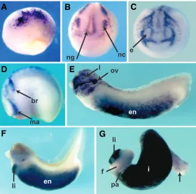
Similar to the expression of other members of the complement system in the Xenopus embryo, properdin mRNA is noticeable early in the neural crest (Fig. 6A, B) and later in the liver precursors and liver bud (Fig. 6C, F). However, it displays a remarkably different expression pattern starting in the tailbud embryo at which time expression is confined to cephalic structures (Fig. 6C). At this stage, strong expression is noted throughout the head, including the eye, otic vesicle, and presumptive hindbrain, but excluding the cement gland (Fig. 6C). In the tadpole, cephalic expression is
B
C
D
E
A
B
C
D
E
F
A
Fig. 5 (Left). Developmental expression of C3bC4bi.(A) Anterior view of a neurula stage embryo shows strong expression in the endoderm (en) lateral and ventral to the neural plate (np). (B)
Transverse section of an early tailbud embryo in the presumptive trunk area. Dorsal is to the top. Expression is in the peripheral endoderm (large arrow) and in the archenteron roof (small arrow). Double arrows indicate early expression in presumptive kidney domains.(C) Tailbud embryo. Anterior is to the left. Endodermal expression is more extensive although not yet uniform. Expression in branchial arches (ba), the otic vesicle (ov), the pronephros (pr), and the pronephric duct (prd) are indicated. (D)Section through the proximal trunk of a tailbud embryo highlighting strong expression in the developing kidney. Dorsal is to the top; (n) notochord, (s) somite. (E) Isolated larval gut reveals strong expression in presumptive intestine (i) and liver (li), but no expression in foregut (f) and distal most segment of the developing intestine.
Fig. 6 (Right). Developmental expression of Properdin. Anterior is to the left and dorsal to the top unless otherwise specified. (A)Anterior view of a neurula stage embryo. Arrow indicates neural crest expression. (B)Late neurula/early tadpole stage embryo. Arrows indicate expression in mandibular, hyoid and mandibular segments of anterior neural crest (from anterior to posterior).(C)Tailbud embryo shows strong cephalic expression. Intense expression in developing eye (e) and hindbrain (hb) are indicated. The ventral arrow highlights beginning expression in the anterior endoderm.
confined to the hindbrain and the lens (Fig. 6D, E). In the developing hindbrain or rhombencephalon, expression is limited to the periventricular tissue (Fig. 6D), and extends caudally to the proximal neural tube (not shown).
Expression pattern of C9
C9 complement factor is the most abundant protein of the membrane attack complex (MAC), which is the termi-nal, cytolytic component of the complement system as it is
with a weaker expression in the developing lens.
In the endoderm, unlike C3, expression in the anterior endoderm is not noted until stage 19-20 (not shown). Endoder-mal expression in the tailbud embryo is noticeable in the peripheral endoderm, confirming the findings of Pollet et al. (Pollet et al., 2005) and similar to the expression of C3bC4b at the same stage. In the isolated larval gut, expression mimics C3 with C9 transcripts observed in the liver and developing intes-tines, but not in the foregut and pancreas. At this stage, unlike C3, which is expressed throughout the gut, the C9 transcripts
Gene Accession No Expression Pattern
C1s BG811630
BE509150
No noticeable expression Notochord expression (*).
CR2 BM261540 Head and somite expression at st 35.
C4 BM180922 Liver expression stage 42.
C5a BF048379 Diffuse, faint neural plate and head expression through st 35
C6 BQ734761 No neural plate staining, strong endodermal expression, similar to C3bC4b, no pronephros expression.
C8 BE509028
BE508140
Liver expression in tailbud embryo (*).
Carboxypeptidase N BM180235 Liver expression at stage 42.
CD46 BG160519 Faint expression in neural plate and head through st 35.
CD59 BG814148 Faint expression in neural plate and head through st 35.
Complement Factor H BC046950 Neural crest expression starting from neurula to tadpole stage. Endodermal expression from st 30 (*). Eye expression from stage 32.
TABLE 1
SUMMARY OF ANALYSIS OF EARLY COMPLEMENT EXPRESSION IN XENOPUS LAEVIS
(*) indicates that the expression pattern was reported by Pollet et al. (Pollet et al. 2005)
Fig. 7. Developmental expression of C9 from gastrula to organ bud stage. Anterior is to the left and dorsal to the top unless otherwise indicated.(A) Dorsal view of stage 10 embryo showing early spotted expression dorsal ectoderm. Anterior is to the top. (B)
Anterior view of stage 17 embryo showing discrete neural crest (nc) and neural groove expression (ng).(C) Anterior view of stage 19-20 embryo. The three segments of the anterior neural crest show discrete expression at this stage. Mandibular segment ex-pression surrounds the presumptive eye field (e). (D)Lateral view of stage 22 embryo shows expression in mandibular (ma) and branchial segments (br) of anterior migrating neural crest. There is discrete hyoid expression noticeable between those two seg-ments. (E)Stage 27 embryo. Neural crest expression is seen in all three segments, as well as in the otic vesicle (ov), the lens (l), and the endoderm (en). (F)Stage 35 embryo shows strong liver and endodermal expression.(G)Isolated gut tube shows liver (li) and intestinal expression sparing the foregut (f), but no distal intestinal (proctodeum) expression (arrow); (lu) lung, (pa) pancreatic bud.
G
B
C
D
E
F
A
understood in immunity. Akin to its MAC partners, C9 is made of multiple building-blocks including thrombospondin, low den-sity lipoprotein receptor, epidermal growth factor, and perforin domains (Perkins, 2005). The C5b-9 (MAC) complex has been shown to participate in cellular proliferation of endothelial cells via the extracellular signal-regulated kinase (Perkins) and to induce cytoskeletal changes in a model of glomerular epithelial injury (Cybulsky et al., 2005, Fosbrink et al., 2006). C9 defi-ciency has been associated with an increased risk of infection and with post-ischemic injury (Liu et al., 1998, Rzepecka-Wozniak et al., 2006, Zoppi et al., 1990). C9 was formerly identified in an expression screen, and the sequence we used to generate the antisense riboprobe was 100% identical to the one previously reported (Pollet et al., 2005). However, previous analysis focused on the tailbud embryo, and we focused on the neurula stage.
are not found in the caudal-most portion of the developing GI tract, or proctodeum, again resembling the findings for C3bC4b.
Expression analysis of other members of the complement system
Using BLAST searches, we identified Xenopus ESTs corre-sponding to other genes of the vertebrate complement system and performed in situ analysis for all of these. For the sake of completion they are listed in Table I. However, only those with an expression pattern of potential developmental interest are ana-lyzed in detail in the figures.
Discussion
In summary, we show the developmental expression patterns of several of the major complement genes during early develop-ment of Xenopus laevis. The expression of each component was remarkable for some degree of tissue- or organ-specificity, often in organs not typically known for a role in immunity. To date, the most convincing data in support of a developmental function for complement is the known role of certain complement factors in hematopoietic cell migration. In fact, both the C1q-C1qR and the C3-C3aR pairs have been shown to participate in the migration and homing of hematopoietic lineages in mammals (Reca et al., 2003, Vegh et al., 2006). Consistent with these functional studies,
both C1qA and C3aR expression is noted in the ventral blood
islands of the developing Xenopus embryo. Ventral blood islands are the site of embryonic blood formation. From a developmental perspective, these findings are important because migration is a critical process in embryonic patterning and organogenesis.
In addition, several of the complement genes analyzed are expressed in other cell-types known for their migratory properties. For example, the neural crest cells, which are characterized by their ability to migrate long distances, express several of the complement genes: C1qA, C1qR, C3, C3aR, Properdin, and C9. Together with the ventral blood island data, this expression pattern raises the question of the potential mechanism. Comple-ment proteins are known both for their ability to bind extracellular matrix proteins, and for their proteolytic activities. Thus, one possibility is that complement proteins bind to extracellular matrix proteins such as fibronectin, thereby facilitating cell movement. Alternatively, if complement proteins are also secreted during early development, they could participate in the extra-cellular release of growth factors either by cleaving inactive precursors or releasing growth factors from the extracellular matrix. Indeed, it was recently shown in Xenopus that the secreted serine protease xHtrA1, has a very similar expression pattern to several of the complement components analyzed, namely in the anterior neural plate, presumptive forebrain, neural folds, and branchial arches (Hou et al., 2007). xHtrA1 causes cleavage of extracellular matrix proteoglycans thereby regulating the diffusion of the secreted ligand FGF4 (Hou et al., 2007).
The second pattern found in this cohort of complement genes is expression in vascular structures. C1qR is expressed in the developing vasculature and is visible in the vessels of the devel-oping liver and lung buds. This expression pattern is significant for two reasons. First, the C1q-C1qR pair has been shown to partici-pate in hematopoietic cell homing in mammalian models. Second, endothelial-endodermal interactions are known to be essential in
the development of highly vascularized organs such as the lung and liver (Del Moral et al., 2006, Lammert et al., 2003). Finally, C1qA and C3bC4b are expressed in the pronephros, another highly vascularized organ.
Third, the expression of C1qA and C3aR in the visceral mesoderm of the gut resembles the expression of the visceral mesoderm transcription factor FoxF1 in the larval gut (Tseng et al., 2004). The visceral mesoderm is the layer of cells surrounding the developing intestinal epithelium which will give rise to the intestinal smooth muscle layer and the mesenchyme. The vis-ceral mesoderm is commonly accepted to drive the elongation of the developing gut (Roberts, 2000). Interestingly, C3aR is ex-pressed in the visceral mesoderm at the same time as C3 is transcribed in the adjacent endoderm, suggesting that these two molecules may function as a pair both in immunity and during the development of the gastrointestinal tract.
Finally, properdin expression is remarkable in the hindbrain and lens of the tailbud embryo. Other molecules with thrombospondin repeats, such as prothrombin and thrombin, have been shown to participate in glial cell proliferation and migration in the extracellular matrix (Krem and Di Cera, 2002), largely in disease states. Besides the homology with these molecules, the significance of our findings is unclear. It is possible that understanding their role in disease could orient research examining their role in development.
Taken together, these data are compelling for a previously unrecognized role for complement components during early pat-terning and organogenesis in lower vertebrates. The following observations support this hypothesis. First, their expression is noted very early in development, long before metamorphosis, the stage of onset of mature immune function in Xenopus. Second, expression of functional pairs occurs in different, sometimes complementary, tissues, rather than in the same tissue. This finding suggests that their role during development may not require expression at the same place and time, which is the typical paradigm in immunity. Instead, reciprocal expression patterns may be necessary to exert novel patterning functions. For ex-ample, C3 is expressed in the endoderm at the same time as C3aR is expressed in the visceral mesoderm, two tissues known to require reciprocal signaling for their development and mainte-nance. Finally, tight spatial and temporal control of the expression of the different complement genes further supports a develop-mental role. For example, although C3 and C9 expression closely resemble each other, C9 expression begins later than C3 in the anterior endoderm, and appears not to extend to the distal most portion of the larval gut.
In conclusion, we have shown in Xenopus the detailed embryo-logical expression pattern of several components of the comple-ment system, all of which were remarkable for some degree of tissue- or organ-specificity in organs not always involved in immunity. The significance of these findings is unclear and warrants further, functional studies.
Materials and Methods
Embryos
Embryos were generated by in vitro fertilization according to conven-tional methods. Embryos were cultured in 0.1xMMR at room temperature or in a 16°C incubator. Developmental stages were determined according to Nieuwkoop and Faber (Nieuwkoop, 1967). Embryos were fixed at different developmental stages according to previously described meth-ods (Harland, 1991).
Isolation of C3
An adult liver-specific cDNA was hybridized first to a stage 13 cDNA library. Multiple copies of C3 were isolated using this approach. The largest clone, 5kb in length,was inserted into pBS.
Identification of other complement clones in Xenopus laevis
Using BLAST searches, we identified expressed sequence tags (ESTs) with significant similarity to the human complement components reported. The corresponding I.M.A.G.E clones were obtained from A.T.C.C. DIG-labeled antisense riboprobes were generated according to conven-tional methods using Ambion MegaScript kit for in vitro transcription from the EST plasmid. pCMV-C1qA (BE507776) SalI, T7, pCMV-C1qR (BC111511) SalI, T7, pCMV-C3bC4bi (BI315342), pCMV-C6 (BC042265), SalI, T7, pCMV-Properdin (BM192350) SalI, T7 pBS-XC3 Kpn1, T7; pCMV-XC9 (BM180706) Kpn1, T7; pCS108-C3aR (CX430718) Sal1, T3.
In situ hybridization
In situ hybridization was performed as previously described (Harland, 1991). 65°C incubation was performed in a water bath. BM Purple (Roche) was used for the chromogenic reaction. In situ hybridization on isolated gut tubes were performed as previously described (Chalmers and Slack, 1998). In situ hybridization on bisected embryos was per-formed to show deep staining (Lee et al., 2001, Sive, 2000). After whole-mount in situ hybridization, pigmented embryos were bleached in a 1% hydrogen peroxide, 5% formamide, 0.5X SSC.
Sections
Following in situ hybridization, embryos were dehydrated in ethanol, embedded in paraffin, and sectioned every 12-16 µm. Eosin was used for counterstaining when appropriate.
Acknowledgements
We would like to thank Dr. Klaus Richter for help with the initial isolation of Xenopus C3 and Neekita Desai for technical assistance. We are also grateful to Drs Wetsel and Lambris for helpful discussions. This work was supported by the Naman Family Fund for Basic Research and HD41648 to VAM. MJ is supported by NIH grants EY012505, EY012163 and the Retina Research Foundation.
References
BALTZINGER, M., MAGER-HECKEL, A.M. and REMY, P. (1999). Xl erg: Expres-sion pattern and overexpresExpres-sion during development plead for a role in endot-helial cell differentiation. Dev Dyn 216: 420-33.
BORDIN, S., GHEBREHIWET, B. and PAGE, R.C. (1990). Participation of c1q and its receptor in adherence of human diploid fibroblast. J Immunol 145: 2520-6. BRÄNDLI A.W. (1999). Towards a molecular anatomy of the Xenopus pronephric
kidney. Int. J. Dev. Biol. 43: 381-395
CATTERALL, C.F., LYONS, A., SIM, R.B., DAY, A.J. and HARRIS, T.J. (1987). Characterization of primary amino acid sequence of human complement control protein factor i from an analysis of cdna clones. Biochem J 242: 849-56. CHALMERS, A.D. and SLACK, J.M. (1998). Development of the gut in Xenopus
laevis. Dev Dyn 212: 509-21.
CHANGKYUN PARK, E., HAYATA, T., CHO, K.W. and HAN, J.K. (2007). Xenopus cdna microarray identification of genes with endodermal organ expression. Dev Dyn 236: 1633-49.
CHEONG, S.M., CHOI, S.C. and HAN, J.K. (2006). Xenopus dab2 is required for embryonic angiogenesis. BMC Dev Biol 6: 63.
COSTA, R.M., MASON, J., LEE, M., AMAYA, E. and ZORN, A.M. (2003). Novel gene expression domains reveal early patterning of the Xenopus endoderm.
Gene Expr Patterns 3: 509-19.
COX, C.M., D’AGOSTINO, S.L., MILLER, M.K., HEIMARK, R.L. and KRIEG, P.A. (2006). Apelin, the ligand for the endothelial g-protein-coupled receptor, apj, is a potent angiogenic factor required for normal vascular development of the frog embryo. Dev Biol 296: 177-89.
CYBULSKY, A.V., TAKANO, T., PAPILLON, J., BIJIAN, K. and GUILLEMETTE, J. (2005). Activation of the extracellular signal-regulated kinase by complement c5b-9. Am J Physiol Renal Physiol 289: F593-603.
DEANGELIS, R.A., MARKIEWSKI, M.M. and LAMBRIS, J.D. (2006). Liver regen-eration: A link to inflammation through complement. Adv Exp Med Biol 586: 17-34.
DEL MORAL, P.M., SALA, F.G., TEFFT, D., SHI, W., KESHET, E., BELLUSCI, S. and WARBURTON, D. (2006). Vegf-a signaling through flk-1 is a critical facilitator of early embryonic lung epithelial to endothelial crosstalk and branch-ing morphogenesis. Dev Biol 290: 177-88.
DEL RIO-TSONIS, K., TSONIS, P.A., ZARKADIS, I.K., TSAGAS, A.G. and LAMBRIS, J.D. (1998). Expression of the third component of complement, c3, in regener-ating limb blastema cells of urodeles. J Immunol 161: 6819-24.
DELOTTO, R. (2001). Gastrulation defective, a complement factor c2/b-like pro-tease, interprets a ventral prepattern in drosophila. EMBO Rep 2: 721-6. DEVIC, E., PAQUEREAU, L., VERNIER, P., KNIBIEHLER, B. and AUDIGIER, Y.
(1996). Expression of a new g protein-coupled receptor x-msr is associated with an endothelial lineage in Xenopus laevis. Mech Dev 59: 129-40.
DIRKSEN, M.L., MATHERS, P. and JAMRICH, M. (1993). Expression of a Xenopus distal-less homeobox gene involved in forebrain and cranio-facial development.
Mech Dev 41: 121-8.
EGGLETON, P., GHEBREHIWET, B., COBURN, J.P., SASTRY, K.N., ZANER, K.S. and TAUBER, A.I. (1994). Characterization of the human neutrophil c1q receptor and functional effects of free ligand on activated neutrophils. Blood 84: 1640-9.
ELLINGSEN, T., STRAND, C., MONSEN, E., BOGWALD, J. and DALMO, R.A. (2005). The ontogeny of complement component c3 in the spotted wolffish (anarhichas minor olafsen). Fish Shellfish Immunol 18: 351-8.
FIJEN, C.A., VAN DEN BOGAARD, R., SCHIPPER, M., MANNENS, M., SCHLESINGER, M., NORDIN, F.G., DANKERT, J., DAHA, M.R., SJOHOLM, A.G., TRUEDSSON, L. et al. (1999). Properdin deficiency: Molecular basis and disease association. Mol Immunol 36: 863-7.
FOSBRINK, M., NICULESCU, F., RUS, V., SHIN, M.L. and RUS, H. (2006). C5b-9-induced endothelial cell proliferation and migration are dependent on akt inactivation of forkhead transcription factor foxo1. J Biol Chem 281: 19009-18. FUSI, F., BRONSON, R.A., HONG, Y. and GHEBREHIWET, B. (1991). Comple-ment component c1q and its receptor are involved in the interaction of human sperm with zona-free hamster eggs. Mol Reprod Dev 29: 180-8.
GHEBREHIWET, B. (1989). Functions associated with the c1q receptor. Behring Inst Mitt 204-15.
GHEBREHIWET, B., HABICHT, G.S. and BECK, G. (1990). Interaction of c1q with its receptor on cultured cell lines induces an anti-proliferative response. Clin Immunol Immunopathol 54: 148-60.
GONGORA, R., FIGUEROA, F. and KLEIN, J. (1998). Independent duplications of bf and c3 complement genes in the zebrafish. Scand J Immunol 48: 651-8. GRAHAM, A., OKABE, M. and QUINLAN, R. (2005). The role of the endoderm in
the development and evolution of the pharyngeal arches. J Anat 207: 479-87. HARLAND, R.M. (1991). In situ hybridization: An improved whole-mount method for
Xenopus embryos. Methods Cell Biol 36: 685-95.
HAUTANEN, A. and KESKI-OJA, J. (1983). Interaction of fibronectin with comple-ment component c3. Scand J Immunol 17: 225-30.
HOPWOOD, N.D. and GURDON, J.B. (1991). Gene activation in the amphibian mesoderm. Dev Suppl 1: 95-104.
HOU, S., MACCARANA, M., MIN, T.H., STRATE, I. and PERA, E.M. (2007). The secreted serine protease xhtra1 stimulates long-range fgf signaling in the early Xenopus embryo. Dev Cell 13: 226-41.
INUI, M. and ASASHIMA, M. (2006). A novel gene, ami is expressed in vascular tissue in Xenopus laevis. Gene Expr Patterns 6: 613-9.
JANSSEN, B.J., HUIZINGA, E.G., RAAIJMAKERS, H.C., ROOS, A., DAHA, M.R., NILSSON-EKDAHL, K., NILSSON, B. and GROS, P. (2005). Structures of complement component c3 provide insights into the function and evolution of immunity. Nature 437: 505-11.
KATO, Y., NAKAO, M., SHIMIZU, M., WARIISHI, H. and YANO, T. (2004). Purification and functional assessment of c3a, c4a and c5a of the common carp (cyprinus carpio) complement. Dev Comp Immunol 28: 901-10.
KISHORE, U. and REID, K.B. (2000). C1q: Structure, function, and receptors.
Immunopharmacology 49: 159-70.
KREM, M.M. and DI CERA, E. (2002). Evolution of enzyme cascades from embryonic development to blood coagulation. Trends Biochem Sci 27: 67-74. KUNNATH-MUGLIA, L.M., CHANG, G.H., SIM, R.B., DAY, A.J. and EZEKOWITZ, R.A. (1993). Characterization of Xenopus laevis complement factor i struc-ture—conservation of modular structure except for an unusual insert not present in human factor i. Mol Immunol 30: 1249-56.
LAMBRIS, J.D. (1993). Chemistry, biology and phylogeny of c3. Complement profiles 1:16.
LAMMERT, E., CLEAVER, O. and MELTON, D. (2003). Role of endothelial cells in early pancreas and liver development. Mech Dev 120: 59-64.
LANGE, S., BAMBIR, S., DODDS, A.W. and MAGNADOTTIR, B. (2004). An immunohistochemical study on complement component c3 in juvenile atlantic halibut (hippoglossus hippoglossus l.). Dev Comp Immunol 28: 593-601. LANGE, S., BAMBIR, S., DODDS, A.W. and MAGNADOTTIR, B. (2004). The
ontogeny of complement component c3 in atlantic cod (gadus morhua l.)—an immunohistochemical study. Fish Shellfish Immunol 16: 359-67.
LEE, M.A., HEASMAN, J. and WHITMAN, M. (2001). Timing of endogenous activin-like signals and regional specification of the Xenopus embryo. Development
128: 2939-52.
LEIVO, I. and ENGVALL, E. (1986). C3d fragment of complement interacts with laminin and binds to basement membranes of glomerulus and trophoblast. J Cell Biol 103: 1091-100.
LI, X., MADISON, B.B., ZACHARIAS, W., KOLTERUD, A., STATES, D. and GUMUCIO, D.L. (2007). Deconvoluting the intestine: Molecular evidence for a major role of the mesenchyme in the modulation of signaling cross talk. Physiol Genomics 29: 290-301.
LIU, L., ALDSKOGIUS, H. and SVENSSON, M. (1998). Ultrastructural localization of immunoglobulin g and complement c9 in the brain stem and spinal cord following peripheral nerve injury: An immunoelectron microscopic study. J Neurocytol 27: 737-48.
LOVOLL, M., FISCHER, U., MATHISEN, G.S., BOGWALD, J., OTOTAKE, M. and DALMO, R.A. (2007). The c3 subtypes are differentially regulated after immunostimulation in rainbow trout, but head kidney macrophages do not contribute to c3 transcription. Vet Immunol Immunopathol.
LOVOLL, M., KILVIK, T., BOSHRA, H., BOGWALD, J., SUNYER, J.O. and DALMO, R.A. (2006). Maternal transfer of complement components c3-1, c3-3, c3-4, c4, c5, c7, bf, and df to offspring in rainbow trout (oncorhynchus mykiss). Immuno-genetics 58: 168-79.
LUO, T., XU, Y., HOFFMAN, T.L., ZHANG, T., SCHILLING, T. and SARGENT, T.D. (2007). Inca: A novel p21-activated kinase-associated protein required for cranial neural crest development. Development 134: 1279-89.
MAHLAPUU, M., ORMESTAD, M., ENERBACK, S. and CARLSSON, P. (2001). The forkhead transcription factor foxf1 is required for differentiation of extra-embryonic and lateral plate mesoderm. Development 128: 155-66.
MARKIEWSKI, M.M., MASTELLOS, D., TUDORAN, R., DEANGELIS, R.A., STREY, C.W., FRANCHINI, S., WETSEL, R.A., ERDEI, A. and LAMBRIS, J.D. (2004). C3a and c3b activation products of the third component of complement (c3) are critical for normal liver recovery after toxic injury. J Immunol 173: 747-54. MASTELLOS, D., GERMENIS, A.E. and LAMBRIS, J.D. (2005). Complement: An
inflammatory pathway fulfilling multiple roles at the interface of innate immunity and development. Curr Drug Targets Inflamm Allergy 4: 125-7.
MASTELLOS, D. and LAMBRIS, J.D. (2002). Complement: More than a ‘guard’ against invading pathogens? Trends Immunol 23: 485-91.
MASTELLOS, D., PAPADIMITRIOU, J.C., FRANCHINI, S., TSONIS, P.A. and LAMBRIS, J.D. (2001). A novel role of complement: Mice deficient in the fifth component of complement (c5) exhibit impaired liver regeneration. J Immunol
166: 2479-86.
MAYOR, R., MORGAN, R. and SARGENT, M.G. (1995). Induction of the prospec-tive neural crest of Xenopus. Development 121: 767-77.
MEYER, D., STIEGLER, P., HINDELANG, C., MAGER, A.M. and REMY, P. (1995). Whole-mount in situ hybridization reveals the expression of the xl-fli gene in several lineages of migrating cells in Xenopus embryos. Int J Dev Biol 39: 909-19.
MEYER, D., WOLFF, C.M., STIEGLER, P., SENAN, F., BEFORT, N., BEFORT, J.J. and REMY, P. (1993). Xl-fli, the Xenopus homologue of the fli-1 gene, is expressed during embryogenesis in a restricted pattern evocative of neural crest cell distribution. Mech Dev 44: 109-21.
MORIKIS, D., HOLLAND, MCH, LAMBRIS, JD. (2005). Structure of the anaphylatoxins c3a and c5a. In Structural biology of the complement system.
Eds. Morikis, D, Holland, MCH, and Lambris JD. Taylor and Francis, Boca Raton, FL, USA
NATAF, S., STAHEL, P.F., DAVOUST, N. and BARNUM, S.R. (1999). Complement anaphylatoxin receptors on neurons: New tricks for old receptors? Trends Neurosci 22: 397-402.
NIEUWKOOP, P.D.A.F., J. (1967). Normal table of Xenopus laevis (daudin). North-Holland, Amsterdam.
PEERSCHKE, E.I. and GHEBREHIWET, B. (1990). Modulation of platelet re-sponses to collagen by clq receptors. J Immunol 144: 221-5.
PEERSCHKE, E.I. and GHEBREHIWET, B. (1990). Platelet c1q receptor interac-tions with collagen- and c1q-coated surfaces. J Immunol 145: 2984-8. PEERSCHKE, E.I., MALHOTRA, R., GHEBREHIWET, B., REID, K.B., WILLIS,
A.C. and SIM, R.B. (1993). Isolation of a human endothelial cell c1q receptor (c1qr). J Leukoc Biol 53: 179-84.
PERKINS, S., FURTADO, PB. (2005). Complement and immunoglobulin protein structure by x-ray and neutron solution scattering and analytical centrifugation. In Structural biology of the complement system. Eds. Morikis, D, Holland, MCH, and Lambris JD. Taylor and Francis, Boca Raton, FL, USA
PETERSEN, S.V., THIEL, S. and JENSENIUS, J.C. (2001). The mannan-binding lectin pathway of complement activation: Biology and disease association. Mol Immunol 38: 133-49.
POHL, B.S. and KNOCHEL, W. (2004). Isolation and developmental expression of Xenopus foxj1 and foxk1. Dev Genes Evol 214: 200-5.
POLLET, N., MUNCKE, N., VERBEEK, B., LI, Y., FENGER, U., DELIUS, H. and NIEHRS, C. (2005). An atlas of differential gene expression during early Xenopus embryogenesis. Mech Dev 122: 365-439.
RECA, R., MASTELLOS, D., MAJKA, M., MARQUEZ, L., RATAJCZAK, J., FRANCHINI, S., GLODEK, A., HONCZARENKO, M., SPRUCE, L.A., JANOWSKA-WIECZOREK, A. et al. (2003). Functional receptor for c3a ana-phylatoxin is expressed by normal hematopoietic stem/progenitor cells, and c3a enhances their homing-related responses to sdf-1. Blood 101: 3784-93. ROBERTS, D.J. (2000). Molecular mechanisms of development of the
gastrointes-tinal tract. Dev Dyn 219: 109-20.
RZEPECKA-WOZNIAK, E., KONIECZNA, M. and BOLECHALA, F. (2006). [myo-cardial ischemia of the driver as a cause of a traffic road accident. Immunohis-tochemical c9 staining method in diagnostics of early myocardial infarction].
SIVE, H.L., GRAINGER, G.R., HARLAND, R.M. (2000). Early development of Xenopus laevis: A laboratory manual. Cold Spring Harbor Laboratory Press, Cold Spring Harbor, New York.
STANLEY, K.K., PAGE, M., CAMPBELL, A.K. and LUZIO, J.P. (1986). A mecha-nism for the insertion of complement component c9 into target membranes. Mol Immunol 23: 451-8.
STREY, C.W., MARKIEWSKI, M., MASTELLOS, D., TUDORAN, R., SPRUCE, L.A., GREENBAUM, L.E. and LAMBRIS, J.D. (2003). The proinflammatory mediators c3a and c5a are essential for liver regeneration. J Exp Med 198: 913-23.
TSENG, H.T., SHAH, R. and JAMRICH, M. (2004). Function and regulation of foxf1
during Xenopus gut development. Development 131: 3637-47.
VEGH, Z., KEW, R.R., GRUBER, B.L. and GHEBREHIWET, B. (2006). Chemotaxis of human monocyte-derived dendritic cells to complement component c1q is mediated by the receptors gc1qr and cc1qr. Mol Immunol 43: 1402-7. WALMSLEY, M., CIAU-UITZ, A. and PATIENT, R. (2002). Adult and embryonic
blood and endothelium derive from distinct precursor populations which are differentially programmed by bmp in Xenopus. Development 129: 5683-95. ZOPPI, M., WEISS, M., NYDEGGER, U.E., HESS, T. and SPATH, P.J. (1990).
Further Related Reading, published previously in the
Int. J. Dev. Biol.
See our recent Special Issue Fertilization, in honor of David L. Garbers and edited by Paul M. Wassarman and Victor D. Vacquier at: http://www.ijdb.ehu.es/web/contents.php?vol=52&issue=5-6
See our recent Special Issue Ear Development edited by Fernando Giraldez and Bernd Fritzsch at: http://www.ijdb.ehu.es/web/contents.php?vol=51&issue=6-7
Two members of the Fxr gene family, Fmr1 and Fxr1, are differentially expressed in Xenopus tropicalis
Lau Blonden, Sandra van ‘t Padje, Lies-anne Severijnen, Olivier Destree, Ben A. Oostra and Rob Willemsen Int. J. Dev. Biol. (2005) 49: 437-441
The Fox gene family in Xenopus laevis:FoxI2, FoxM1 and FoxP1 in early development
Barbara S. Pohl, Antje Rössner and Walter Knöchel Int. J. Dev. Biol. (2005) 49: 53-58
Using Xenopus as a model system for an undergraduate laboratory course in vertebrate development at the University of Bordeaux, France.
Michelle Olive, Pierre Thiebaud, Marc Landry, Michel Duvert, Alain Verna, Wilfrid Barillot and Nadine Theze Int. J. Dev. Biol. (2003) 47: 153-160
Isolation and characterization of a Xenopus gene (XMLP) encoding a MARCKS-like protein.
H Zhao, Y Cao and H Grunz Int. J. Dev. Biol. (2001) 45: 817-826
Fox (forkhead) genes are involved in the dorso-ventral patterning of the Xenopus mesoderm.
H El-Hodiri, N Bhatia-Dey, K Kenyon, K Ault, M Dirksen and M Jamrich Int. J. Dev. Biol. (2001) 45: 265-271
Effects of follistatin and BMP4 proteins on early dorso-ventral patterning in chick.
D J Connolly, K Patel, S Withington and J Cooke Int. J. Dev. Biol. (2000) 44: 129-140
Sequence and translation initiation properties of the Xenopus TGFbeta5, PDGF-A, and PDGF-alpha receptor 5' untranslated regions.
A W van der Velden, A Los, H O Voorma and A A Thomas Int. J. Dev. Biol. (2000) 44: 851-859
Evidence that platelet derived growth factor (PDGF) action is required for mesoderm patterning in early amphibian (Xenopus laevis) embryogenesis.
J S Ghil and H M Chung
Int. J. Dev. Biol. (1999) 43: 329-334
Evidence for non-axial A/P patterning in the nonneural ectoderm of Xenopus and zebrafish pregastrula embryos.
E M Read, A R Rodaway, B Neave, N Brandon, N Holder, R K Patient and M E Walmsley
Int. J. Dev. Biol. (1998) 42: 763-774
Genes and mechanisms involved in early embryonic development in Xenopus laevis.
M Méchali, G Almouzni, Y Andéol, J Moreau, S Vriz, M Leibovici, J Hourdry, J Géraudie, T Soussi and M Gusse
Int. J. Dev. Biol. (1990) 34: 51-59