Embryonic heat shock reveals
latent hsp90 translation in zebrafish (Danio rerio)
MICHELLE H. CONNOLLY* and BRIAN K. HALL
Department of Biology, Dalhousie University, Halifax, B3H 4J1, Canada
ABSTRACT There is increasing evidence that more genetic variation is present among metazoans than is normally expressed in the phenotype, due in part to the canalization of development. Among teleosts (as in other vertebrates), this genetic variation is often expressed as phenotypic change in response to environmental cues. Using embryonic zebrafish (Danio rerio), this ‘hidden’ variation is explored in the context of environmental stress by investigating the activity of heat shock protein-90 (HSP90), a cytosolic chaperone that interacts with transcription factors to mediate multiple developmental pathways. Following a 37°C heat shock during early somitoge-nesis (1 hour heat shock, targeting 2-14 somite stages), phenotypic variability was expressed in the lower trunk and tail bud regions, where somite development was reduced or ceased prematurely. In situ hybridization showed that hsp90 was localized to this caudal region 16 hours after heat shock, indicating its potential to coordinate somitic fate. By following transcription and translation of this chaperone, we show that 24 hours following heat shock zebrafish embryos express a protein signature which reflects the RNA message. However, by 48 hours, message and protein are uncoupled; while endogenous gene expression is downregulated, heat-shocked embryos express a discrete segmented protein pattern within the trunk, suggesting regulation of transcription and of translation in response to environmental stress.
KEY WORDS:
hsp90
α
, translation, heat shock, somite, variation
Introduction
Environmental stresses such as heat shock can induce the expression of a highly conserved family of genes encoding for heat shock proteins (HSPs; Morimoto, 1994). Although heat shock (stress) proteins were originally identified on the basis of their increased synthesis in response to exposure to heat or other stresses, they also are expressed in cells under normal non-stressed conditions (Latchman, 1999).
Hsps ensure the proper conformational folding of proteins, thereby enabling targeted proteins to fulfill their function. Whereas HSP activity is required under normal cellular conditions, expres-sion is especially critical in stressed cells, when exposure to environmental perturbations such as high temperature results in the production of abnormally folded proteins (Young et al., 2001). Specifically, HSP90, a cytoplasmic stress protein, interacts with denatured proteins to protect from irreversible aggregation and degradation. HSP90 acts as a chaperone in vitro preventing unproductive protein interactions without becoming part of the final protein structure (Weich et al., 1992).
*Address correspondence to: Michelle H. Connolly. Department of Biology, 1355 Oxford Street, Dalhousie University, Halifax, B3H 4J1, Canada. Fax: +1-902-494-3736. e-mail: mhconnol@dal.ca
Accepted: 19th June 2007. Published online: 6th November 2007
0214-6282/2008/$35.00
© UBC Press Printed in Spain
HSPs are highly conserved among eukaryotes. In particular, the activity of HSP90 appears to be essential for survival; inacti-vation of the hsp90 gene is lethal in Drosophila and yeast (Latchman, 1999). Constitutive expression early in development coupled with their induction to high levels following a stressful event, suggests that HSP90 plays a vital role in maintaining developmental stability (Picard, 2002).
HSP90 as a regulator of growth and development
HSP90 is unusual among chaperones due to its highly selec-tive interaction with key regulators of growth, differentiation and apoptosis (Mayer and Bukau, 1999; Hooven et al., 2004). In addition, HSP90 plays a fundamental role in maintaining over 150 known client proteins such as steroid receptors and tyrosine kinases in an inducible configuration along multiple signal trans-duction pathways by maintaining proteins in stable conforma-tional states (Rutherford et al., 2007). While other chaperones are
Abbreviations used in this paper: hpf, hours post-fertilization; HSP, heat shock protein; s, somite.
known to bind to newly synthesized proteins preventing aggrega-tion and promoting proper folding in the confines of the cytosol, HSP90 subsequently intervenes by interacting with early folding intermediates (Picard, 2002). In addition, in the event of protein damage, HSP90 acts upstream from other heat shock proteins to maintain protein integrity.
Hsp90 is inefficiently translated in the absence of heat stress Hsp90 translation in Drosophila is relatively inefficient under normal cellular conditions but is preferentially translated following heat shock, due in part to the elements that distinguish the 5'-UTR of the gene (Ahmed and Duncan, 2004). This hsp90 upregulation helps restore the activity of stress-damaged proteins at a time when virtually all other cellular processes have been subdued or even inhibited by the stress response.
By contrast, little is known about the translation of heat shock proteins among vertebrates. It is perhaps taken for granted that mRNA will inevitably translate into its associated protein product. Notably however, recent research indicates that strong mRNA expression does not necessarily convey strong protein expres-sion; the message does not always correlate with the product. A study using rat embryos revealed that the protein expression of another HSP, HSP70, varied in regions that shared identical RNA expression patterns, especially evident in the embryonic brain which expressed high levels of hsp70 following hyperthermia, but expressed relatively little of the protein product (Krueger et al., 1999). These findings suggest that heat-stressed rats regulate HSP70 synthesis postranscriptionally. Therefore, we deduced that an examination of HSP90 expression in a teleost such as the zebrafish (Danio rerio), which is particularly amenable to heat shock could broaden our understanding of this disproportionate gene-to-protein transition that may in fact characterize all molecu-lar chaperones.
Danio as a vertebrate model
Zebrafish (Danio rerio) are small freshwater teleosts native to the rivers of South East Asia (Kimmel et al., 1995). Danio rerio is
an ideal species in which to study heat shock proteins as the sequences of hsp47, hsp70 and hsp90 gene families are closely related to their counterparts in mammals (Krone et al., 1997). Furthermore, these fish are optically transparent during early development, making them optimal candidates for in situ hybrid-ization studies.
Danio and heat shock
Zebrafish develop normally within 23-33oC, yet they are
usu-ally maintained in the laboratory between 28-29oC (Kimmel et al.,
1995). While there is a proportional increase in the rate of development above this range, exposure of embryos to 34-35oC
for 2-3 hours results in higher death rates and increased produc-tion of developmental abnormalities (Krone et al., 1997). Zebrafish blastulae are more susceptible to heat shock than later embryonic stages, but the embryos gain resistance as they progress through blastula and gastrula stages, perhaps because of the concurrent activation of hsp90 during the mid-blastula stage (Krone et al., 1997).
Past studies indicate that as little as a one-hour heat shock at 34oC can induce the accumulation of multiple HSPs, including
hsp90 mRNA, in zebrafish embryos (Krone and Sass, 1994). This induction is maximized in embryos that experience a 37ºC heat shock for the same amount of time. Embryos showed anomalous development and increased lethality following a 40ºC heat shock, suggesting that the upper limit for protein induction lies below this temperature. Notably, upon molecular inspection, embryos incu-bated at 40ºC showed very weak induction of hsp mRNA, confirm-ing that the heat-protective mechanisms of HSP90 in zebrafish embryos are not as effective above 37ºC (Krone et al., 1997).
HSP90 expression in zebrafish
Among vertebrates there are two HSP90 isoforms, HSP90α and HSP90β. Krone and Sass (1994) found that zebrafish hsp90β mRNA is constitutively expressed within the developing central nervous system. By contrast, hsp90α mRNA was detected by whole-mount in situ hybridization within presomitic paraxial
soderm, somites and pectoral fin primordia. This study also revealed that among zebrafish, the alpha isoform is highly upregulated in response to heat shock while its beta counterpart is only weakly inducible in response to heat stress, suggesting that these isoforms are independently regulated during embry-onic development.
In light of recent studies that examine the upregulation of this gene in response to heat stress, salinity and handling stress in salmon (Palmisano et al., 2000), it has become increasingly important to address the active component of this molecular chaperone in order to understand the cellular and physiological responses in cultured fish. While Krone and Sass (1994) and Krone et al. (1997, 2003) substantiated the expression pattern of hsp90 among zebrafish following heat shock, their studies were limited to very short-term embryonic response using in situ examination of hsp90α and hsp90β expression patterns one hour
Fig. 2. Somitic stages targeted for embryonic heat shock. Stages are colour-coded with respect to the onset of heat shock (HS) treatment (5-8S, 6/7S, 9/10S and 12/13S, respectively). Also shown are the expected developmental stages of control embryos 1-3 hours after experimental embryos were exposed to heat shock. The latter stages were those expected to be attained by controls two hours after the completion of heat shock treatment, at which point embryos were collected for immunohistochemistry analyses. Note: embryos develop two somites per hour from 6-30S stages.
following heat shock. Here, we examine hsp90α ex-pression among zebrafish embryos using a previously developed molecular probe (Krone and Sass, 1994), where hsp90α expression was examined for an addi-tional 72 hours following heat shock, creating an ex-tended depiction of the hsp90α signature among zebrafish embryos. Our study suggests a refined as-sessment of this chaperone’s gene-to-protein transition among zebrafish, as we present the first investigation of whole-mount HSP90 protein expression for any teleost, contributing to our understanding of the vertebrate stress response in an aquatic environment.
Results
Phenotypic change associated with heat shock Zebrafish embryos were heat-shocked at discrete stages of development from mid-gastrula through to mid-somitogenesis (5-16 hours post fertilization; hpf) to determine the developmental stages that are sensitive to environmental perturbation as revealed by pheno-typic variation.
Developmental changes among embryos heat-shocked during the gastrula period (which is marked by the onset of primary germ layers) were extreme and
Fig. 3. Developmental effects 24 hours after heat shock. Phenotypic variability occurred in the caudal trunk and tail bud region among embryos that were heat-shocked during early somitogenesis (2/3S stages), where the paraxial somites condensed toward the developing tail bud. Here, 12.56% of embryos (25/199) showed noticeable morphological changes 24 hours after a given heat shock at 2/3S (A-C). These changes are categorized as gross abnormalities (3%), where embryos show overall reduced development (A); abnormal (6%), where embryos show re-duced eye/trunk development and no pigment development (B); and mildly affected embryos (3%) where embryos developed a bend to the tip of their trunk (C). By contrast, control embryos have an evenly segmented trunk and a fully developed median fin fold (D,D’). When embryos are heat-shocked at 5/6S stage, 5.42% (13/240) showed some level of anomalous development, with 1.67% expressing gross abnor-malities; 2.5% showing abnormal development (as described above); and 1.25% showing mild effects that were particular to the posterior trunk and tail (E,E’). Death rates 24 hours after treatment were 16.1% and 6.25% respectively. Scale bars: 0.30 mm in (A,B); 0.50 mm in (C,D,E); 0.20 mm in (D’,E’).
affected major embryonic regions including the heart, trunk and eyes. These embryos did not survive more than 48 hours after heat shock and did not develop an air bladder. Therefore, subse-quent work focused on later developmental stages within the segmentation period (10-24 hours post fertilization), which is characterized by segmentation of the paraxial mesoderm into discrete somites (Fig. 1). This period is also associated with the migration of trunk neural crest as well as the development of the brain, optic primordia, lens and otic placodes (Kimmel, 1995).
Phenotypic change was consistently noted in the caudal trunk and tail bud region in embryos that were heat-shocked during early somitogenesis (5-8 somite stage; Fig. 2), where somite development was reduced or ceased prematurely (Fig. 3). Devel-opmental effects 24 hours after heat shock (Fig. 3B-E’) were consistently located within the developing trunk, notably within the expression boundaries of hsp90 following heat stress (see
E'
B
C
D
E
D'
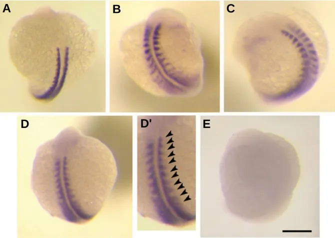
Fig. 4. Endogenous hsp90ααααα expression among zebrafish embryos. Zebrafish embryos were hybridized to DIG-labeled antisense (A-D) and sense (negative control) (E) hsp90αRNA probes. Under control conditions, zebrafish express an hsp90 signature within the developing somites (A). By contrast, upon heat shock, hsp90αwas upregulated and extended away from somitic boundaries (B-D). Embryos heat-shocked during early somitogenesis (5-8S stage) showed the greatest upregulation of hsp90α, with peak expression 2.5 hours later (B, dorsal view; C, lateral view). Development slowed or even ceased at the time of heat shock; heat-shocked embryos developed a maximum of eleven somites 2.5 hours after heat shock (D,D’) rather than the full complement of 12-15 (see Fig. 2). Scale bars: 0.30 mm in (A-D, E); 0.17 mm in (D’).
below). In particular, the paraxial somites were condensed toward the developing tail bud, thereby disturbing their subsequent contribution to trunk and tail muscle. Therefore, early segmenta-tion of the paraxial mesoderm into somites is influenced by heat shock, an association that was further investigated by in situ hybridization.
Upregulation of hsp90ααααα following heat shock
In order to determine the extent of the HSP90 stress response in an aquatic vertebrate, one-hour heat shocks, targeting discrete developmental stages, were applied to embryonic zebrafish. Under normal physiological conditions, zebrafish express an hsp90 signature within the developing somites. At the onset of the segmentation period, endogenous expression of hsp90α was limited to developing somites and, notably, was absent from the head region (Fig. 4). By 24 hours post-fertilization, endogenous hsp90α became increasingly concentrated in the caudal part of the embryos, namely at the tip of the developing tail (Fig. 5, arrow). However upon heat shock, hsp90α was upregulated and extended away from somitic boundaries (Fig. 4B-D’). Embryos
* Note: This antibody also shares a high sequence identity with Hsp90β. However, Hsp90αand Hsp90β have distinct expression patterns in Danio, Hsp90α being expressed within the somites and Hsp90βwithin the developing nervous system (Krone et al., 1997), permitting the unambiguous localization of either isoform in those regions. In addition, Western Blot analysis confirmed the specificity of this antibody in zebrafish (see Supplementary Fig. 1).
exposed to heat shock during early somitoge-nesis (5-8S stage) showed the greatest upregulation of hsp90α, with peak expression 2.5 hours after treatment. Here, the expres-sion pattern extended farther away from the central axis than among controls and became less defined where the somites had yet to form (Fig. 4B). Development slowed or even ceased at the time of heat shock; heat-shocked em-bryos expressed a maximum of eleven somites 2.5 hours after heat shock (Fig. 4, inset) rather than the full complement of 12-15 (Fig. 2). Further investigation revealed that heat shock led to a developmental lag that reflected the period of heat shock, consisting of two to three fewer somites. However, most embryos re-covered from this developmental delay within 24 hours.
Embryos continued to show increased hsp90α expression 13-16 hours after heat shock, when gene expression extended throughout the trunk from the anterior-most somite to the tip of the tail (Fig. 5B). By contrast, control embryos showed expression that was limited to the ten caudal-most somites. As zebrafish develop a total of approximately 30 somites, this represents a three-fold in-crease in the range of expression of this gene following heat stress.
Notably, hsp90α was absent within the head of both heat-shocked and control fish until the onset of eye lens development at about 24 hpf (Fig. 5). Hsp90α was not detected in heat-shocked embryos 24-72 hours after treatment among all stages under investigation (data not shown), suggesting that hsp90α mRNA is downregulated within 24 hours of environmental stress.
Immunohistochemistry staining for Hsp90ααααα
HSP90α protein expression was detected among zebrafish using an antibody that was originally developed from an Arabidopsis template that shares 100% sequence identity with zebrafish HSP90α* (Santa Cruz SC-12833, HSP90α). Here, we present to
our knowledge, the first whole-mount investigation of HSP90 for any teleost.
Overall, the protein mirrored the expression pattern high-lighted by in situ hybridization (Fig. 6). However, endogenous protein expression was limited until about 15/16S stage among fish heat-shocked at 9/10S (see Fig. 2 for corresponding stages), when expression intensified within adaxial cells – slow muscle precursor cells that lie adjacent to the notochord (Fig. 6C, arrow). HSP90 remained restricted to this region throughout somitogen-esis and was completely absent within the head. By contrast, embryos heat-shocked at all stages examined (6/7S, 9/10S and 12/13S) showed upregulation of the protein within the paraxial mesoderm. In addition, embryos heat-shocked at 9/10S stage showed reduced expression within the adaxial cells and concur-rent upregulation within the developing somites within two hours of heat shock (Fig. 6D). Expression found within the eye primordia of embryos heat-shocked at 9/10S stage (Fig. 6F) may have been
B
C
D
D'
E
due to cross-reaction with the beta isoform*. Notably, upregulation
was pronounced in the tail bud of fish two hours after heat shock at 12/13S stage (Fig. 6L) and extended throughout the trunk 14 hours after treatment (Fig. 6J).
Similarly, fourteen hours following a 6/7S heat shock, embryos expressed a protein pattern that is reminiscent of the mRNA message. Yet by 48 hours there was a striking difference between the mRNA and protein expression in 54% of heat-shocked em-bryos (N=11; Fig. 7). At a time when mRNA has been downregu-lated, the protein product was expressed in a very discrete and segmental fashion. We find five to six sequential segments expressing high levels of the protein followed caudally by an equal number of segments that did not, followed more caudally by renewed expression toward the end of the trunk (Fig. 7D). Thus, hsp90α is characterized by latent translation among fish heat-shocked during early somitogenesis.
Discussion
The interplay between the genotype and phenotype remains one of the unsolved riddles in biology. How and to what extent the genetic code is translated into developmental form has been Fig. 5. Hsp90ααααα expression following heat shock. Control (A) and 10S heat-shocked embryos 13 hours after treatment (B). Note the unchanged expression in the lens of the developing eye in contrast to the greatly expanded expression throughout the trunk from the anterior-most somite through to the tip of the tail 13 hours after heat shock (B, arrow). By contrast, control embryos show expression that is limited to the ten caudal-most somites (A, arrow). Scale bar: 0.33 mm.
Fig. 6. Immunohistochemistry staining for HSP90ααααα. Endog-enous Hsp90α(A,C,E,G,I,K) was limited during early somitogenesis (A), becoming increasingly promi-nent among adaxial cells by mid-somitogenesis; see arrow in (C). By contrast, embryos heat-shocked at all stages examined (6/7S (B); 9/10S (D) and 12/13S (F,H,J,L)) showed upregulation of the protein within the paraxial mesoderm two hours after heat shock. In addition, embryos heat-shocked at 9/10S stage showed reduced expression within the adaxial cells concomitant with in-creased expression within the paraxial mesoderm; see arrow-heads in (D). Expression also in-creased throughout the trunk and tail bud among fish heat-shocked at 12/13S stage [(F), lateral view; (H) dorsal view; (L) ventral medial view, star ] and remained extended throughout muscle segments 14 hours after treatment (J). Scale bar in (L), 0.14 mm for (A,B); 0.13 mm for (C,D); 0.16 mm for (E-H); 0.42 mm for (J,I); 0.16 mm for (K,L).
investigated over the last century, leaving much to debate. Is there a 1:1 relationship so that whatever is writ in the genes will be directly portrayed by the phenotype? Or is there some inherent flexibility within the genetic code, allowing for environmental influences to mold the final developmental outcome? Recent work has shown that HSP90 may represent the gate key that relays the genotype to the phenotype (Queitsch et al., 2002; Rutherford, 2003), suggesting the latter holds true.
HSP90 also conveys an extraordinary potential in terms of understanding lens and muscle development in fish (Hooven et al., 2004; Sass et al., 1996). Its direct application as a bioindicator among aquatic systems (Rendell and Currie, 2005) and a target
B
A
G
B
C
D
E
F
H
I
J
K
L
for cancer therapies (Kamal et al., 2004) has made HSP90 an essential component among many scientific circles. Therefore, many fields of study will benefit by examining the activity of this protein during embryonic development. Here we discuss the molecular response of this chaperone to embryonic heat shock in terms of both gene and protein expression in zebrafish.
Phenotypic variation following heat shock mirrors hsp90ααααα expression
Zebrafish embryos were exposed to heat shock during early to mid-somitogenesis to explore phenotypic variability in terms of hsp90α expression patterns. Embryos exposed to a 37°C heat shock for one hour exhibited reduced growth as well as increased variation in the posterior trunk, regions that are also characterized by upregulation of hsp90 following heat shock (Figs. 3-5).
While heat shock during early somitogenesis led to a
short-lived developmental lag with respect to control embryos, it also contributed to an extensive upregulation of hsp90 transcripts throughout the developing trunk (Fig. 4). Past work indicates that endogenous expression of hsp90α is associated with fast and slow muscle cell development as this chaperone is co-expressed and downregulated with myoD, an early muscle determinant (Sass et al., 1996). Therefore, it follows that endogenous hsp90α activity reflects the developmental rate of muscle precursors, while the comparatively diffused nature of the heat shock signal following heat shock (Fig. 4, Fig. 5) suggests that the gene is activated in other regions. Further analyses will be required to determine the long-term effects of the HSP90 stress response on somitic derivatives.
HSP90ααααα protein expression following heat shock
Here we present, to our knowledge, the first application of whole-mount immunohistochemistry for HSP90α in zebrafish. HSP90α protein expression was detected in zebrafish using an antibody that was developed from an Arabidopsis template. We followed the expression of HSP90α among the same embryonic stages examined for the mRNA message. Embryos expressed a strong HSP90α baseline pattern within adaxial cells beginning at about 15S stage, although the onset of protein expression begins as early as 12/13S stage (Fig. 2, Fig. 6). In all stages examined, heat shock lead to a prominent shift with respect to endogenous HSP90 expression, where HSP90 expression decreased within the adaxial cells and became more pronounced in the paraxial mesoderm (Fig. 6).
While the overall HSP90 protein signature reflects the expres-sion pattern highlighted by in situ hybridization, it did not parallel the response to heat shock among embryos undergoing early somitogenesis (Fig. 6), suggesting that there may be a threshold level of transcripts for translation to ensue. The endogenous expression of HSP90 also extends beyond the temporal limits of the mRNA signature, where the protein is expressed within the caudal trunk by the second day of development and retreats gradually toward the tip of the tail by 60 hpf (data not shown). By contrast, 48 hours after heat shock at 6/7S, embryos showed a segmental expression pattern within the myotome (Fig. 7). This expression pattern was found in 6/11 individuals, suggesting that HSP90α is upregulated at this stage in response to developmen-tal damage rather than being part of the developmendevelopmen-tal program. Indeed, heat shock factor-1 (HSF-1) is also upregulated in dam-aged tissue, where HSP90 uses the former as an intermediate target to maintain protein integrity (Pirkkala et al., 2001). Notably, the time required to produce 5-6 somites reflects the length of a single cell cycle in Danio (see below; Roy et al., 1999), suggesting that HSP90 plays a functional role within cell cycle pathways, linking myogenic and cyclic pathways via cellular homeostasis.
HSP90 and the cell cycle
In the context of the cell cycle, the distance within and between HSP90α expression boundaries 48 hours after a heat shock – namely 5-6 consecutive segments – is consistent with the period-icity of somitic disturbance outlined by Roy et al., (1999). In particular, a brief (30 minute) heat shock at 39-40°C can lead to enlarged (wider than normal) somites with disrupted or incom-plete boundaries and more rarely, multiple, abnormally formed boundaries. While Roy et al. (1999) described a five-somite Fig. 7. Immunohistochemistry staining for HSP90ααααα. Control (A,C) and
6/7S heat-shocked embryos (B,D,D’). Fourteen hours following heat shock (B), embryos express a protein pattern that is reminiscent of the mRNA message. By contrast, 48 hours after a heat shock targeting the same developmental stage, 54% of heat-shocked embryos (N=11) are characterized by a segmental expression pattern (D,D’). Scale bar in (D’), 0.50 mm for (A,B); 0.36 mm for (C,D) and 0.45 mm for (D’).
B
C
D
periodicity with respect to somitic disturbance, the timeline in-volved (2.5 h; one somite per 0.5 h) does not correspond to the average cell cycle during somitogenesis in zebrafish, which is reported to be four hours (Kane, 1999). However, the cell cycle during gastrulation, prior to the onset of somitogenesis, has been measured as 2.5 hours. Here, a single heat shock can have multiple repercussions, creating a periodic pattern reminiscent of that seen in chick embryos (Primmett et al., 1989).
By contrast, hsp90 (= hsp89) expression in chick does not seem to correlate with the location of segmental anomalies. According to Primmett et al., (1989), hsp expression in chick did not exhibit a periodic distribution in cross sections of somitic tissue; staining intensity did not differ among regions with somite anomalies and those without, nor was there a periodic upregulation of hsp that mimicked somitic disturbance. Therefore it was con-cluded that hsp induction was not directly responsible for segmen-tal anomalies in chick embryos exposed to cell cycle inhibitors. However, the results presented here, support a re-evaluation of hsp activity in chick or perhaps re-examination of the role of HSPs in mediating developmental change in vertebrates.
Temperature-induced differentiation in Leishmania is medi-ated by HSP90
The antibody work presented here indicates that there is greater upregulation of mRNA with respect to the protein, sug-gesting that not all the mRNA is translated. This disparity is shared with invertebrates that express HSP90 as a component of their life cycle.
Larreta et al. (2004) investigated hsp90 expression and trans-lation in Leishmania infantum, a flagellated protozoan that para-sitizes mammals via a sand fly intermediate host. Leishmania is characterized by a dimorphic life cycle that is induced by the body temperature of its secondary host. The parasite experiences a heat shock when exposed to the secondary host, a relationship that enables evaluation of the heat shock response under natural conditions.
Here hsp90 gene expression is regulated post-transcription-ally, where heat shock (here a temperature increase from 26°C to 37°C upon transfer between hosts) led to increased stability of hsp90 mRNA. Under basal cellular conditions hsp90 mRNA is readily degraded by nucleases. However, upon transfer to its warm-blooded host, the message is not degraded due to reduced nuclease activity, suggesting that protein expression is mediated by a negative feedback mechanism (Argaman et al., 1994).
More recently, Buckley et al. (2006) showed that a similar disparity occurs among gobies (Gillichthys mirabilis), where the concentration of hsp90 transcripts following stress was found to be a weak predictor of associated protein levels. Here, cDNA microarray analyses and immunohistochemistry indicate that gene and protein expression patterns can differ with respect to the relative timing and magnitude of protein production. Therefore, caution should be exercised in using only mRNA levels to assess the condition of an organism under environmental stressful con-ditions, a finding that is particularly applicable if HSPs are to be used as bioindicators of stress in nature. Moreover, the stress response should be examined from the level of the protein, as heat shock protein activity appears to extend beyond the temporal boundaries of its mRNA precursor. Further studies at the protein level will aid in bridging our understanding of the gene to protein
transition of this chaperone, especially in response to environ-mental stress.
Conclusions
Hsp90 is upregulated in response to heat shock in zebrafish embryos, with peak expression two to three hours following heat stress. HSP90 protein expression reflects the overall mRNA signature, however is limited with respect to the message during early development, becoming increasingly upregulated toward mid-somitogenesis. Following heat stress, HSP90 is expressed beyond the temporal limits of the RNA message, where it is upregulated an additional 24 hours following the downregulation of its RNA precursor.
The data presented are consistent with two interpretations; HSP90 may be upregulated in response to disrupted protein configurations, where the segmented expression that is seen 48 hours after heat shock may highlight the muscle segments that continue to show a threshold level of protein damage, thus influencing local HSP90 expression levels. Alternatively, HSP90 may serve as a regulatory mechanism for threshold-mediated developmental pathways in Danio as exemplified by Leishmania species, allowing for the expression of otherwise hidden pheno-typic variation (Rutherford, 2003). Yet it is premature to decide whether one or both hold true among vertebrates. Therefore an investigation of the long-term effects of embryonic heat shock in terms of heritable phenotypic change is currently underway, allowing a re-evaluation of the influence of environmental stress on developmental form.
Materials and Methods
Embryo maintenance
Adult zebrafish were purchased from a local pet store and maintained according to standard procedures (Westerfield, 1995). Embryos were collected from natural spawning over a bed of marbles, staged according to Kimmel et al. (1995) and raised for two weeks in 200-500 ml glass
tumblers containing double carbon-filtered tap water before being trans-ferred to 5-gallon aquaria. All fish were reared at 28.5°C and fed a mixture of prepared and live foods (Artemia franciscana) twice daily.
Heat shock procedures
Heat shock (one hour at 37ºC) was applied to a range of discrete embryonic stages from mid-gastrula to mid-somitogenesis, correspond-ing to approximately 5-16 hours post-fertilization (hpf; Kimmel et al.,
1995). Embryos were transferred to a water-filled glass tumbler that was submerged in a pre-heated water bath. The temperature within the glass vessel was continuously monitored with a digital thermometer and main-tained within +/- 0.5ºC of the targeted temperature. Following heat shock,
embryos were incubated at a standard temperature of 28.5ºC. In prepa-ration for molecular analyses, a subset of heat-shocked embryos were fixed overnight in 4% paraformaldehyde (PFA) at 2, 4, 12 and/or 16 hours after heat shock, manually dechorionated, dehydrated in methanol and stored at –20ºC.
Whole-mount in situ hybridization
Digoxigenin-11-UTP-labeled sense and antisense RNA probes (Boehringer-Mannheim) were synthesized by in vitro transcription, where
cDNA encoding an hsp90α isoform (Krone and Sass, 1994) was used as a template. In situ hybridization was conducted following standard
proce-dures (Puschel et al., 1992, as modified by Sass et al., 1996). Embryos
post-fixation in PFA, transferred to 100% MeOH and stored at –20ºC.
Whole-mount antibody staining and assay for HSP90 activity Following standard fixation in 4% paraformaldehyde (PFA), embryos were rehydrated in a PBST/MeOH series. Embryos older than 14 hpf (10 somites stage) were permeabilized in ice-cold acetone, washed in dis-tilled water and rinsed in PBST. All embryonic stages were incubated for two hours in a blocking medium consisting of 10% rabbit serum and 1% BSA. A goat polyclonal anti-HSP90α primary antibody (1:800, Santa Cruz, CA; SC-12833) was applied overnight at 4ºC in blocking solution. This primary antibody was pre-incubated with embryonic zebrafish tissue prior to its application to whole-mount antibody detection. Following antibody removal, embryos were washed several times with PBST and incubated for 1.5 hours in a 1:500 dilution of a rabbit anti-goat IgG AP-conjugated secondary antibody (Santa Cruz, CA; SC-2771). A chro-mogenic reaction (BCIP/NBT) was used to visualize the antibody.
Acknowledgments
We thank Dr. Tamara Franz-Odendaal, Dr. Gregory Handrigan and Leona Chu for their technical assistance. We would also like to thank Dr. Patrick Krone for kindly providing hsp90αcDNA. This work was funded by a Lett award to M.H.C. and an NSERC discovery grant (A5056) and funds from the Killam Trusts and the Canada Council to B.K.H.
References
AHMED, R. and DUNCAN R.F. (2004). Translational regulation of Hsp90 mRNA: AUG-proximal 5'-untranslated region elements essential for preferential heat shock translation. J. Biol. Chem. 279: 49919-49930.
ARGAMAN M., ALY R. and SHAPIRA M. (1994). Expression of heat shock protein 83 in Leishmania is regulated post-transcriptionally. Mol. Biochem. Parasitol.
64: 95-110.
BUCKLEY B. A., GRACEY A. Y. and SOMERO G. N. (2006). The cellular response to heat stress in the goby Gillichthys mirabilis: a cDNA microarray and protein-level analysis. J. Exp. Biol. 209: 2660-2677.
HOOVEN T.A., YAMAMOTO Y. and JEFFERY W.R. (2004). Blind cavefish and heat shock protein chaperones: a novel role for hsp90α in lens apoptosis. Int. J. Dev. Biol. 48: 731-738.
LARRETA R., SOTO M., QUIJADA L., FOLGUEIRA C., ABANADES D.R., ALONSO C., REQUENA J.M. (2004). The expression of HSP83 genes in Leishmania infantum is affected by temperature and by stage-differentiation and is regu-lated at the levels of mRNA stability and translation. Mol. Biol. 5: 1-18.
LATCHMAN D.S. (1999). Stress proteins: an overview. In Handbook of Experimen-tal Pharmacology, 136: Stress Proteins (Ed. D.S. LATCHMAN). Springer, NY,
pp. 1-7.
KAMAL A., THAO L., SENSINTAFFAR J., ZHANG L., BOEHM M., FRITZ L.C. and BURROWS F.J. (2004). A high-affinity conformation of Hsp90 confers tumor selectivity on Hsp90 inhibitors. Nature 425: 407-410.
KANE D.A. (1999). Cell cycles and development in the embryonic zebrafish. In
Methods in Cell Biology, 59: The Zebrafish: biology (Eds. Detrich H. W.,
Westerfeild M. and Zon L. I.) Academic Press, New York, pp. 11-26. KIMMEL C.B., BALLARD W.W., KIMMEL S.R., ULLMANN B. and SCHILLING‘T.F.
(1995). Stages of embryonic development of the zebrafish. Dev. Dyn. 203:
253-310.
KRONE P.H., SASS J.B. (1994). Hsp90α and Hsp90β genes are present in
zebrafish and are differentially regulated in developing embryos. Biochem. Biophys. Res. Commun. 204: 746-752.
KRONE P.H., SASS J.B. and LELE Z. (1997). Heat shock protein gene expression during embryonic development of the zebrafish. Cell. Mol. Life Sci. 53: 122-129.
KRONE P.H., EVANS T.G. and BLECHINGER S.R. (2003). Heat shock gene expression and function during zebrafish embryogenesis. Sem. Cell Dev. Biol.
14: 267-74.
KRUEGER A.M.R., ARMSTRONG J.N., PLUMIER J.-C., ROBERTSON H.A. and CURRIE R.W. (1999). Cell specific expression of Hsp70 in neurons and glia of the rat hippocampus after hyperthermia and kainic acid-induced seizure activ-ity. Mol. Brain Res. 71: 265-278.
MAYER M.P., BUKAU B. (1999). Molecular chaperones: the busy life of Hsp90.
Current Biology. 9: R322-325.
MORIMOTO R.I. (1994). Regulation of the heat shock transcriptional response: cross talk between a family of heat shock factors, molecular chaperones and negative regulators. Genes Dev. 12: 3788-3796.
PALMISANO A.N., WINTON J.R. and DICKHOFF W.W. (2000). Tissue-specific induction of Hsp90 mRNA and plasma cortisol response in chinook salmon following heat shock, sea water challenge and handling challenge. Mar. Bio-technol. 2: 329-338.
PICARD D. (2002). Heat shock protein 90, a chaperone for folding and regulation.
Cell Mol. Life Sci. 59: 1640-1648.
PIRKKALA L., NYKANEN P. and SISTONEN L. (2001). Roles of the heat shock transcription factors in regulation of the heat shock response and beyond.
FASEB J. 15: 1118-1131.
PRIMMETT D.R.N., NORRIS W.E., CARLSON G.J., KEYNES R.J. and STERN C.D. (1989). Periodic segmental anomalies induced by heat shock in the chick embryo are associated with the cell cycle. Development. 105: 119-130.
PÜSCHEL, A.W., GRUSS, P. and WESTERFIELD, M. (1992). Sequence and expression pattern of pax-6 are highly conserved between zebrafish and mouse. Development 114: 643-651.
QUEITSCH C., SANGSTER T.A. and LINDQUIST S. (2002). Hsp90 as a capacitor of phenotypic variation. Nature 417: 618-624.
RENDELL J.L. and CURRIE S. (2005). Intracellular localization of hsp90 is influenced by developmental stage and environmental estrogens in rainbow trout, Oncorhynchus mykiss. Physiol. Biochem. Zool. 78: 937-946.
ROY M.N., PRINCE V.E. and HO R.K. (1999). Heat shock produces periodic somitic disturbance in the zebrafish embryo. Mech. Dev. 85: 27-34.
RUTHERFORD S.L., KNAPP J.R. and CSERMELY P. (2007), Hsp90 and develop-mental networks. In Advances in experimental medicine and biology, vol. 594: Hsp90 and developmental networks (Eds. Csermely P. and L. Vigh). Springer
Science, New York, pp. 190-196.
RUTHERFORD S.L. (2003). Between genotype and phenotype: protein chaper-ones and evolvability. Nat. Rev. Genet. 4: 263- 274.
SASS J.B., WEINBERG E.S. and KRONE P.H. (1996). Specific localization of zebrafish hsp90 α mRNA to myoD-expressing cells suggests a role for hsp90α
during normal muscle development. Mech. Dev. 54: 195-204.
VAGLIA J.L. and HALL B.K. (2000). Patterns of migration and regulation of trunk neural crest cells in zebrafish (Danio rerio). Int. J. Dev. Biol. 44: 867-881.
WIECH H., BUCHNER J., ZIMMERMANN R., JAKOB U. (1992). Hsp90 chaperone protein folding in vitro. Nature 358: 169-170.
WESTERFIELD M. (1995). The Zebrafish Book. University of Oregon Press. Eugene, Oregon.
Related, previously published Int. J. Dev. Biol. articles
See our Special Issue Ear Development edited by Fernando Giraldez and Bernd Fritzsch at: http://www.ijdb.ehu.es/web/contents.php?vol=51&issue=6-7
See our Special Issue Mammalian Reproduction & Development edited by Brigid Hogan at: http://www.ijdb.ehu.es/web/contents.php?vol=45&issue=3
Blind cavefish and heat shock protein chaperones: a novel role for hsp90alpha in lens apoptosis
Thomas A. Hooven, Yoshiyuki Yamamoto and William R. Jeffery Int. J. Dev. Biol. (2004) 48: 731-738
Restricted expression of the zebrafish hsp90alpha gene in slow and fast muscle fiber lineages.
J B Sass, C C Martin and P H Krone Int. J. Dev. Biol. (1999) 43: 835-838
Hyperthermia in the chick embryo: HSP and possible mechanisms of developmental defects.
D Buckiová, L Kubínová, A Soukup, R Jelínek and N A Brown Int. J. Dev. Biol. (1998) 42: 737-740
Cell-cycle-dependent nuclear translocation of HSP70 in amphibian embryonic cells.
N Moreau, C Prudhomme and N Angelier Int. J. Dev. Biol. (1998) 42: 633-636
Does the chaperone heat shock protein hsp70 play a role in the control of developmental processes?
N Angelier, N Moreau, M L Rodriguez-Martin, M Penrad-Mobayed and C Prudhomme
Int. J. Dev. Biol. (1996) 40: 521-529
Expression of paternal and maternal mitochondrial HSP70 family, hsc74, in preimplantation mouse embryos.
P Dvorak, D Dvorakova, A Yoshiki, T Ohashi, K Kitamura and M Kusakabe
Int. J. Dev. Biol. (1995) 39: 511-517
Immunolocalization of HSP 70-related proteins constitutively expressed during Xenopus laevis oogenesis and development.
C Herberts, N Moreau and N Angelier Int. J. Dev. Biol. (1993) 37: 397-406
Supplementary Data
Supplementary Figure 1. Specificity of anti-Hsp90 antiserum. Western blot for HSP90 protein expression. The α and β isoforms could not be resolved, but the antibody is specific for zebrafish HSP90. Protein was loaded at three different concentrations, i.e. 10 µg (lanes 1-3); 15 µg (lanes 4-6) and 20 µg (lanes 7-9). Protein expression was evaluated two hours after heat shock at 72 hpf (lanes 1, 4, 7); control embryos (lanes 2, 5, 8); muscle tissue from mature fish (lanes 3, 6, 9).