S H O R T R E P O R T
Open Access
Development of an optimized method for the
detection of airborne viruses with real-time PCR
analysis
Panos G Ziros, Petros A Kokkinos, Euaggelia Legaki and Apostolos Vantarakis
*Abstract
Background:Airborne viruses remain one of the major public health issues worldwide. Detection and
quantification of airborne viruses is essential in order to provide information regarding public health risk assessment.
Findings:In this study, an optimized new, simple, low cost method for sampling of airborne viruses using Low Melting Agarose (LMA) plates and a conventional microbial air sampling device has been developed. The use of LMA plates permits the direct nucleic acids extraction of the captured viruses without the need of any preliminary elution step. Molecular detection and quantification of airborne viruses is performed using real-time quantitative (RT-)PCR (Q(RT-)PCR) technique. The method has been tested using Adenoviruses (AdVs) and Noroviruses (NoVs) GII, as representative DNA and RNA viruses, respectively. Moreover, the method has been tested successfully in outdoor experiments, by detecting and quantifying human adenoviruses (HAdVs) in the airborne environment of a wastewater treatment plant.
Conclusions:The great advantage of LMA is that nucleic acids extraction is performed directly on the LMA plates, while the eluted nucleic acids are totally free of inhibitory substances. Coupled with QPCR the whole procedure can be completed in less than three (3) hours.
Keywords:airborne viruses, air sampling, air pollution, human adenovirus, norovirus, and wastewater treatment plant
Background
Viruses are pathogenic to humans and animals. There is a growing concern regarding exposure to bioaerosols since they represent major health and economic risks to human and animal populations. Exposure to airborne viruses in different environments is responsible for various health problems and disorders worldwide [1]. Moreover, there is an increased concern of using highly pathogenic airborne microorganisms, as bioterrorism agents. Aerosolization of viral pathogens occurs in pro-cesses such as spray irrigation of wastewater and opera-tion of sewage treatment plants, but also from humans and animals as a result of coughing, sneezing, breathing, especially in indoor air environments [1-3]. The question
of airborne transmission is especially important for the waste treatment plant workers and for healthcare work-ers where patients tend to congregate during for example influenza season [4]. Many highly pathogenic viruses, such as SARS coronavirus (SARS-CoV), which causes severe acute respiratory syndrome, a highly infectious disease with a significant mortality, have the potential of being converted from droplet to airborne transmission and for that reason health care workers are particularly vulnerable [5]. Airborne spread of the SARS epidemic has been suggested for the transmission of the disease in Hong Kong in 2003 and several epidemiological studies have proposed an airborne transmission for various pathogenic viruses [6-10].
To control and prevent exposure to airborne viruses, efficient monitoring through accurate sampling is funda-mental. Many types of samplers have been used over the * Correspondence: [email protected]
Environmental Microbiology Unit, Department of Public Health, School of Medicine, University of Patras, Rion, GR 26504, Greece
years, including liquid impingers, solid impactors, filters, electrostatic precipitators, and many others [8]. In this study, we used low melting agarose (LMA) as a capture medium instead of agar based media, since preliminary spiking experiments using Tryptic Soy Agar (TSA) resulted in very pure recovery efficiencies (5-10% recov-eries, data not shown), due to inhibition of the final Q (RT-)PCRs. Agar itself has been reported to cause inhibi-tion of the PCR assay [11]. On the other hand, the use of LMA has been shown to improve the PCR amplification from templates which were naturally contaminated by various PCR inhibitors [12].
Methods
Preparation of LMA plates and air sampling
Airborne viruses were captured on LMA plates (0.5% LMA) in acidic conditions (0.05 M Glycine, pH 3.5). LMA plates were prepared by dissolving 0.5 gr of Low Melting Agarose (Invitrogen) in 100 ml of Glycine buffer, pH 3.5. The LMA solution was autoclaved for 20 min and after being cooled down to 40°C in a water bath, (2 ml) was transferred to 60 mm Petri dishes. The LMA plates were left at room temperature in order to solidify and stored at 4°C until used. Air was sampled at a pro-grammable flow rate using the Surface Air System (SAS International Pbi, Italy), an impactor sampler with con-tact agar plates. These plates were filled with 2 ml of LMA instead of agar, as described above.
Spiking experiments
Spiking experiments were performed using Human Ade-novirus 35 (HAdV-35) and Human Norovirus GII (NoV) (kindly donated by Annika Allard, Umeå University, Sweden). Virus titres of approximately 105copies/ml for HAdV, and 106copies/ml for NoV were used. In order to test the efficiency of virus recovery from LMA plates, three (3) different sets of samples were spiked with serial dilutions of 100μl viral suspensions in PBS and spread on the surface of the plates, using a 2μl eppendorf pip-ette. LMA plates were seeded with (i) HAdV containing
1 × 10⁴GC (ii) HAdV containing 1 × 10³GC (iii) HAdV containing 1 × 10² GC (Table 1). Before NA extraction, all samples were placed on SAS air sampler and air sam-pling was performed under sterile conditions for 20 min (at constant flow rate of 150 lt per minute) in a level two cell culture laminar flow. NA extraction was performed directly from the LMA plates and virus recovery was esti-mated with QPCR for HAdV. In a second set of spiking experiments, an RNA virus was tested. To this end, NoV GII was used in three different sets of samples spiked with 1 × 10⁴, 1 × 10³and 1 × 10²GC of NoV, as shown in Table 2. NA extraction was performed directly from the LMA plates and virus recovery was estimated with QRT-PCR for NoV GII.
Nucleic acid extraction
Nucleic acid (NA) extraction from LMA plates was per-formed using the NucliSENS Magnetic extraction Reagents (bioMerieux, France) according to kit instruc-tions, with the following minor modifications. In detail, 4 ml of NucliSENS lysis buffer were added to each LMA plate. Upon addition of lysis buffer LMA was easily dis-solved. Then the lysate was transferred to 15 ml falcon tube, briefly vortexed and incubated for 10 min at 55°C in a water bath. Afterwards, 50μl of well-mixed magnetic silica were added to the lysate and the instructions of the manufacturer were followed until the final step of NA elu-tion. NAs were finally extracted in 50μl of elution buffer, and extracts were stored at -20°C until analyzed by QPCR. It is worthy of note that any commercial or homemade nucleic acid extraction reagent using a lysis buffer based on GuSCN can be used.
Real time Q(RT-)PCR analyses
The quantitative real-time PCR (QPCR) for human Ade-novirus based on the method described by Hernroth and colleagues [13], was performed in a final volume of 25μl, using the TaqMan Universal PCR master mix (Invitro-gen). The sensitivity of this QPCR HAdV assay has been estimated to be of 1-10 genome copies [13,14]. The
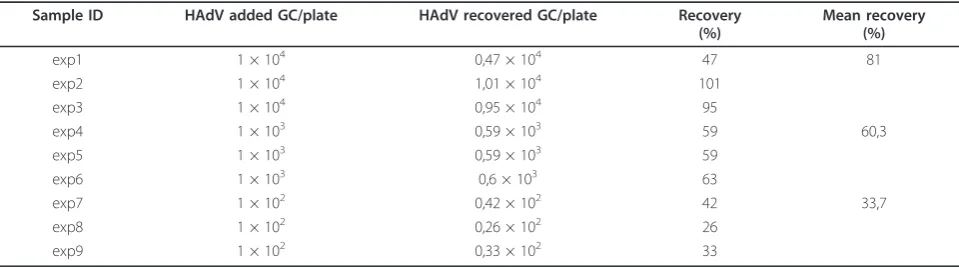
Table 1 Recovery values of HAdV 35 seeded in LMA plates
Sample ID HAdV added GC/plate HAdV recovered GC/plate Recovery
following oligonucleotide primers and conditions were
used. Forward primer: AdF (5’- CWT ACA TGC ACA
TCK CSG G-3’), at a final concentration of 0.9 μM,
Reverse primer: AdR (5’- CRC GGG CRA AYT GCA
CCA G-3’), at a final concentration of 0.9μM,
Adeno-virus TaqMan Probe: AdP1 (5’- FAM- CCG GGC TCA
GGT ACT CCG AGGCGT CCT-BHQ-3’), at a final
con-centration of 0.225μM. The temperature-time program
was as follows: 2 min at 50°C, 10 min at 95°C as a hot start, and 45 cycles of 15 s at 95°C for denaturation, 1 min at 60°C for denaturation, annealing-extension. Stan-dard curves used in the QPCR were generated by using serial dilutions of purified viral genomic DNA from ade-novirus 35 (HAdV-35) viral stocks.
The quantitative real-time RT-PCR (QRT-PCR) for human Norovirus GII (detection limit < 10 copies per reaction) based on a method described previously [15], was performed in a final volume of 20μl, using the RNA
UltraSense™ One-Step Quantitative RT-PCR System
(Invitrogen). The following primers and conditions were
used. Forward primer: QNIF2 (5’- ATG TTC AGR TGG
ATG AGR TTC TCW GA -3’), Reverse primer: COG2R
(5’- TCG ACG CCA TCT TCA TTC ACA -3’),
Noro-virus GG II Probe: QNIFS (5’- FAM- AGC ACG TGG
GAG GGC GAT CG -BHQ1-3’). The temperature-time
program was as follows: 15 min at 50°C for the RT reac-tion, 10 min at 95°C as a hot start, and 45 cycles of 15 s at 95°C for denaturation, 1 min at 60°C for denaturation, annealing-extension. Standard curves used in the QRT-PCR were generated by using serial dilutions of purified viral genomic RNA from NoV GII viral stocks.
Internal amplification controls (IAC) for human Ade-novirus (YORKBIO Cat# IAC-HADV, UK) and Noro-virus GII (YORKBIO Cat# IAC-NVGII, UK) were used in order to test the eluted nucleic acids for the presence of inhibitory substances. The following TaqMan probe
was used, IAC probe: IACP (5’-VIC-CCA TAC ACA
TAG GTC AGG -MGBNFQ-3’), while the primers and
conditions were the same to those for HAdV and NoV-GII QPCRs, respectively.
Standard precautions were applied in all Q(RT-)PCR assays, such as use of separate work areas and pipettes for pre- and post-amplification steps.
Outdoor experiments
In order to test the LMA method of air sampling under field conditions, air sampling experiments were performed in an urban area of the city of Patras, located in south-western Greece, and in the sewage treatment plant of Patras’University (SWPT). Samplings at the SWPT were performed during a two months period of summer 2010 at the plant’s Aerobic Wastewater Mechanical Aeration Treatment System. Each air sampling was performed for 20 min, which corresponds to 3000 lt of air at a distance of 1 m from the sewage tank, except for two samples which where collected outside the SWTP facilities, at a distance of approximately 50 m from the sewage tank (Table 3). This SWTP operates periodically every 3 hours for 30 min. Samplings were performed under both condi-tions, during operation or immediately after the end of an operation cycle (Table 3). After sampling, the LMA plates were processed on the same day for NA extraction. NA extracts (50μl) were stored at -20 C until analyzed.
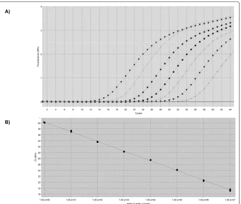
QPCR for HAdV detection and quantification were performed under the same experimental conditions described previously, except for the construction of the standard curve which was generated by using serial dilu-tions (range 1 to 107) of known amounts of a linearized plasmid containing the entire hexon region of HAdV 41 (kindly donated by Annika Allard, Umeå University, Sweden) (Figure 1).
Results
Efficiency of viral recovery from LMA plates
HAdV recovery from LMA plates was estimated with QPCR. Mean values of three independent experiments for the determination of viral recovery were 81% (for
the samples spiked with 1 × 10⁴ GC), 60.3% (for the
samples spiked with 1 × 10⁴ GC), and 33.7% (for the
samples spiked with 1 × 10²GC) (Table 1).
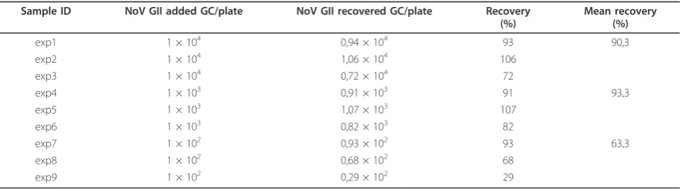
Table 2 Recovery values of NoV GII seeded in LMA plates
Sample ID NoV GII added GC/plate NoV GII recovered GC/plate Recovery
Table 3 Detection and quantification of Human Adenovirus in the air of SWTP
Sample ID SWTP operation Adenovirus load per 3000 lt of air Distance from the tank (m)
A3559 yes 384 1
A3560 yes 116 1
A3561 no 39 1
B3568 no 6,5 1
B3569 no ND 1
A3602 yes 75 1
A3603 yes 79 1
A3604 no 6,7 1
C3622 no 69 1
C3624 yes 1017 1
C3623 no 83 1
H3761 yes 34 50
H3762 no ND 50
Detection and quantification of Human Adenovirus in the air of a SWTP using the LMA method and QPCR. Sample IDs with the same initiating letter were sampled during the same day. (ND: Not detected)
A)
B)
NoV GII recovery from LMA plates was estimated with QRT-PCR. The mean viral recovery values of three independent experiments were 90.3% (for the samples spiked with 1 × 10⁴ GC), 93.3% (for the samples spiked
with 1 × 10⁴ GC), and 63.3% (for the samples spiked
with 1 × 10²GC) (Table 2).
The NA extracts from the field experiments were tested for inhibition against control samples of pure NA elution buffer, with the use of previously described IACs. No statistically significant difference was observed between the LMA samples and the control samples. Regarding HAdV, the mean Ct value for LMA samples was 34.4 (SD 0.44), while the Control Ct was 34.4 (SD 0.39). For the NoVGII analyses, the mean Ct values for the LMA samples was 30.53 (SD 0.27), while the Control Ct value was 30.29 (SD 0.38).
Detection of viruses in outdoor experiments
As it is shown in Table 3, HAdV were detected in ele-ven (11) out of thirteen (13) samples collected at the SWTP. At the Aerobic Wastewater Mechanical Aeration Treatment System the results indicated a considerable airborne viral pollution, with HAdV detected in 84.6% of the tested samples. Interestingly, 100% of the samples were found to be positive when the SWTP was in operation. HAdV were not detected in anyone of the samples (six samples) originating from the urban area of the city. QRT-PCR for Norovirus GII detection were also performed in the same samples. No positive NoV GII samples were detected, neither to the urban area, nor to the wastewater treatment plant.
Discussion and conclusions
The results of the present study showed that Low Melt-ing Agarose (LMA) can be efficiently used as a capturMelt-ing medium of airborne viruses in solid impactor air sam-plers. Usually, sampling with this type of air samplers is performed using agar based media [8]. The drawback of agar plates is that viruses have to be eluted from the agar plates before the subsequent nucleic acid extraction step, which results in viral losses. Moreover, inhibitory sub-stances presented in the agar media tend to remain dur-ing the subsequent NA isolation and finally inhibit the Q (RT-)PCR reactions, thus considerably reducing the detection limit of airborne viruses. In contrary, the great advantage of LMA is that NA extraction is performed directly on the LMA plates, while the eluted NA is totally free of inhibitory substances. Coupled with QPCR the whole procedure can be completed in less than three (3) hours.
In accordance to the findings of others studies, which support that the wastewater treatment plants are sources of considerable airborne contamination, which may pollute the environment and constitute an
important biological hazard for workers [2,16,17], the present study revealed a significantly high level of viral contamination during the operation of the studied SWTP.
We intend to apply this optimized method for the detection of airborne viruses in indoor and outdoor environments.
List of abbreviations
HAdV: human adenovirus; NoV: norovirus; LMA: low melting agarose; SWTP: sewage treatment plant; QPCR: quantitative real time PCR; GC: genome copies; NA: nucleic acids.
Authors’contributions
PZ was responsible for setting up the study, performed the samplings at the SWTP, and participated in the molecular analyses and in the writing of the manuscript. PK performed the samplings at the urban areas, and
participated in the writing and revision of the manuscript; EL performed the nucleic acid extractions, while AV was responsible for coordinating the study, and drafted the manuscript. All authors read and approved the final manuscript.
Competing interests
The authors declare that they have no competing interests.
Received: 28 June 2011 Accepted: 27 July 2011 Published: 27 July 2011
References
1. Douwes J, Thorne P, Pearce N, Heederik D:Bioaerosol health effects and exposure assessment: progress and prospects.Ann Occup Hyg2003,
47(3):187-200.
2. Carducci A, Tozzi E, Rubulotta E, Casini B, Cantiani L, Rovini E, Muscillo M, Pacini R:Assessing airborne biological hazard from urban wastewater treatment.Water Research2000,34(4):1173-1178.
3. Srikanth P, Sudharsanam S, Steinberg R:Bio-aerosols in indoor environment: composition, health effects and analysis.Indian J Med Microbiol2008,26(4):302-12.
4. Lindsley WG, Blachere FM, Thewlis RE, Vishnu A, Davis KA, Cao G, Palmer JE, Clark KE, Fisher MA, Khakoo R, Beezhold DH:Measurements of airborne influenza virus in aerosol particles from human coughs.PLoS One2010,
5(11):e15100..
5. Hui DS, Chan PK:Severe acute respiratory syndrome and coronavirus.
Infect Dis Clin North Am2010,24(3):619-638.
6. Hammond GW, Raddatz RL, Gelskey DE:Impact of atmospheric dispersion and transport of viral aerosols on the epidemiology of influenza.Rev Infect Dis1989,11(3):494-497.
7. Marks PJ, Vipond IB, Regan FM, Wedgwood K, Fey RE, Caul EO:A school outbreak of Norwalk-like virus: evidence for airborne transmission.
Epidemiol Infect2003,131(1):727-736.
8. Verreault D, Moineau S, Duchaine C:Methods for sampling of airborne viruses.Microbiol Mol Biol Rev2008,72(3):413-444.
9. Yu IT, Li Y, Wong TW, Tam W, Chan AT, Lee JH, Leung DY, Ho T:Evidence of airborne transmission of the severe acute respiratory syndrome virus.
N Engl J Med2004,350(17):1731-1739.
10. Yu IT, Wong TW, Chiu YL, Lee N, Li Y:Temporal-spatial analysis of severe acute respiratory syndrome among hospital inpatients.Clin Infect Dis 2005,40(9):1237-1243.
11. Gibb AP, Wong S:Inhibition of PCR by agar from bacteriological transport media.J Clin Microbiol1998,36(1):275-276.
12. Moreira D:Efficient removal of PCR inhibitors using agarose-embedded DNA preparations.Nucleic Acids Res1998,26(13):3309-3310.
13. Hernroth BE, Conden-Hansson AC, Rehnstam-Holm AS, Girones R, Allard AK:
Environmental factors influencing human viral pathogens and their potential indicator organisms in the blue mussel, Mytilus edulis: the first Scandinavian report.Appl Environ Microbiol2002,68(9):4523-4533. 14. Bofill-Mas S, Calgua B, Clemente-Casares P, La Rosa G, Iaconelli M,
adenoviruses in European recreational waters.Food Environ Virol2010,
2(2):101-109.
15. Kageyama T, Kojima S, Shinohara M, Uchida K, Fukushi S, Hoshino FB, Takeda N, Katayama K:Broadly reactive and highly sensitive assay for Norwalk-like viruses based on real-time quantitative reverse transcription-PCR.J Clin Microbiol2003,41(4):1548-1557.
16. Brandi G, Sisti M, Amagliani G:Evaluation of the environmental impact of microbial aerosols generated by wastewater treatment plants utilizing different aeration systems.J Appl Microbiol2000,88(5):845-852. 17. Fracchia L, Pietronave S, Rinaldi M, Giovanna Martinotti M:Site-related
airborne biological hazard and seasonal variations in two wastewater treatment plants.Water Res2006,40(10):1985-1994.
doi:10.1186/1743-422X-8-369
Cite this article as:Ziroset al.:Development of an optimized method for the detection of airborne viruses with real-time PCR analysis. Virology Journal20118:369.
Submit your next manuscript to BioMed Central and take full advantage of:
• Convenient online submission
• Thorough peer review
• No space constraints or color figure charges
• Immediate publication on acceptance
• Inclusion in PubMed, CAS, Scopus and Google Scholar
• Research which is freely available for redistribution