M E T H O D O L O G Y
Open Access
Development of cost-effective real-time PCR test:
to detect a wide range of HBV DNA concentrations
in the western amazon region of Brazil
Alcione de Oliveira dos Santos
1,2,3*, Luan Felipo Botelho Souza
1,2,3, Lourdes Maria Borzacov
2,
Juan Miguel Villalobos-Salcedo
1,2,3and Deusilene Souza Vieira
1,2,3Abstract
Background:Currently there is a significant risk of infection with hepatitis B virus (HBV) during blood transfusion in high epidemic area. This is due to the pre-seroconversion window period, immunovariant viral strains and the presence of occult HBV infection (OBI). The aim of this study was to develop an in-house real-time PCR-based method, which was both ultra-sensitive and efficient offering an alternative method for nucleic acid testing (NAT). Methods:A precore fragment with 109 bp was cloned and serial diluted to standard curve construction. The calibration of the HBV - DNA values was performed against OptiQuant® HBV-DNA Quantification Panel, Acrometrix Europe B.V.).
Results:From our in-house plasmid we prepared serial dilutions ranging from 2 × 10³–2 × 10⁹copies/ml. The threshold was adjusted automatically during analysis and the data collected were analyzed by linear regression (r2= 0.99). The limit of detection for the assay with pHBVRO standards was 2000/ml in a total reaction volume of 30μl. We found a strong correlation between the two methods (r2= 0.9965 and p < 0.0001). The regression line give us the following equation: Log 10 (IU/mL) = 0.9038Log 10 (copies/mL)−1.0643, suggesting that 1 IU/mL = 15 copies/mL.
Conclusions:Therefore, we can affirm that the qHBVRO PCR can detect HBV DNA in individuals with hepatitis B at any stage of the disease showing high capacity for NAT screening in hepatitis b donors. This results of sensitivity could provide an advance for automation in blood banks and increasing safety of patients who receive blood transfusions.
Introduction
The hepatitis B virus (HBV) is one of the most common human pathogens and can cause hepatitis and aggressive and advanced liver disease, including cirrhosis and hepato-cellular carcinoma [1]. Despite the availability of a vaccine, the implementation of preventive measures and serological screening in blood banks remains a major public health problem worldwide [2]. HBV can be transmitted peri-natally, percutaneously, sexually or by horizontal transmis-sion, especially among children, presumably through open cuts or sores [3].
Early detection of HBV surface antigen (HBsAg) signifi-cantly reduces the risk of infection through blood transfu-sions [4]. However, there are two situations in the course of infection where this early detection is currently ineffective: First, during the acute phase of infection, there is a window period where HBsAg may be undetectable in serum [5]. In another situation, occult infection which is defined as the presence of HBV DNA in the liver (with or without detect-able serum HBV DNA) may be present during the persist-ent of infection in subjects who test negative for hepatitis B surface antigen (HBsAg). These subjects often have very low viral load (< 200UI/ml) [6]. Reducing the risk of trans-mission in these situations will require increased sensitivity the detection of HBV surface antigen (HBsAg), screening for antibodies to HBV core antigen (anti-HBc), and contin-ued testing and implementation of NATs [7,8].
* Correspondence:[email protected]
1Fundação Oswaldo Cruz, Rondônia-FIOCRUZ, Porto Velho, Rondonia, Brazil 2
Centro de Pesquisa em Medicina Tropical de Rondônia-CEPEM, Porto Velho, Rondonia, Brazil
Full list of author information is available at the end of the article
Real-time polymerase chain reaction (qRT-PCR) has enabled the development of improved diagnostic tests offering greater speed while maintaining excellent levels of sensitivity and specificity [9-11]. qRT-PCR-based detection methods have been developed for the diagnosis of HBV and other pathologies in clinical la-boratories [12-14].
To successfully monitor viral load, it is important to diagnose viral replication, establish the prognosis of liver disease, to assess the risk of disease progression, to iden-tify patients who need antiviral therapy and to monitor the virologic response to treatment. Currently there are several types of detection and quantitation assays in use, with varying levels of success [15,16]. The aim of this study was to develop an in-house real-time PCR-based method, which was both ultra-sensitive and effective, of-fering a new NAT alternative.
Materials and methods Clinical samples
This study included 134 patients with chronic HBV in-fection who were treated at the Viral Hepatitis Clinic Specialized Center of Research in Tropical Medicine in Rondonia (CEPEM). A control group of 30 donors, who all tested negative por ELISA for human immunodefi-ciency virus (HIV) 1 and 2, HBsAg, HBc and anti-HCV, and who attended the blood bank of the State of Rondonia (FHEMERON) was included in the study. We also included 10 and 26 serum samples from individuals with chronic HCV and co-infection with HBV/HDV respectively.
Ethical consent
This study was approved by the Brazilian Institutional Ethics Committee of the Centro de Pesquisa em Medicina Tropical (CEPEM), with process number 107/10. Written, informed consent was obtained from each patient for the publication of this manuscript and any accompanying images.
DNA extraction
Viral DNA extraction was performed using the QIAamp DNA Mini Kit (Qiagen, Hilden, Germany) and 200μl of serum according to the manufacturer’s instructions. Three samples with viral load known were tested: first with high viral load and the others medium and low viral load. After this were diluted with final volume 200 ul, 100 ul and 50 ul. Besides, HBV DNA was extracted from 22 samples of individuals with same profile serological: total isolated anti-HBc. These samples were diluted in 50 uL and 200 uL to optimize the final volume of extraction for samples with low viral load. Subsequently the samples were submitted to reaction for sensitivity analysis. Precipi-tated DNA was resuspended in elution buffer and stored
at−20°C until further use. To avoid false-positive results, we followed strict procedures for nucleic acid amplifica-tion [17].
In-house testing
Primer concentrations were optimized using a concen-tration gradient ranging from 100–900 nM and SYBR® Universal PCR Master Mix (Applied Biosystems, Foster City, CA, USA). TaqMan® probe concentrations were simi-larly optimized using a concentration gradient ranging from 50–300 nM.
Ultra-sensitive real-time PCR
The assay was performed on an ABI 7500 platform (Applied Biosystems) with 30 μl reaction volumes containing 15 μl TaqMan® Universal Master Mix (Applied Biosystems), 3μl
HBVRO1 forward primer (5′
-AGGAGGCTGTAGGCAT-AAATTGG 3′), 3μl reverse primer (5′-GCACAGCTTGG AGGCTTG-3′), 0.6 μl probe (5′-FAM TCACCTCTGCC TAATC-3′-MGB, 6μl extracted DNA and 2.4μl of water.
Construction of the standard curve
To construct the standard curve, we initially used conven-tional PCR with amplification of a 109 bp fragment in the pre-core region according Kavita 2006 adapted, selecting five samples with known viral load. Approximately 50 ng of DNA was used per reaction with a final volume of 50μl. Amplification was performed on an ABI Prism 7500 Veriti (Applied Biosystems) with an initial denaturation temperature of 94°C for 5 min, followed by 40 cycles of 94°C for 1 min, 58°C for 45 sec, 72°C for 1 min and a final extension of 10 min at 72°C. The selected fragment was purified, ligated to the p-GEM-T Easy® vector (Promega, New York, USA), cloned into a prokaryotic system and subsequent linearization withPstI (Invitrogen™Life Tech-nologies, Carlsbad, CA, USA). Absolute quantitation was used to determine the exact number of DNA molecules for estimating viral load.
Inter- and intra-assay variation and reproducibility of real-time PCR
To determine intra-experimental variation, we tested the reproducibility of six HBV-positive sera with different viral loads, in duplicate, in the same reaction setup. The same set of samples was used in three experiments per-formed on different days, to estimate inter-experimental variation in the estimation of viral load. Reproducibility was estimated by calculating the coefficient of variation (CV), which is calculated as the ratio of the standard de-viation and the mean of the replicates.
HBV DNA quantification
(AcroMetrix Europe BV). Specifically, a serial dilution was prepared from the standards included in the kit, ranging from 2 × 10²– 2 × 10⁶IU/ml. For our in-house plasmid, pHBVRO, a serial dilution was prepared with a range 2 × 10³–2 × 10⁹copies/ml. The concentration was measured spectrophotometrically both by using a NanoDrop® ND-1000 (Thermo Scientific NanoDrop Products, Wilming-ton, Delaware), and the measurements were recorded in units of nanograms per microliter, which was converted into copies per microliter by using the following equation: ([x ng/μL × 10-9] / [p-GEM-T Easy® vector and 109 HBV DNAbps × 660]) × 6.022e23= y copies/μL. Using linear regression a standard curve was constructed, which was used to convert copies/ml to standard international units (IU/ml).
Quantification panel
To compare the performance of our in-house method (qHBVRO) with that of the commercial kit, OptiQuant® HBV-DNA Quantification Panel (AcroMetrix® Europe BV), we tested 100 serum samples collected from pa-tients chronically infected with HBV.
Statistical analysis
The correlation between the AcroMetrix® test kit and the qHBVRO assay was calculated using GraphPad 5.0 (Graph-Pad software) and a two-tailed Pearson′s correlation test with a confidence interval of 95%. The units for measuring viral load (copies/ml and IU/ml) were transformed to log base 10.
Analytical specificity
We tested 30 samples from blood donors, 10 serum sam-ples from mono-infected HCV patients, 28 samsam-ples from patients co-infected with HBV/HDV and 15 samples that were judged indeterminate for HBV surface antigen by the Serology Laboratory of Viral Hepatitis Clinic - IPEPATRO. All samples were submitted to qHBV PCR to determine viral load.
Analytical sensitivity
We selected 15 sera, which were considered indetermin-ate for HBsAg by ELISA when tested in duplicindetermin-ate and which had absorbance values within the gray zone, or ± 10% cut-off confidence interval. These samples were subjected to three separate assays, with each sample per-formed in duplicate to evaluate the performance of our assay in detecting uncertain samples.
Results
Analytical sensitivity and efficiency
[image:3.595.56.292.89.262.2]From our in-house plasmid we prepared serial dilutions ranging from 2 × 10³ – 2 × 10⁹ copies/ml. The concen-tration of primers used was 300 nM, for both primers, and 100 nM for the probe. The threshold was adjusted automatically during analysis and the data collected were analyzed by linear regression (r2= 0.99), Figure 1. The limit of detection for the assay with pHBVRO standards was 2000/ml in a total reaction volume of 30 μl. Two positive samples with known viral load were used as in-ternal controls. HBV DNA extraction was patterned on elution of 200 μl because there was no significant vari-ation in sensitivity of method in different volumes of patients with intermediate and high viral load. But the 22 samples of patients with serological profile“anti-HBc
Table 1 Intra-experimental variation in the qHBVRO assay for serum samples qHBV RO - intra assay
Sample 1st run (IU/mL) 2nd run (IU/mL) 3th run (IU/mL) Avarage inter assay SD CV
1 4.6 × 10⁶ 4.6 × 10⁶ 4.6 × 10⁶ 4.6 × 10⁶ 0.027 × 10⁶ 0,01
2 2.8 × 10⁴ 2,7 × 10⁴ 2.7 × 10⁴ 2.7 × 10⁴ 0.025 × 10⁴ 0,01
3 5.0 × 10² 4.3 × 10² 4.6 × 10² 4.6 × 10² 0.36 × 10² 0,08
4 3.2 × 10² 2.9 × 10² 2.7 × 10² 3.0 × 10² 0.26 × 10² 0,09
5 3.3 × 10⁵ 3.1 × 10⁵ 3.0 × 10⁵ 3.2 × 10⁵ 0.16 × 10⁵ 0,05
[image:3.595.58.540.624.733.2]6 4.8 × 10⁴ 4.0 × 10⁴ 4.3 × 10⁴ 4.4 × 10⁴ 0.38 × 10⁴ 0,09
total isolated” eluted in 50 and 200 uL, 15 samples were negative on both elution, 5 samples were positive only in elution of 50 uL within which 2 samples were positive in both elution.
Inter- and intra-assay variation and reproducibility of real-time PCR
The six HVB-DNA positive sera tested showed no statis-tical differences between repeats. The coefficient of vari-ation was similar in both high and low viral load samples (0.01-0.16%), indicating the same efficiency of amplification for varying viral loads. There was no statistical difference in intra-and inter-assay variation (CV), which confirmed the reproducibility of the assay (Tables 1 and 2).
Validation of the qHBVRO method
Using linear regression, we found a strong correlation between the qHBVRO assay and the AcroMetrix® HBV-DNA kit (r2= 0.998 and p <0.0001) as shown in Figure 2. Viral load values between the AcroMetrix® HBV-DNA and the qHBVRO assay were compared for 134 patients by Pearson’s correlation. We found a strong correlation between the two methods (r2= 0.9965 and p < 0.0001). The regression line give us the following equation:
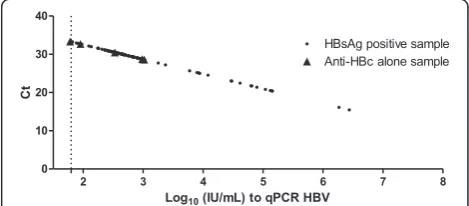
Log 10 (IU/mL) = 0.9038Log 10 (copies/mL)−1.0643, suggesting that 1 IU/mL = 15 copies/mL (Figure 3).
Analytical specificity and detection performance of indeterminate for ELISA samples and anti-HBc isolated samples
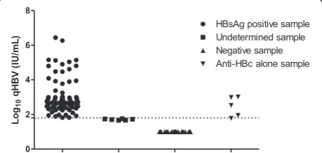
Of the 15 indeterminate for ELISA samples tested, six were positive for HBV DNA by both methods (Table 3). All samples that were classified as negative by ELISA were confirmed as such as shown. Of the 22 samples isolated anti-HBc eluted in 50 uL, 5 were positive (Figure 4).
Discussion
[image:4.595.56.540.101.211.2]Currently, there is a significant risk of infection with hepa-titis B virus (HBV) during blood transfusions. This is due to the pre-seroconversion window period, immunovariant viral strains and the presence of occult HBV infection (OBI) [8,18,19]. Combined detection of HBsAg and anti-HBc constitutes an important strategy in donor screening which excludes the vast majority of OBIs [20-22]. How-ever, in many countries, especially in areas of low HBV prevalence (< 3%) these strategies are inefficient especially during the pre-seroconversion window period. However, in countries with high prevalence of positivity for
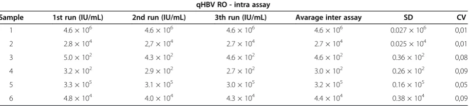
[image:4.595.57.291.534.703.2]Figure 2WHO 1st International Standard (HBV ® kit Acrometrix DNA) as determined by linear regression.
[image:4.595.304.539.562.665.2]Figure 3Analytical sensitivity of qPCR HBV demonstrated with 97 samples: 91 samples positive for HBsAg and 6 samples total anti-HBc isolated.These 6 samples were DNA-HBV positive detected within 50 clinical samples tested in patients with profile total anti-HBc isolated. The dashed line is the limit of analytical detection of qPCR HBV.
Table 2 Inter-experimental variation in the qHBVRO assay for serum samples
qHBV RO - inter assay
Sample 1st run (IU/mL) 2nd run (IU/mL) 3th run (IU/mL) Avarage inter assay SD CV
1 4.6 × 10⁶ 5.0 × 10⁶ 4.6 × 10⁶ 4.7 × 10⁶ 0.02 × 10⁶ 0,05
2 2.7 × 10⁴ 3.4 × 10⁴ 27 × 104 2.9 × 10⁴ 0.004 × 10⁴ 0,14
3 4.3 × 10² 4.6 × 10² 4.0 × 10² 4.3 × 10² 0.3 × 10² 0,07
4 3.2 × 10² 2.7 × 10² 3.0 × 10² 3.0 × 10² 0.2 × 10² 0,08
5 3.3 × 10⁵ 3.0 × 10⁵ 2.8 × 10⁵ 3.0 × 10⁵ 0.2 × 10⁵ 0,08
anti-HBc, using this marker could involve both the exclusion of several potential donors leading to a de-crease of blood products in various regions - such as the failure to block potential donors with OBI - keeping present the risk of post-transfusion HBV [18,23]. The qHBVRO test presents an excellent alternative to HBV detection and quantification because it allows significant reduction in the risk of transmission during the window period as well as improved detection of occult HBV.
In this study we developed a qRT-PCR assay for the identification and quantification of HBV DNA with an ef-ficiency of 94.06% and good correlation with the currently used commercial test: WHO 1st International Standard (HBV ® kit Acrometrix DNA), r = 0.998, p < 0.0001. The qHBVRO PCR test was 100 fold more sensitive, allowing detection of up to two thousand copies per ml of serum in HBV-infected individuals. Our method also proved to be more sensitive than other in-house qRT-PCRs [24-26], and could detect occult hepatitis B infections as well as cases which were inconclusive by ELISA, using only 6μl of DNA extracted from 200 μl of serum in final reaction volume of 30μl for qHBVRO. However, was observed that in cases of samples with low viral load is important to consider a smaller volume in the elution of DNA. It is the case of 22 samples tested from individuals with isolated anti-HBc where 5 were positive in 50 uL elution and only 2 were positive on PCR qHBV. These results reinforce the importance that in case of occult infection or immunological
window period, the concentration of DNA is an important factor to consider.
The high analytical specificity of the test, using sam-ples from individuals that tested positive, negative and indeterminate for HBsAg by ELISA demonstrates that qHBVRO PCR can detect HBV DNA in individuals with hepatitis B at any stage of the disease, qualifying it as an important alternative NAT. The qHBVRO assay is highly reproducible, with low intra- and inter-experimental variation of between 0-1% (CV), whereas other in-house tests, which are considered to have good reproducibility, show experimental variation of 4.94–10.59% [27-31].
Our method proved to be efficient, sensitive, specific and reproducible in the detection of occult HBV, and could therefore be used for nucleic acid testing (NAT) in blood banks to prevent HBV transmission by blood trans-fusion. The advantages of NAT relating to cost and effect-iveness compared with serological diagnostics have been widely debated [28-30]. It has been suggested that NAT offers advantages in many instances including occult infec-tions, in the confirmation of viremia, for screening blood-and organ-donors, discriminating between patients with chronic or acute infection which has been resolved, diagnosis of perinatal infection, solving indeterminate serological results, monitoring patients on antiviral therapy and to identify the virus in immunocomprom-ised individuals [31-33].
Conclusion
[image:5.595.56.545.100.198.2]In conclusion, the real-time PCR assay qHBVRO is ap-propriate for the quantification of HBV DNA in serum samples. This test is reproducible and proved be sensi-tive detecting samples with low viral load. Therefore, we can affirm that the qHBVRO PCR can detect HBV DNA in individuals with hepatitis B at any phase of disease showing good NAT screening for hepatitis B. Samples of patients anti-HBc positive isolated were selected and submitted to qHBVRO test to enhancing sensitivity the this results. This developed test may be automated and used in blood banks, increasing safety of patients who receive blood transfusions.
Table 3 Data summary for indeterminate samples
1st run (Ct*) 2nd run (Ct*) 3th run (Ct*) Mean SD CV Log10IU/ml DO* Cut-off
1 37.31 37.56 37.74 37.54 0.21595 1% 0.69 0.086 0.062
2 38.90 38.98 39.1 38.99 0.10066 0% 0.31 0.075 0.079
3 38.69 38.83 38.95 38.82 0.13013 0% 0.35 0.070 0.063
4 37.47 37.54 37.87 37.63 0.21362 1% 0.66 0.062 0.065
5 39.05 39.1 39.45 39.20 0.21794 1% 0.25 0.060 0.068
6 38.94 39.03 39.26 39.08 0.16503 0% 0.28 0.069 0.068
DO*: Optical Density by spectrophotometry (ELISA). Ct*: Amplification cycle in the assay qHBVRO.
[image:5.595.58.290.585.695.2]Competing interests
The authors declare that they have no financial or competing interest with this article.
Authors’contributions
AOS participated in the design of the study, drafted the manuscript and in its design and coordination. LFBS participated in the PCR amplification and sequencing process. DSV participated in the design of the study. LMB participated in the elaboration of the manuscript. JMS participated in the design of the study. All authors read and approved the final manuscript.
Acknowledgments
This work was supported by IPEPATRO, CEPEM, Universidade Federal de Rondônia and Fundação Oswaldo Cruz- Rondônia. Our thanks to Larissa Deadame F Nicolete by collaboration in the statistical analyzes.
Author details
1
Fundação Oswaldo Cruz, Rondônia-FIOCRUZ, Porto Velho, Rondonia, Brazil.
2Centro de Pesquisa em Medicina Tropical de Rondônia-CEPEM, Porto Velho,
Rondonia, Brazil.3Universidade Federal de Rondonia-UNIR, Porto Velho, Rondonia, Brazil.
Received: 3 June 2013 Accepted: 14 January 2014 Published: 28 January 2014
References
1. Ganem D, Prince AM:“Hepatitis B virus infection–natural history and clinical consequences”.N Engl J Med2004,350:1118–1129.
2. Kitab B, Essaid EL Feydi A, Afifi R, Trepo C, Benazzouz M, Essamri W, Zoulim F, Chemin I, Alj HS,et al:“Variability in the precore and core promoter regions of HBV strais in Morocco: characterization and impact on liver diasease progression.PLoS One2012,7:e42891.
3. Bauer T, Sprinzl M, Protzer U:Immune control of hepatitis B virus.Dig Dis 2011,29:423–433.
4. Kuhns MC, Kleinman SH, McNamara AL, Rawal B, Glynn S, Busch MP:“Lack of correlation between HBsAg and HBV DNA levels in blood donors who test positive for HBsAg and anti-HBc: implications for future HBV screening policy”.Transfusion2004,44:1332–1339.
5. Datta S, Banerjee A, Chandra PK, Chakraborty S, Basu SK, Chakravarty R:
“Detection of a premature stop codon in the surface gene of hepatitis B virus from an HBsAg and antiHBc negative blood donor”.J Clin Virol 2007,40:255–258.
6. Raimondo G, Navarra G, Mondello S, Costantino L, Colloredo G, Cucinotta E, Di Vita G, Scisca C, Squadrito G,et al:“Occult hepatitis B virus in liver tissue of individuals without hepatic disease”.J Hepatol2008,48:743–746. 7. Saldanha J:Validation and standardisation of nucleic acid amplification
technology (NAT) assays for the detection of viral contamination of blood and blood products.J Clin Virol2001,20:7–13.
8. Candotti D, Allain JP:“Transfusion-transmitted hepatitis B virus infection”.
J Hepatol2009,51:798–809.
9. Foy CA, Parkes HC:“Emerging homogeneous DNA-based technologies in the clinical laboratory”.Clin Chem2001,47:990–1000.
10. Arya SC, Agarwal N:“Hepatitis B vaccination of health care workers in Saudi Arabia”.Am J Infect Control2005,33:613–614. author reply 612–613. 11. Gunson RN, Abraham E, Carman WF:“Contamination with PCR detectable
virus in a virus isolation quality assurance panel”.J Virol Methods2006,
137:150–151.
12. Pas SD, Fries E, De Man RA, Osterhaus AD, Niesters HG:Development of a quantitative real-time detection assay for hepatitis B virus DNA and comparison with two commercial assays.J Clin Microbiol2000,
38:2897–2901.
13. Weiss MJ:“Beware! Uncle Sam has your DNA: legal fallout from its use and misuse in the U.S”.Ethics Inf Technol2004,6:55–63.
14. Ronsin C, Pillet A, Bali C, Denoyel GA:“Evaluation of the COBAS AmpliPrep-total nucleic acid isolation-COBAS TaqMan hepatitis B virus (HBV) quantitative test and comparison to the VERSANT HBV DNA 3.0 assay”.J Clin Microbiol2006,44:1390–1399.
15. Berger A, Preiser W, Doerr HW:“The role of viral load determination for the management of human immunodeficiency virus, hepatitis B virus and hepatitis C virus infection”.J Clin Virol2006,20:23–30.
16. Mackay IM, Arden KE, Nitsche A:“Real-time PCR in virology”.Nucleic Acids Res2002,30:1292–1305.
17. Kwok S, Higuchi R:Avoiding false positives with PCR.Nature1989,
339:237–238.
18. Liu CJ, Kao JH, Chen DS:“Kinetics of hepatitis B virus reactivation after chemotherapy: more questions than answers”.Gastroenterology2006,
131:1656–1657.
19. Panigrahi R, Majumder S, Gooptu M, Biswas A, Datta S, Chandra PK, Banerjee A, Chakrabarti S, Bandopadhyay D,et al:“Occult HBV infection among anti-HBc positive HIV-infected patients in apex referral centre, Eastern India”.Ann Hepatol2012,11:870–875.
20. Allain JP:“Occult hepatitis B virus infection: implications in transfusion”.
Vox Sang2004,86:83–91.
21. Fang CT:“Blood screening for HBV DNA”.J Clin Virol2006,36(1):30–32. 22. Keyvani H, Agah S, Kabir A, Alavian SM:“Prevalence and risk factors of
isolated anti-HBc antibody and occult hepatitis B infection in hemodialysis patients: a nationwide study”.Ann Hepatol2013,
12:213–219.
23. Chakrabarti S, Bandopadhyay D:“Occult HBV infection among anti-HBc positive HIV-infected patients in apex referral centre, Eastern India”.
Ann Hepatol2012,11:870–875.
24. Paraskevis D, Beloukas A, Haida C, Katsoulidou A, Moschidis Z,
Hatzitheodorou H, Varaklioti A, Sypsa V, Hatzakis A:Development of a new ultra sensitive real-time PCR assay (ultra sensitive RTQ-PCR) for the quantification of HBV-DNA.Virol J2010,7:57.
25. Paraskevis D, Haida C, Tassopoulos N, Raptopoulou M, Tsantoulas D, Papachristou H, Sypsa V, Hatzakis A:Development and assessment of a novel real-time PCR assay for quantitation of HBV DNA.J Virol Methods 2012,103:201–212.
26. Lole KS, Arankalle VA:“Quantitation of hepatitis B virus DNA by real-time PCR using internal amplification control and dual TaqMan MGB probes”.
J Virol Methods2006,135:83–90.
27. Pawlotsky JM, Bastie A, Hezode C, Lonjon I, Darthuy F, Remire J, Dhumeaux D:
“Routine detection and quantification of hepatitis B virus DNA in clinical laboratories: performance of three commercial assays”.J Virol Methods2000,
85:11–21.
28. Noborg U, Gusdal A, Pisa EK, Hedrum A, Lindh M:“Automated quantitative analysis of hepatitis B virus DNA by using the Cobas Amplicor HBV monitor test”.J Clin Microbiol1999,37:2793–2797.
29. Yoshikawa A, Gotanda Y, Itabashi M, Minegishi K, Kanemitsu K, Nishioka K:
HBV NAT positive [corrected] blood donors in the early and late stages of HBV infection: analyses of the window period and kinetics of HBV DNA.Vox Sang2005,88:77–86.
30. Leung VK, Lee CK, Chau TN, Cheung WI, Lo FH, Lai KB, Lin CK:“A probable case of transfusion-transmitted hepatitis B virus infection in an immunosuppressed recipient caused by an occult HBV-infected donor with negative ID-NAT”.Transfus Med2010,20:276–277.
31. Tani Y, Aso H, Matsukura H, Tadokoro K, Tamori A, Nishiguchi S, Yoshizawa H, Shibata H:Significant background rates of HBV and HCV infections in patients and risks of blood transfusion from donors with low anti-HBc titres or high anti-HBc titres with high anti-HBs titres in Japan: a prospective, individual NAT study of transfusion-transmitted HBV, HCV and HIV infections.Vox Sang2012,102:285–293.
32. Chatterjee K:An improved nat assay with enhanced sensitivity for hbv for donor blood screening.J Lab Physicians2012,4:127.
33. Liu CJ, Chen DS, Chen PJ:“Epidemiology of HBV infection in Asian blood donors: emphasis on occult HBV infection and the role of NAT.
J Clin Virol2006,36:33–44.
doi:10.1186/1743-422X-11-16