R E S E A R C H
Open Access
Comparison of ipsilateral breast tumor
recurrence after breast-conserving surgery
between ductal carcinoma in situ and
invasive breast cancer
Young Jin Choi
1, Young Duck Shin
2and Young Jin Song
1*Abstract
Background:We aimed to evaluate the differences in the rates and predictive factors for ipsilateral breast tumor recurrence (IBTR) after breast-conserving surgery (BCS) between ductal carcinoma in situ (DCIS) and invasive breast cancer. And, we evaluated the impact of IBTR on overall survival and distant metastasis.
Methods:We retrospectively reviewed 322 consecutive patients with DCIS or invasive breast cancer who underwent BCS between 2004 and 2010. We evaluated the rates of IBTR of DCIS and invasive breast cancer. Univariate and multivariate analyses were performed to determine the predictive factors for IBTR, and survival rates were analyzed with Kaplan-Meier estimates.
Results: With a median follow-up period of 57 months, 5 (10 %) out of 50 DCIS patients and 14 (5.1 %) out of 272 invasive cancer patients had developed IBTR. Factors associated with IBTR on univariate and multivariate analyses were positive resection margin status in DCIS and omission of radiotherapy in invasive cancer, respectively. The hormone receptor negativity was strong independent predictive factors for IBTR in both DCIS and invasive breast cancer. Although the differences of survival curve did not reach statistical significance, the 5-year overall survival and distant metastasis-free survival of invasive cancer patients who suffered IBTR were inferior to those without (84 vs. 98 % and 63.3 vs. 96.5 %, respectively). Advanced initial stage, lymph node metastasis and experience of IBTR were associated with poor overall survival and distant metastasis on univariate and multivariate analyses.
Conclusions:The hormone receptor negativity was revealed as independent predictive factor for IBTR after BCS in both DCIS and invasive cancer. Experience of IBTR was independent prognostic factor for poor overall outcome in patients with invasive breast cancer. Aggressive local control and adjuvant therapy should be made in hormone receptor-negative patients who receive treatment with BCS.
Keywords:Ipsilateral breast tumor recurrence (IBTR), Breast-conserving surgery (BCS), Ductal carcinoma in situ (DCIS), Breast carcinoma, Margin, Radiotherapy, Survival
* Correspondence:[email protected]
1Department of Surgery, Chungbuk National University School of Medicine,
410 Sungbong-ro, Heungdeok-gu, Cheongju 361-763, South Korea Full list of author information is available at the end of the article
Background
Breast cancer is the second most common newly diag-nosed malignancy in Korean women. The number of early breast cancer detections has increased due to the development of screening programs, and the rates of breast-conserving surgery (BCS) have also increased. The rate of BCS was as high as 62 % at 2012, according to the nationwide database of the Korean Breast Cancer Society.
Breast-conserving treatment, defined as lumpectomy or quadrantectomy followed by radiation therapy, is the standard surgical method in the treatment of invasive breast cancer. The results of 20 years of follow-up from the National Surgical Adjuvant Breast and Bowel Project (NSABP) B-06 trial showed no significant differences in disease-free survival, distant disease-free survival, or overall survival between the BCS and mastectomy groups [1]. However, the incidence of ipsilateral breast tumor recurrence (IBTR) after BCS is 5–10 % during 5 years of follow-up, and the risk continues over a longer period of follow-up [1, 2]. Positive margin status, human epidermal growth factor receptor 2 (HER-2) positivity, young age, in situ lesions around the tumor, triple-negative subtype, and lymphovascular invasion (LVI) of tumors are known as predictive factors for the develop-ment of IBTR after BCS [3–7]. Nevertheless, the ration-ale for performing BCS is that the overall survival (OS) was not different compared to the mastectomy group.
BCS has also been the standard surgical method in pa-tients with ductal carcinoma in situ (DCIS). The 5-year rate of IBTR after BCS in DCIS has been reported to be about 5–10 %. Young age, positive or close resection margins, and omission of endocrine therapy and radio-therapy have been associated with an increased risk of IBTR after BCS in DCIS patients [8–10]. Negative surgi-cal margins should be obtained for patients with DCIS after BCS regardless of radiotherapy. Numerous studies have found that margin thresholds greater than 10 mm are warranted for the treatment of patients with DCIS who undergo BCS [9, 11]. There is a difference in the appropriate margin threshold between DCIS and inva-sive cancer, and 2014 American Society of Clinical On-cology (ASCO) guidelines recommend the use of “no ink on tumor”as the standard for an adequate margin in invasive cancer [12].
There have been many controversies regarding the ef-fect of IBTR on distant recurrence and survival. Many investigators have reported that IBTR after BCS is asso-ciated with subsequent mortality. Studies showed that early IBTR within 2 years after BCS was associated with a higher risk of distant relapse and mortality [13–15]. Clinicopathologic features of the recurrent tumor in-cluding LVI, high-grade histology, high Ki-67 index, close/positive margins, and estrogen receptor (ER)
negativity are predictive factors for poor OS after IBTR [16–18]. Fisher et al. also found a significant association between IBTR after BCS and OS based on the results of 15 years of follow-up of the NSABP B-06 trial, but they pointed that IBTR represents a marker rather than a cause of distant disease [19].
In the current study, we reviewed the medical records of 322 consecutive patients with stage 0–IIIA breast can-cer who underwent BCS in a single center with a median follow-up period of 57 months. The first aim of this study was to identify the rates of IBTR after BCS in DCIS and invasive breast cancer and to evaluate the dif-ferences in the predictive factors for IBTR between DCIS and invasive breast cancer after BCS. The second aim of this study was to evaluate the association of IBTR and OS and distant metastasis in this study population.
Methods
Study patients
We retrospectively reviewed the records of 322 consecu-tive patients with stage 0 to IIIA breast cancer who underwent BCS at the Department of Surgery, Chung-buk National University Hospital, Cheongju, South Korea, from January 2004 to December 2010. Bilateral tumors were observed in two patients, and 324 invasive and in situ breast cancers were treated with BCS. We excluded patients who received primary chemotherapy. Because we treated patients with lobular carcinoma in situ (LCIS) as closed surveillance after pathologic con-firm by excision or core needle biopsy only, the patients with LCIS were also excluded. We reviewed the medical records of each patient for clinical data including age at diagnosis, follow-up status, and information regarding outcome. Pathological parameters were evaluated, in-cluding tumor size, lymph node (LN) metastasis, estro-gen receptor (ER) status, progesterone receptor (PR) status, nuclear grade, HER 2 and Ki-67 status, and re-sults of BCS including resection margins and presence of intraductal component. The breast cancer stage was classified according to the American Joint Committee on Cancer (AJCC) TNM criteria (7th edition).
patients underwent additional re-excision surgery and the surgical margins turned out to be negative. Resection margins were classified to positive (cancer cells on inked margin), close (≤1 mm from the margin), and negative (>1 mm from the margin).
ER and PR status were determined by immunohisto-chemistry, and tumors with 1 % or more positively stained tumor cells were classified as positive. Any ER-or PR-positive tumER-ors were considered as hER-ormone re-ceptor (HR) positive. HR negativity was defined as both ER-negative and PR-negative. HER2 status was con-sidered positive if immunohistochemistry was 3+ or fluorescence in situ hybridization (HER-2/neu to chro-mosome 17 ratio) was >2.0. Proliferation activity was assessed by immunostaining with the Ki-67 antibody (Dako, Carpinteria, CA, USA). Ki67 expression was scored as the percentage of positive invasive tumor cells with any nuclear staining and recorded as mean percent-age of positive cells. The Ki-67 index was evaluated by one pathologist and the proportion of proliferating cells was determined by counting at least 500 tumor cells. Ki67 index≥14 % was considered as positive.
Hormonal therapy was performed in 236 patients (73.3 %). Tamoxifen was the most commonly used agent (136 patients, 57.8 %), followed by aromatase inhibitors (100 patients, 42.2 %). Adjuvant chemotherapy was per-formed according to the National Comprehensive Can-cer Network (NCCN) consensus available at the time of treatment. The majority of patients (96.3 %) received ad-juvant radiotherapy. Radiation therapy was administered with a total dose of 5040 cGy to the whole breast (28 × 180 cGy). A boost dose with a total of 900 cGy (5 × 180 cGy) was applied to the tumor bed. Radiation ther-apy to the whole breast and boost doses were adminis-tered using photons.
An IBTR was defined as a recurrent in situ or invasive carcinoma that occurred after BCS in either the skin or parenchyma of the ipsilateral breast without clinical-radiologic evidence of regional or distant disease. Distant failure was defined as all cancers that occurred at sites other than ipsilateral or contralateral breast or regional sites. The OS end-point included all deaths.
Statistical analysis
Statistical analyses were performed using the SPSS soft-ware statistical package (Version 12 for Windows, SPSS Inc., Chicago, IL, USA). Rates of IBTR, distant failure, and OS were calculated using the Kaplan-Meier method. The significance of differences of the curves for numer-ous risk factors was determined using the log-rank test. Logistic regression was used to evaluate the association between covariates of interest and the probability of IBTR. Multivariate analyses were performed using Cox proportional hazards model. Cox proportional hazards
model was used for univariate and multivariate analyses to determine the hazard ratios and significance of poten-tial risk factors for OS and distant metastasis-free sur-vival. Statistical significance was defined as a p value <0.05.
Results
The median age of the study population at the time of diagnosis was 51 years (range, 23 to 79). Details on the distribution of the clinicopathological factors in the study cohort are displayed in Table 1. Fifty patients (15.5 %) had stage 0 disease, 233 patients (72.4 %) had stage I to IIA disease, and 39 patients (12.1 %) had stage IIB to IIIA disease. Multiple lesions were identified in 29 patients (9.0 %). At first operation, 13 patients were re-vealed as having positive margin. Of those, seven pa-tients refused reoperation and six papa-tients underwent re-excision; the patients who underwent re-excision were classified as having negative resection margins. Pa-tients with having close resection margin after first oper-ation did not undergo reoperoper-ation. Subsequently, seven patients (2.2 %, four patients with DCIS and three with invasive cancer) were classified as having positive resec-tion margins, and five patients (1.6 %) were identified as having close resection margins of less than 1 mm. Radi-ation therapy was performed in 310 patients (96.3 %), and hormonal therapy was performed in 236 patients (73.3 %). Chemotherapy was administered to 198 pa-tients (61.5 %).
The median follow-up period was 57 months (range 12–133 months). Overall, 19 (5.9 %) out of 322 patients had developed IBTR. Five (10 %) out of 50 patients with DCIS and 14 (5.1 %) out of 272 patients with invasive cancer had developed IBTR. The 5-year IBTR-free sur-vival of patients with DCIS and invasive cancer was 90.1 and 94.8 %, respectively. Contralateral recurrences were identified in six out of 272 invasive cancer patients and regional recurrence involving ipsilateral axilla and supra-clavicular LN were identified in 4 out of 272 invasive cancer patients.
margin status showed no significant association with IBTR in invasive cancer patients.
At the time of analysis, all patients with DCIS were alive without distant metastasis. Out of 272 invasive can-cer patients, 17 patients (6.3 %) were suffering from dis-tant metastasis and 8 patients (2.9 %) had died. The 5-year OS and distant metastasis-free survival of 272 inva-sive cancer patients were 98.7 and 95.6 %, respectively.
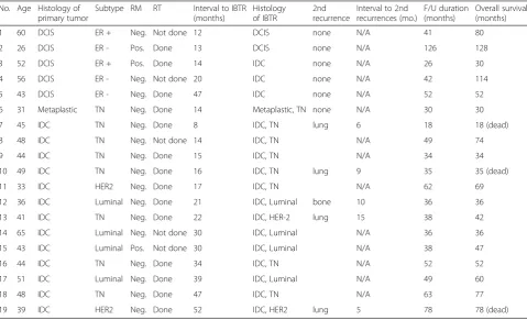
Table 4 showed the course of disease of the patients with IBTR. The treatment of IBTR was mastectomy in all patients. Although three out of five IBTRs in DCIS patients were invasive recurrences, all patients with DCIS were alive without distant metastasis. Rather, all 13 invasive cancer patients who suffered IBTR experi-enced invasive IBTR. Among 272 invasive breast cancer, five (38.5 %) out of 13 patients who suffered IBTR developed distant metastasis and 3 (23.1 %) had died. The 5-year OS of patients with IBTR was inferior to those without (84 vs. 98 %). And, the 5-year distant metastasis-free survival of patients with IBTR was in-ferior to those without (63.3 vs. 96.5 %). However, the dif-ferences in outcome did not reach statistical significance.
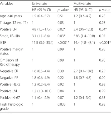
The univariate and multivariate analyses of potential prognostic factors for OS and distant metastasis-free survival in invasive cancer patients are shown in Tables 5 and 6. LN positivity, advanced stage, and experience of Table 1Patient, tumor, and treatment characteristics of 322
patients
Characteristics N(%)
Age, years
Median, range 51 (23~79)
T stage
Tis 50 (15.5 %)
T1 (≤2 cm) 189 (58.7 %)
T2 (>2 and≤5 cm) 83 (25.8 %)
N stage
N0 239 (78.1 %)
N1 55 (18.0 %)
N2 12 (3.9 %)
AJCC stage
0 50 (15.5 %)
I–IIA 233 (72.4 %)
IIB–IIIA 39 (12.1 %)
Histology
Ductal in situ 50 (15.5 %)
Invasive ductal 244 (75.8 %)
Invasive lobular 7 (2.2 %)
Others 21 (6.5 %)
Hormonal receptor
Positive 238 (82.1 %)
Negative 49 (16.9 %)
HER2
Negative 235 (73.0 %)
Positive 40 (12.4 %)
Ki-67
Negative 139 (43.2 %)
Positive 173 (53.7 %)
Nuclear grade
Low 38 (11.8 %)
Intermediate 170 (52.8 %)
High 114 (35.4 %)
Resection margin status
Negative 310 (96.3 %)
Close 5 (1.6 %)
Positive 7 (2.2 %)
Hormonal therapy
Done 236 (73.3 %)
Not done 86 (26.7 %)
Chemotherapy
Done 198 (61.5 %)
Not done 124 (38.5 %)
Table 1Patient, tumor, and treatment characteristics of 322 patients(Continued)
Radiotherapy
Done 310 (96.3 %)
Not done 12 (3.7 %)
Table 2Univariate and multivariate analyses of predictive risk factors for IBTR in DCIS (n= 50)
Variables Univariate Multivariate
OR (95 % CI) pvalue HR (95 % CI) pvalue Age <40 years 2.0 (0.2–21.6) 0.56 1.9 (0.2–17.8) 0.54
Tumor size >2 cm 1.1 (0.6–4.2) 0.23 1.2 (0.1–5.3) 0.29
Positive margin status
4.7 (0.5–14.2) 0.02* 15.6 (2.7–66.3) 0.007*
Multiplicity 2.5 (0.4–4.4) 0.44 2.5 (0.2–2.5) 0.42
High nuclear grade 2 (0.1–9.1) 0.63 1.2 (0.1–11.5) 0.67
EIC positive 1 0.99 1 0.99
Negative ER 9.0 (1.2–65.6) 0.03* 6.7 (1.2–40.5) 0.03*
Negative PR 10.5 (1.1–105.0) 0.04* 8.9 (0.9–79.4) 0.05
Positive HER2 1 0.99 1 0.34
Comedo necrosis 2.5 (0.1–16.3) 0.40 2.2 (0.4–13.6) 0.36
Omission of radiotherapy
3.6 (0.5–25.8) 0.19 3.4 (0.6–20.1) 0.18
IBTR were associated with poor OS and distant meta-stasis on univariate analysis. LN positivity (p= 0.03) and experience of IBTR (p< 0.001) remained as highly sig-nificant in multivariate analysis for predicting OS and distant metastasis-free survival.
Discussion
The purpose of this study was to evaluate the differences in the rates and predictive risk factors for IBTR between DCIS and invasive breast cancer, from 322 consecutive patients with primary breast cancer undergoing BCS at a single center. We also analyzed their prognostic influ-ence of IBTR on patients’ overall survival and distant metastasis.
In the current study, 19 (5.9 %) patients, 5 (10 %) out of 50 with DCIS and 14 (5.1 %) out of 272 with invasive cancer, had developed IBTR. These results are compar-able to those of previous studies regarding IBTR after BCS [1, 2, 7]. In those studies, the incidence of IBTR after BCS for invasive cancer was found to vary between 5 and 10 % over 5 years, and the risk continues over a longer period of follow-up. However, our results showed some high rates of IBTR in comparison to a previous study using a Korean database, which reported an the overall 5-year rate of IBTR as low as 1.6 % in invasive cancer [4]. In that study, patients who did not re-ceive adjuvant radiotherapy and those with positive surgical margins were excluded. Because we analyzed consecutive breast cancer patients who underwent BCS in a single center, five patients with positive/ close margins and three patients who did not undergo radiotherapy were included. We therefore assume these factors to be the reason for our higher Table 3Univariate and multivariate analyses of predictive risk
factors for IBTR in invasive cancer (n= 272)
Variables Univariate Multivariate
OR (95 % CI) pvalue HR (95 % CI) pvalue Age <40 years 1.3 (0.8–6.7) 0.06 2.9 (0.9–9.2) 0.07
Tumor size >2 cm 1.1 (0.5–2.1) 0.85 0.9 (0.5–1.9) 0.94
Positive margin status
1 0.99 1 0.99
Multiplicity 0.78 (0.1–6.3) 0.82 1.2 (0.2–9.8) 0.81
High nuclear grade 1 0.99 2.5 (0.8–7.6) 0.86
EIC positive 1.5 (0.3–6.9) 0.59 1.4 (0.3–6.7) 0.08
Negative ER 7.9 (2.4–26.1) 0.001* 7.5 (2.3–23.8) 0.001*
Negative PR 8.2 (2.2–30.0) 0.002* 7.7 (2.2–27.8) 0.002*
Positive HER2 2.0 (0.3–16.1) 0.50 1.1 (0.7–17.3) 0.95
Positive LVI 1.4 (0.2–10.8) 0.77 1.3 (0.3–11.2) 0.72
Positive Ki-67 4.8 (1.1–21.7) 0.04* 5.0 (1.0–20.8) 0.04*
Omission of Radiotherapy
5.6 (0.3–17.2) 0.03* 50.0 (12.8–194.5) <0.001*
*p< 0.05
Table 4Analyses of 19 patients who had experienced IBTR No. Age Histology of
primary tumor
Subtype RM RT Interval to IBTR (months)
Histology of IBTR
2nd recurrence
Interval to 2nd recurrences (mo.)
F/U duration (months)
Overall survival (months)
1 60 DCIS ER + Neg. Not done 12 DCIS none N/A 41 80
2 26 DCIS ER - Pos. Done 13 DCIS none N/A 126 128
3 52 DCIS ER + Pos. Done 14 IDC none N/A 26 30
4 56 DCIS ER - Neg. Not done 20 IDC none N/A 42 114
5 43 DCIS ER - Neg. Done 47 IDC none N/A 52 52
6 31 Metaplastic TN Neg. Done 14 Metaplastic, TN none N/A 30 30
7 45 IDC TN Neg. Done 8 IDC, TN lung 6 18 18 (dead)
8 48 IDC TN Neg. Not done 14 IDC, TN N/A 49 74
9 44 IDC TN Neg. Done 15 IDC, TN N/A 34 34
10 49 IDC TN Neg. Done 16 IDC, TN lung 9 35 35 (dead)
11 33 IDC HER2 Neg. Done 17 IDC, TN N/A 62 69
12 36 IDC Luminal Neg. Done 21 IDC, Luminal bone 10 36 36
13 41 IDC TN Neg. Done 22 IDC, HER-2 lung 15 38 42
14 65 IDC Luminal Neg. Not done 30 IDC, Luminal N/A 36 36
15 43 IDC Luminal Pos. Not done 30 IDC, Luminal N/A 38 47
16 44 IDC TN Neg. Done 34 IDC, TN N/A 52 52
17 51 IDC Luminal Neg. Done 39 IDC, Luminal N/A 49 60
18 48 IDC TN Neg. Done 47 IDC, TN N/A 63 77
19 39 IDC HER2 Neg. Done 52 IDC, HER2 lung 5 78 78 (dead)
IBTR rate. The 5-year rate of IBTR after BCS in DCIS has been reported to be about 5–10 % [8, 9, 20]. Our study showed comparable findings, with a rate of IBTR after DCIS of 10 % with a median follow-up period of 57 months.
In this study, we wanted to know whether or not there was a difference, in predictive factors for IBTR between DCIS and invasive cancer. In patients with DCIS, multi-variate analysis revealed that positive margin status and ER negativity remained as independent predictors for IBTR. In contrast, omission of radiotherapy, HR negativity, and high Ki67 score were the independent predictive factors for IBTR in invasive cancer. Al-though we had a small study population, multiplicity, young age, and extensive intraductal component (EIC), LVI, and HER-2 positivity showed little or no clinical significance as predictive factors for IBTR in this study. Larger studies with longer follow-up are needed to understand more detailed predictive factors for IBTR.
However, in this study, HR negativity was the strong independent predictive factor for IBTR in both DCIS and invasive cancer. This is a consistent finding with others who showed patients with triple-negative breast cancers behave less favorably than luminal breast can-cers and almost as poorly as HER2/neu over-expressing cancers in terms of locoregional recurrence following BCS [3]. Demicheli et al. analyzed recurrence peak after BCS according to ER status and showed IBTR risk peak delay observed in ER-positive tumors than ER-negative tumors after BCS. They suggest that the microenviron-ment within the residual breast tissue may enforce more stringent constraints upon ER-positive breast tumor cell growth than other tissues, prolonging the latency of IBTR. This local environment is, however, apparently less constraining to ER-negative cells [21]. Additionally, there were many studies about the effect of subtype of IBTR on prognosis. Ishitobi et al. [18] showed that IBTR with luminal A subtype showed the most favorable outcome in terms of distant recurrence than others and breast cancer subtype was an inde-pendent predictor associated with distant recurrence after IBTR. In other study, locoregional failure showed significantly larger effect on mortality and distant dis-ease in ER-negative patients than in ER-positive pa-tients [17]. Aggressive local control and adjuvant therapy should be considered for patients with HR-negative breast cancer who undergo BCS.
In our study population, all three invasive breast cancer patients who did not undergo radiotherapy developed IBTR. Despite our small study population and the small number of events, these results are consistent with the findings of NSABP B-06, which showed that patients treated with radiation remained with low IBTR as compared to those receiving no radi-ation through 9 years of follow-up, regardless of age, nodal status, and tumor size [22]. Radiotherapy should be per-formed in all patients of invasive cancer who treated with BCS.
Table 5Univariate and multivariate analyses of potential risk factors for overall survival in patients with invasive cancer (n= 272)
Variables Univariate Multivariate
HR (95 % CI) pvalue HR (95 % CI) pvalue Age <40 years 0.6 (0.2–2.4) 0.51 1.2 (1.5–9.7) 0.88
T stage, T2 (vs. T1) 1.8 (0.9–3.2) 0.12 2.1 (0.3–5.7) 0.54
Positive LN 3.6 (1.5–11.3) 0.02* 6.9 (1.2–38.4) 0.03*
Stage, IIB-IIIA 4.8 (1.2–19.6) 0.03* 4.9 (1.6–29.3) 0.02*
IBTR 21.9 (4.7–101.9) <0.001* 31.8 (6.1–165.3) <0.001*
Positive margin status
1 0.85 1 0.93
Omission of Radiotherapy
1 0.78 1 0.92
Negative ER 1.6 (0.5–4.4) 0.40 3.1 (0.7–12.3) 0.12
Negative PR 1.8 (0.7–4.9) 0.26 3.4 (0.8–14.3) 0.09
Positive HER2 1.2 (0.1–8.3) 0.92 1 0.95
Positive LVI 1.2 (0.2–1.3) 0.84 2.3 (0.3–18.8) 0.43
Positive Ki-67 1.1 (0.4–2.9) 0.87 1.5 (0.3–5.9) 0.64
High histologic grade
1 0.84 1 0.97
*p< 0.05
Table 6Univariate and multivariate analyses of potential risk factors for distant metastasis in patients with invasive cancer (n= 272)
Variables Univariate Multivariate
HR (95 % CI) pvalue HR (95 % CI) pvalue Age <40 years 1.5 (0.4–5.7) 0.51 1.2 (0.3–4.2) 0.78
T stage, T2 (vs. T1) 1 0.83 1 0.98
Positive LN 4.8 (1.3–17.7) 0.02* 3.4 (0.9–12.3) 0.04*
Stage, IIB–IIIA 3.1 (1.1–8.4) 0.03* 3.83 (1.4–10.8) 0.07
IBTR 11.5 (3.9–33.4) <0.001* 14.4 (4.8–43.1) <0.001*
Positive margin status
1 0.99 1 0.95
Omission of Radiotherapy
1 0.99 1 0.90
Negative ER 1.6 (0.5–4.4) 0.39 2.7 (0.1–10.6) 0.25
Negative PR 1.8 (0.6–4.9) 0.22 1.8 (0.7–4.8) 0.90
Positive HER2 1.2 (0.2–8.4) 0.92 1 0.98
Positive LVI 1.2 (1.0–10.1) 0.84 1 0.99
Positive Ki-67 1.1 (0.4–2.9) 0.87 1.2 (0.4–3.0) 0.65
High histologic grade
1 0.833 1 0.98
Likewise, postoperative radiotherapy after BCS has known as the standard treatment in DCIS patients. How-ever, there remains variation in the use of radiotherapy in favorable type of DCIS. In a series published by Silver-stein et al., patients with small lesions, with favorable his-tologies, and of low to intermediate grade with widely negative margins (>1 cm) treated by BCS alone reported an IBTR rate as low as 6 % at 5 years [23]. Rudloff et al. [24] reported that radiotherapy reduced IBTR only in cases of close margins below 1 cm and the effect of radio-therapy on IBTR risk was influenced by the margin width and the volume of disease near the margin. Kim et al. [9] analyzed the outcome of local excision alone in patients with small size (<1 cm) DCIS and reported that radio-therapy is necessary in patients with resections margin of <1.0 cm. But in recent prospective studies, in good-risk subset of patients with DCIS, with a median follow-up of 7 years, the local failure rate was decreased significantly with the addition of RT [25]. In our study, two out of nine patients with DCIS who omitted radiotherapy devel-oped IBTR. This was the similar finding with recent stud-ies, and careful selection should be taken for radiotherapy omission in patients with DCIS after BCS.
Although there were three invasive cancer patients with positive margins after BCS, marginal status had not reach statistical significance in terms of predicting IBTR in patients with invasive cancer. This is probably due to the limited number of patients and events with short follow-up period. Recent ASCO guidelines showed the use of“no ink on tumor”should be the standard for an adequate margin in invasive cancer. A positive margin, defined as ink on invasive cancer or DCIS, is associated with at least a twofold increase in IBTR; this increased risk in IBTR is not nullified by delivery of a boost, deliv-ery of systemic therapy, or favorable biology [12]. Fur-ther detailed comparison of close and positive margin after BCS in invasive cancer patients should be needed in further larger study. In contrast, although there were small number of events and patients with DCIS, positive marginal status was an independent predictive factor for IBTR in DCIS. Through meta-analysis, Wang et al. [11] reported that negative margins should be obtained for DCIS patients regardless of radiotherapy, and a negative margin threshold greater than 10 mm is the best option compared with other margin thresholds. According to the 2015 national comprehensive cancer network (NCCN) guideline, in patients with DCIS, margins greater than 10 mm are widely accepted as negative and margins less than 1 mm are considered inadequate. There should be more careful attention to achieve negative margin in pa-tients with DCIS who underwent BCS.
Probably due to the limited number of patients and the short follow-up period in our study, we could not demon-strate statistically significant differences on survival curve
according to IBTR. But, among 272 invasive cancer, the 5-year OS of patients with IBTR was inferior to those without (84 vs. 98 %). And, the 5-year distant metastasis-free survival of patients with IBTR was inferior to those without (63.3 vs. 96.5 %). On univariate and multivariate analyses, LN metastasis and IBTR revealed as independ-ent predictive factor for OS and distant metastasis-free survival in invasive cancer. Many investigators have reported that IBTR, especially early IBTR after BCS, is as-sociated with subsequent mortality. In addition, clinico-pathologic features of the recurrent tumor including LVI, high-grade histology, high Ki-67 index, close/positive margins, and ER negativity are known as predictive fac-tors of poor OS after IBTR [16–18]. Investigations on lar-ger study populations with lonlar-ger follow-up periods should be conducted to assess the more detailed clinical impact of IBTR on overall patient outcomes. This infor-mation can be helpful in optimizing local and systemic therapy at the time of original diagnosis for patients undergoing conservative treatment breast cancer. When we treat patient with BCS, identification of individual pa-tient’s clinical and pathologic factors that predict for IBTR and efforts to lowering IBTR should be needed.
Our analysis is a single institution retrospective study of small number of patients with a relatively short median follow-up of 57 months. A longer follow-up period is needed to estimate the long-term outcome. In addition, selection bias is expected due to the heterogeneities of the study population in the clinical and pathological charac-teristics and treatment methods.
Conclusions
The ER and PR negativity was revealed as an independ-ent predictive factor for IBTR after BCS in both DCIS and invasive breast cancer. Meanwhile, positive margin status in DCIS and the omission of radiotherapy in inva-sive cancer, respectively, were the independent prognos-tic factors for IBTR. LN metastasis and experience of IBTR were found to be associated with poor overall sur-vival and distant metastasis in invasive cancer patients after BCS. More efforts should be made to reduce IBTR when treating patient with BCS. And, aggressive local control and adjuvant therapy should be considered in HR-negative patients with advanced stage who receive treatment with BCS.
Ethics approval and consent to participate
Abbreviations
DCIS:ductal carcinoma in situ; EIC: extensive intraductal component; EIC: extensive intraductal component; ER: estrogen receptor; HER2: human epidermal growth receptor 2; HR: hormonal receptor; IBTR: ipsilateral breast tumor recurrence; IDC: invasive ductal carcinoma; LVI: lymphovascular invasion; PR: progesterone receptor; RM: resection margin; RT: radiotherapy.
Competing interests
The authors declare that they have no competing interests
Authors’contribution
CYJ and SYJ participated in the design of this study, and they both performed the breast-conserving surgery. SYD and CYJ performed the statistical analysis. CYJ and SYJ collected the medical datas and drafted the manuscript. SYD helped to draft the manuscript. All authors read and approved the final manuscript.
Funding
This work was supported by the intramural research grant of Chungbuk National University in 2013.
Author details
1Department of Surgery, Chungbuk National University School of Medicine,
410 Sungbong-ro, Heungdeok-gu, Cheongju 361-763, South Korea.
2Department of Anesthesiology, Chungbuk National University School of
Medicine, 410 Sungbong-ro, Heungdeok-gu, Cheongju 361-763, South Korea.
Received: 26 November 2015 Accepted: 21 April 2016
References
1. Fisher B, Anderson S, Bryant J, Margolese RG, Deutsch M, Fisher ER, et al. Twenty-year follow-up of a randomized trial comparing total mastectomy, lumpectomy, and lumpectomy plus irradiation for the treatment of invasive breast cancer. N Engl J Med. 2002;347:1233–41.
2. Karasawa K, Mitsumori M, Yamauchi C, Gomi K, Kataoka M, Uematsu T, et al. Treatment outcome of breast-conserving therapy in patients with positive or close resection margins: Japanese multi institute survey for radiation dose effect. Breast Cancer. 2005;12:91–8.
3. Lowery AJ, Kell MR, Glynn RW, Kerin MJ, Sweeney KJ. Locoregional recurrence after breast cancer surgery: a systematic review by receptor phenotype. Breast Cancer Res Treat. 2012;133:831–41.
4. Kim HJ, Han W, Yi OV, Shin HC, Ahn SK, Koh BS, et al. Young age is associated with ipsilateral breast tumor recurrence after breast conserving surgery and radiation therapy in patients with HER2-positive/ER-negative subtype. Breast Cancer Res Treat. 2011;130:499–505.
5. Cefaro GA, Genovesi D, Trignani M, DI Nicola M. Human epidermal growth factor receptor-2 positivity predicts locoregional recurrence in patients with T1-T2 breast cancer. Anticancer Res. 2014;34:1207–12.
6. Jobsen J, van der Palen J, Riemersma S, Heijmans H, Ong F, Struikmans H. Pattern of ipsilateral breast tumor recurrence after breast-conserving therapy. Int J Radiat Oncol Biol Phys. 2014;89:1006–14.
7. Mechera R, Viehl CT, Oertli D. Factors predicting in-breast tumor recurrence after breast-conserving surgery. Breast Cancer Res Treat. 2009;116:171–7. 8. Sweldens C, Peeters S, van Limbergen E, Janssen H, Laenen A, Patil S, et al.
Local relapse after breast-conserving therapy for ductal carcinoma in situ: a European single-center experience and external validation of the Memorial Sloan-Kettering Cancer Center DCIS nomogram. Cancer J. 2014;20:1–7. 9. Kim H, Noh JM, Choi DH, Lee J, Nam SJ, Lee JE, et al. Excision alone for
small size ductal carcinoma in situ of the breast. Breast. 2014;23:586–90. 10. Vicini FA, Shaitelman S, Wilkinson JB, Shah C, Ye H, Kestin LL, et al.
Long-term impact of young age at diagnosis on treatment outcome and patterns of failure in patients with ductal carcinoma in situ treated with breast-conserving therapy. Breast J. 2013;19:365–73.
11. Wang SY, Chu H, Shamliyan T, Jalal H, Kuntz KM, Kane RL, et al. Network meta-analysis of margin threshold for women with ductal carcinoma in situ. J Natl Cancer Inst. 2012;104:507–16.
12. Buchholz TA, Somerfield MR, Griggs JJ, El-Eid S, Hammond ME, Lyman GH, et al. Margins for breast-conserving surgery with whole-breast irradiation in stage I and II invasive breast cancer: American Society of Clinical Oncology
endorsement of the Society of Surgical Oncology/American Society for Radiation Oncology consensus guideline. J Clin Oncol. 2014;32:1502–6. 13. Whelan T, Clark R, Roberts R, Levine M, Foster G. Ipsilateral breast tumor
recurrence postlumpectomy is predictive of subsequent mortality: results from a randomized trial. Investigators of the Ontario Clinical Oncology Group. Int J Radiat Oncol Biol Phys. 1994;30:11–6.
14. Haffty BG, Reiss M, Beinfield M, Fischer D, Ward B, McKhann C. Ipsilateral breast tumor recurrence as a predictor of distant disease: implications for systemic therapy at the time of local relapse. J Clin Oncol. 1996;14:52–7. 15. Yau TK, Soong IS, Chan K, Chan M, Cheung P, Lau HW, et al. Clinical
outcome of breast conservation therapy for breast cancer in Hong Kong: prognostic impact of ipsilateral breast tumor recurrence and 2005 St. Gallen risk categories. Int J Radiat Oncol Biol Phys. 2007;68:667–72.
16. Panet-Raymond V, Truong PT, Alexander C, Lesperance M, McDonald RE, Watson PH. Clinicopathologic factors of the recurrent tumor predict outcome in patients with ipsilateral breast tumor recurrence. Cancer. 2011; 117:2035–43.
17. Anderson SJ, Wapnir I, Dignam JJ, Fisher B, Mamounas EP, Jeong JH, et al. Prognosis after ipsilateral breast tumor recurrence and locoregional recurrences in patients treated by breast-conserving therapy in five National Surgical Adjuvant Breast and Bowel Project protocols of node-negative breast cancer. J Clin Oncol. 2009;27:2466–73.
18. Ishitobi M, Okumura Y, Arima N, Yoshida A, Nakatsukasa K, Iwase T, et al. Breast cancer subtype and distant recurrence after ipsilateral breast tumor recurrence. Ann Surg Oncol. 2013;20:1886–92.
19. Fisher ER, Anderson S, Tan-Chiu E, Fisher B, Eaton L, Wolmark N. Fifteen-year prognostic discriminants for invasive breast carcinoma: National Surgical Adjuvant Breast and Bowel Project Protocol-06. Cancer. 2001;91:1679–87. 20. Tamura N, Tsuda H, Yoshida M, Hojo T, Akashi-Tanaka S, Kinoshita T, et al.
Clinicopathological predictive factors for ipsilateral and contralateral events following initial surgery to treat ductal carcinoma in situ. Breast Cancer. 2016; 23:510-8.
21. Demicheli R, Ardoino I, Boracchi P, Coradini D, Agresti R, Ferraris C, et al. Recurrence and mortality according to estrogen receptor status for breast cancer patients undergoing conservative surgery. Ipsilateral breast tumour recurrence dynamics provides clues for tumour biology within the residual breast. BMC Cancer. 2010;10:656.
22. Fisher B, Wickerham DL, Deutsch M, Anderson S, Redmond C, Fisher ER. Breast tumor recurrence following lumpectomy with and without breast irradiation: an overview of recent NSABP findings. Semin Surg Oncol. 1992;8:153–60. 23. Silverstein MJ, Lagios MD, Groshen S, Waisman JR, Lewinsky BS, Martino S,
et al. The influence of margin width on local control of ductal carcinoma in situ of the breast. N Engl J Med. 1999;340:1455–61.
24. Rudloff U, Brogi E, Reiner AS, Goldberg JI, Brockway JP, Wynveen CA, et al. The influence of margin width and volume of disease near margin on benefit of radiation therapy for women with DCIS treated with breast-conserving therapy. Ann Surg. 2010;251:583–91.
25. McCormick B, Winter K, Hudis C, Kuerer HM, Rakovitch E, Smith BL, et al. RTOG 9804: a prospective randomized trial for good-risk ductal carcinoma in situ comparing radiotherapy with observation. J Clin Oncol. 2015;33:709–15.
• We accept pre-submission inquiries
• Our selector tool helps you to find the most relevant journal
• We provide round the clock customer support
• Convenient online submission
• Thorough peer review
• Inclusion in PubMed and all major indexing services
• Maximum visibility for your research
Submit your manuscript at www.biomedcentral.com/submit