M E T H O D O L O G Y
Open Access
Development of an isothermoal
amplification-based assay for rapid visual
detection of an Orf virus
Yang Yang
1, Xiaodong Qin
1, Guangxiang Wang
1, Jiaxin Jin
1,2, Youjun Shang
1and Zhidong Zhang
1*Abstract
Background:Orf virus (ORFV) is the causative agent of a severe infectious skin disease (also known as contagious ecthyma) in goats, sheep and other small ruminants. Importantly, ORFV also infect humans which causes a public health concern in the context of changing environment and increase in human populations. The rapid detection is critical in effective control of the disease and urgently needed.
Results:A novel“point of care”molecular amplification assay for rapid visual detection of ORFV was developed based on isothermoal recombinase polymerase amplification (RPA) technology in combination with a simpler lateral flow immunoassay strip (ORFV RPA- LFD assay). The developed ORFV RPA- LFD assay was able to detect ORFV in less than 25 min. This assay was highly sensitive, with detection limit of as low as 80 copies/reaction, and highly specific, with no cross-reactions with capripox virus, foot-and-mouth disease virus and peste des petits ruminants virus. Furthermore, the ORFV RPA- LFD assay has good correlation with qPCR assay for detection of ORFV present in clinical samples.
Conclusions:The developed ORFV RPA-LFD assay was a sensitive and specific method for rapid detection of ORFV, and has great potential as an onsite molecular diagnostic tool in control of Orf.
Keywords:Orf virus, Recombinase polymerase amplification, RPA, Lateral flow, Rapid test, Isothermal nucleic acid amplification
Background
Orf, also known as contagious ecthyma, is a skin dis-ease caused by Orf virus (ORFV), which is classified in the genus Parapoxvirus of the family Poxviridae. Orf principally affects goats and sheep with world-wide distribution and significant financial import-ance. The clinical symptoms of Orf are characterized by the formation of papules, vesicles and growing scabs on the lips and muzzle of infected animals. ORFV is also a zoonotic parapoxvirus endemic to most countries in the world and is principally associ-ated with small ruminants [1]. Humans of infection with ORFV is thought to be under diagnosed and causes a public health concern in the context of
changing environment and increase in human popu-lations because it can resemble skin lesions associated with potentially life-threatening zoonotic infections such as tularemia, cutaneous anthrax, and erysipeloid. Therefore, rapid and definitive diagnosis is critical and highly needed for confirmation and epidemiological investigations [1].
At the present, molecular amplification assays, in particular real-time quantitative PCR (qPCR) and loop-mediated isothermal amplification (LAMP) are sensitive for detection of ORFV [2–5]. However, due to the high complexity of assays or dependency of the specialized and rather expensive equipments, none of these tests can be easily adapted to be a Point of care test or are widely used in resource-limited settings where the dis-ease is mostly endemic [4, 6, 7]. In recent years, a novel and rapid isothermal molecular diagnostic approach, termed recombinase polymerase amplification(RPA), has been developed as an alternative to PCR assay and
* Correspondence:[email protected]
1
State Key Laboratory of Veterinary Etiological Biology, Key Laboratory of Grazing Animal Diseases of Ministry of Agriculture, Lanzhou Veterinary Research Institute, Chinese Academy of Agriculture Sciences, Lanzhou, Gansu, China Full list of author information is available at the end of the article
LAMP [8]. In this assay, just two target specific oligo-nucleotide primers are used to bind the template DNA with the assistance of a recombinase in combination with strand-displacement DNA synthesis. Importantly, an amplification of DNA targets can be achieved in less than 30 min at temperatures just above room tem-perature. Since its initial development in 2006, a differ-ent detection format of RPA assays including probe based and lateral flow dipstick (LFD) detection has been successfully developed for rapid detection of various pathogens [8–22]. To meet our need for rapid assay on side and resource-limited settings, a RPA assay in com-bination with a simpler LFD is a desirable option. Most recently, we successfully developed fluorescent probe-based RPA assay for detection of ORFV with high sensi-tivity and specificity [23]. In the present study, a PRA assay in combination with a simpler LFD (designated as ORFV RPA-LFD assay) has been developed and evalu-ated for rapid detection of ORFV. To the best of our knowledge, it is for the first time a rapid molecular amp-lification assay was developed for on site detection of ORFV. After determination of the sensitivity and specifi-city of the assay, clinical samples from sheep were tested and compared with results from the corresponding qPCR assay.
Methods
Virus and cells
All viruses used in this study were preserved in our laboratory: ORFV/Vaccine/CHA, ORFV/Gansu/CHA, ORFV/HB/CHA; Capripox virus CHA vaccine strain, Capripox virus/Henan/CHA; peste des petits ruminants virus (PPRV) Nigeria 75/1; foot-and-mouth disease virus (FMDV)/O/CHA, FMDV/A/CHA and FMDV/Asia 1/CHA.
Sample preparation
Twenty four field samples (n= 24) were collected from goats with suspected orfv infection and eight nasal swabs(swab up the nostrils of the animals concerned) collected from eight experimentally infected sheep as previously described [23]. To prepare ORFV-spiked tis-sues lysates, ORFV-free tistis-sues samples of skin, lymph-atic nodes, liver, lungs, stomach and kidney (n= 24, three each tissue) were collected from four healthy sheep. ORFV is an epitheliotropic virus, therefore sam-ple of skin tissue was selected. In addition, samsam-ples of other tissues were also chosen because various types of tissues are often received in the field diagnostics for dif-ferential diagnosis, therefore, it would be critical to ac-cess the compatibility of the developed assay with different tissues matrix. 10 % (w/vol) tissue suspensions were prepared as previously described [23], and then
tissue lysates were spiked with 104 TCID50 of ORFV/ HB/CHA and stored at -80 °C until used.
DNA extraction
Total DNA was extracted from samples using high pure viral nucleic acid kit (Roche) according to the manufacturer’s instructions and eluted in a final
vol-ume of 50 μL. Extracted DNA was stored at -80 °C
until further use.
Real-time qPCR assay
Real-time qPCR assay was performed with SYBR® Select Master Mix on Aglient Technologies Stratagene Mx3005P thermocycler (Life technologies) as previ-ously described [3, 23].
ORFV RPA- LFD assay
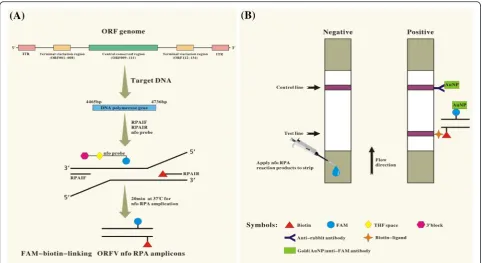
Sequences of primers and probe were previously de-scribed [23] and designed using parameters according to the TwistDx nfo RPA instruction manual. The reverse primer RPA1R with a 5'-biotine label. The probe consists of an upstream stretch (30 nt) carrying a 5’-FAM anti-genic label, which is connected via a THF spacer to an adjacent downstream oligonucleotide (15 nt) carrying a C3-spacer (polymerase extension blocking group) at its 3’ end. All oligonucleotides in this study were synthe-sized by Sangon Biotech (Shanghai, China).
Exo RPA reactions were performed in a 50 μL vol-ume with the TwistAmp nfo kit (TwistDx, UK) con-tained 420 nM nfo RPA primers, 30 nM RPA probes and 1x rehydration buffer. The reaction were incu-bated for a typical 20 min at optimized temperature (37 °C), if not indicated otherwise, in a water bath (Fig. 1a).
Results
Optimization of the ORFV RPA-LFD conditions
To determine the optimal temperature for the RPA reac-tion, the ability of the ORFV RPA–LFD assay was tested to amplify 80 copies of ORFV DNA as template at a range of temperatures from 15 °C to 50 °C. Initially, we assessed this range of temperature under incubation time of 20 min. As shown in Fig. 2, no amplification products were observed in reactions incubated at <30 and≥50 °C. There were weak test line at 30 °C, and there were no differences in amplification at 35, 37, 39,
40 and 45 °C (Fig. 2a). Thus, 37 °C was selected arbitrar-ily as the ORFV RPA-LFD assay standard temperature. We next tested the performance of the ORFV RPA-LFD assay at 37 °C incubated for 1, 5, 10, 20, 25, 30 and 35 min. As shown in Fig. 1b, no amplified products were observed in reactions incubated for less than 5 min and weak amplified product observed for 10 min. When incubation time were increased from 15 to 30 min, the assay performance was improved and there were no dif-ferences in amplification in reactions incubated between 15 to 30 min, thus 20 min was selected arbitrarily as
Fig. 1Schematic representation of ORFV RPA-LFD assay principle for the detection of ORFV.aAmplifying FAM-biotin-linking ORFV nfo RPA amplicons, in the presence of target template (nt position 4465 bp–4736 bp on ORFV DNA polymerase gene), recombinase nfo endonuclease driven primers (RPA1F/Biotin labeled RPA1R) and FAM labeled probes produced FAM-biotin-linking ORFV nfo RPA.bDetecting the RPA amplicons by LFD assay. The amplicons are mixed with the appropriate buffer. Dipping the mixture on LFD strips (Milenia Biotec, Giessen, Germany), the RPA amplicons travel in a buffer stream to be trapped at the test line by biotin-ligands, resulting in an appearance of red-pink color indicative of a positive result. Non-captured gold particles move through the test line to be fixed at the control line by anti-rabbit antibodies, and then produce color serving as a flow control for the strip. In the absence of ORFV target amplicons, color will appear at a control line only
standard incubation time of the ORFV RPA-LFD assay in this study.
Detection limit of the ORFV RPA-LFD assay
The detection limit of the ORFV RPA-LFD assay was determined using a dilution series of the DNA plasmid standards pOrfv/RP1 (corresponding to 101to 106 gen-ome copies/reaction) (Fig. 3a) as previously described [23]. Additionally, the amplified products in the ORFV RPA-LFD reaction was also tested by subsequent visualization with agarose gel electrophoresis (Fig. 3b). The results showed that the ORFV RPA-LFD gave clear positive signal at 80 copies/reaction while the agarose gel electrophoresis gave a clear band at 200 copies/reac-tion. This result indicated the lateral flow dipsticks-based detection has a higher sensitivity than the agarose gel-based detection.
Specificity of the ORFV RPA-LFD assay
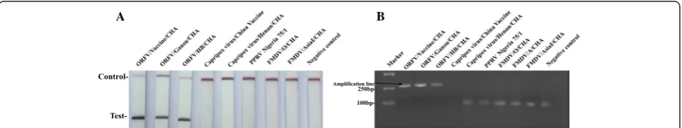
In testing specificity of the ORFV RPA-LFD assay at the optimal conditions described above, the reactions were performed using DNA or RNA from ORFV other important viruses of small ruminants which cause simi-lar clinical signs. As clearly showed in Fig, no cross-reactions were observed in the viruses examined. With all other viruses, red-purple color line was only observed at the control line on the LFD strips (Fig. 4a), which was confirmed by the agarose gel electrophoresis (Fig. 4b). The result indicated that ORFV RPA-LFD assay were specific for detection of ORFV.
Performance of the ORFV RPA-LFD assay on samples
Initially, the feasibility of the ORFV RPA-LFD assay for diagnosis was tested validated with known positive samples (n= 9) and negative samples (n= 9) which was confirmed by ORFV qPCR assay. As showed in Fig. 5, the ORFV RPA-LFD assay was able to be 100 % correct to identify all samples. Its feasibility was then further validated with samples (n= 53) collected from goats with suspected orfv infection, eight nasal swabs collected from experimentally infected sheep and five samples ob-tained from healthy goats. Of 53 samples collected from suspected goats of the Orf, 17 samples were found to be positive by ORFV RPA-LFD assay, the results were identical to the results of real-time ORFV qPCR assay (CT value ranging from 15.3 to 32.8). Furthermore, all of the nasal swabs (n= 8) collected from experimentally infected sheep and ORFV-spiked tissues lysates (n= 24)
were positive while samples (n= 5) obtained from
healthy goats were negative by ORFV RPA-LFD assay. Based on a total of 90 samples examined, the sensitivity and the specificity of ORFV RPA-LFD assay for identification of ORFV was 100 and 100 % res-pectively when compared to real-time ORFV qPCR (Additional file 1: Table S1).
Discussion
In this study, we development a RPA in combination with LFD described for rapid visual detection of ORFV, which is minimally instrumented and has the potential to used as a point of care diagnostic tool. The results have showed that the developed ORFV RPA-LFD assay
A B
Fig. 3Reaction sensitivity of the ORFV RPA-LFD assay. A serial dilution of the ORFV DNA standard plasmids. NC represent negative control.aIn the lateral flow format the sensitivity was 80 copies of the ORFV DNA standard plasmids.bPositive RPA reaction products (273 bp) can be detect on a stained agarose gel (2 %)
A B
has an enough wide range of temperatures which the assay can tolerate because there were no differences in amplification with the range from 30 to 45 °C. It is an important feature for a point of care assay which may be used under uncontrolled temperature environment in the field. More importantly, this result can be read out as early as 20 min (15 min reaction time plus 5 min on LFD).
Recently, another isothermal amplification method for detection of ORFV was developed based on LAMP tech-nology [6]. In contrast to the PPA, LAMP requires a lar-ger set of six primers, a higher temperature (62 °C) and a longer run time. The sensitivity of RPA assay usually presents equal to LAMP assay area, but its specificity is higher that the LAMP assay. The developed ORFV RPA-LFD assay results demonstrated an optimal specificity. Testing results of the ORFV and other related virus showed an analytical specificity of 100 % compared to the qPCR assay and no cross-reaction with non-ORFV vi-ruses. The result has also showed that the assay has high sensitivity for detection of ORFV (80 copies/reaction), which is higher than 200 copies/reaction detected by a fluorescent probe-based RPA assay [23]. Nonetheless, this level of sensitivity should be sufficient to detect ORFV present in samples collected from infected animals. Fur-ther assessment of the assay’s performance with clinical samples has demonstrated satisfactory performance in term of specificity and sensitivity compared to the qPCR assay. Based on results in this study, the ORFV RPA-LFD assay has great potential as a point-of-care molecular diag-nostic assay for detection of ORFV because of its simpli-city, minimal dependency of any specified instrument (portability) and short reaction times. In the developed ORFV RPA-LFD assay, simple instrumentation is needed for both RPA reaction itself and the analysis on the LFD, resulting in overall lower diagnostic costs, which is ideal for onsite testing. A visible band on the LFD strip gives a clear positive/negative answer, which can be easily identified by the naked eyes without any training. In the context of the ever-expanding epidemic of many viral infection in animals and humans over the past
years, there is an urgent need for affordable
molecular detection assay with good sensitivity and specificity for viral diagnostic in developing countries, where no such available assays are currently afford-able or accessible. Such availability of more affordafford-able
assays would also fill this gap because of
decentralization of testing in developing countries.
Conclusions
The developed ORFV RPA-LFD assay is a sensitive and specific method for rapid visual detection of ORFV, and it has great potential as a point-of-care molecular diag-nostic assay. However, the effectiveness of this assay for diagnosis of Orf must be fully evaluated with a larger number of ovine samples before this assay can be con-sidered for routine diagnostic tool.
Additional file
Additional file 1: Table S1.Comparison of ORFV RPA-LFD assay with qPCR assay on ORFV spiked samples and clinical samples. (DOC 26 kb)
Abbreviations
LAMP:loop-mediated isothermal amplification; ORFV: Orf virus; qPCR: quantitative PCR; RPA: recombinase polymerase amplification; RPA-LFD: recombinase polymerase amplification in combination with a simpler lateral flow immunoassay strip.
Competing interests
The authors declare that they have no competing interests.
Authors’contributions
YY, XTQ and ZZ conceived and designed the project. YY performed the study and wrote manuscript, GXW and YJS participated in preparation of virus and samples, XTQ participated in analysis of data. JXJ participated in preparation of the manuscript. XTQ and ZZ revised the manuscript. ZZ is the leader of the project. All authors read and approved the final manuscript.
Acknowledgements
This work was supported by Innovation Fund of Chinese Academy of Agricultural Sciences (CAAS), China, and the National Natural Science Foundation of China (No. 31572522).
Author details 1
State Key Laboratory of Veterinary Etiological Biology, Key Laboratory of Grazing Animal Diseases of Ministry of Agriculture, Lanzhou Veterinary Research Institute, Chinese Academy of Agriculture Sciences, Lanzhou, Gansu, China.
2School of Life Science, Lanzhou University, Lanzhou, Gansu, China.
A B
Received: 1 December 2015 Accepted: 11 March 2016
References
1. Nougairede A, Fossati C, Salez N, Cohen-Bacrie S, Ninove L, Michel F, Aboukais S, Buttner M, Zandotti C, de Lamballerie X, Charrel RN. Sheep-to-human transmission of Orf virus during Eid al-Adha religious practices, France. Emerg Infect Dis. 2013;19:102–5.
2. Mondal B, Bera AK, Hosamani M, Tembhurne PA, Bandyopadhyay SK. Detection of Orf virus from an outbreak in goats and its genetic relation with other parapoxviruses. Vet Res Commun. 2006;30:531–9.
3. Gallina L, Dal Pozzo F, Mc Innes CJ, Cardeti G, Guercio A, Battilani M, Ciulli S, Scagliarini A. A real time PCR assay for the detection and quantification of orf virus. J Virol Methods. 2006;134:140–5.
4. Torfason EG, Gunadottir S. Polymerase chain reaction for laboratory diagnosis of orf virus infections. J Clin Virol. 2002;24:79–84.
5. Chan KW, Hsu WL, Wang CY, Yang CH, Lin FY, Chulakasian S, Wong ML. Differential diagnosis of orf viruses by a single-step PCR. J Virol Methods. 2009;160:85–9.
6. Wang G, Shang Y, Wang Y, Tian H, Liu X. Comparison of a loop-mediated isothermal amplification for orf virus with quantitative real-time PCR. Virol J. 2013;10:138.
7. Venkatesan G, Bhanuprakash V, Balamurugan V. Development and comparative evaluation of loop mediated isothermal amplification (LAMP) assay for simple visual detection of orf virus in sheep and goats. Mol Cell Probes. 2015;29:193–5. 8. Piepenburg O, Williams CH, Stemple DL, Armes NA. DNA detection using
recombination proteins. PLoS Biol. 2006;4:e204.
9. Euler M, Wang Y, Otto P, Tomaso H, Escudero R, Anda P, Hufert FT, Weidmann M. Recombinase polymerase amplification assay for rapid detection of Francisella tularensis. J Clin Microbiol. 2012;50:2234–8. 10. Euler M, Wang Y, Nentwich O, Piepenburg O, Hufert FT, Weidmann M.
Recombinase polymerase amplification assay for rapid detection of Rift Valley fever virus. J Clin Virol. 2012;54:308–12.
11. Krolov K, Frolova J, Tudoran O, Suhorutsenko J, Lehto T, Sibul H, Mager I, Laanpere M, Tulp I, Langel U. Sensitive and rapid detection of Chlamydia trachomatis by recombinase polymerase amplification directly from urine samples. J Mol Diagn. 2014;16:127–35.
12. Kersting S, Rausch V, Bier FF, von Nickisch-Rosenegk M. Multiplex isothermal solid-phase recombinase polymerase amplification for the specific and fast DNA-based detection of three bacterial pathogens. Mikrochim Acta. 2014;181:1715–23.
13. Abd El Wahed A, El-Deeb A, El-Tholoth M, Abd El Kader H, Ahmed A, Hassan S, Hoffmann B, Haas B, Shalaby MA, Hufert FT, Weidmann M. A portable reverse transcription recombinase polymerase amplification assay for rapid detection of foot-and-mouth disease virus. PLoS One. 2013;8:e71642.
14. Crannell ZA, Rohrman B, Richards-Kortum R. Equipment-free incubation of recombinase polymerase amplification reactions using body heat. PLoS One. 2014;9:e112146.
15. Chao CC, Belinskaya T, Zhang Z, Ching WM. Development of recombinase polymerase amplification assays for detection of orientia tsutsugamushi or rickettsia typhi. PLoS Negl Trop Dis. 2015;9:e0003884.
16. Euler M, Wang Y, Heidenreich D, Patel P, Strohmeier O, Hakenberg S, Niedrig M, Hufert FT, Weidmann Mc. Development of a panel of recombinase polymerase amplification assays for detection of biothreat agents. J Clin Microbiol. 2013;51:1110–7.
17. Lutz S, Weber P, Focke M, Faltin B, Hoffmann J, Muller C, Mark D, Roth G, Munday P, Armes N, et al. Microfluidic lab-on-a-foil for nucleic acid analysis based on isothermal recombinase polymerase amplification (RPA). Lab Chip. 2010;10:887–93.
18. Hakenberg S, Hugle M, Weidmann M, Hufert F, Dame G, Urban GA. A phaseguided passive batch microfluidic mixing chamber for isothermal amplification. Lab Chip. 2012;12:4576–80.
19. Rohrman BA, Richards-Kortum RR. A paper and plastic device for performing recombinase polymerase amplification of HIV DNA. Lab Chip. 2012;12:3082–8. 20. Shen F, Davydova EK, Du W, Kreutz JE, Piepenburg O, Ismagilov RF. Digital
isothermal quantification of nucleic acids via simultaneous chemical initiation of recombinase polymerase amplification reactions on SlipChip. Anal Chem. 2011;83:3533–40.
21. Kersting S, Rausch V, Bier FF, von Nickisch-Rosenegk M. Rapid detection of Plasmodium falciparum with isothermal recombinase polymerase amplification and lateral flow analysis. Malar J. 2014;13:99.
22. Jaroenram W, Owens L. Recombinase polymerase amplification combined with a lateral flow dipstick for discriminating between infectious Penaeus stylirostris densovirus and virus-related sequences in shrimp genome. J Virol Methods. 2014;208:144–51.
23. Yang Y, Qin XD, Wang GX, Zhang Y, Shang YJ, Zhang Z. Development of a Fluorescent Probe-Based Recombinase Polymerase Amplification Assay for Rapid Detection of Orf Virus. Virology J. 2015; 12:206. doi:10.1186/s12985-015-0440-z.
• We accept pre-submission inquiries
• Our selector tool helps you to find the most relevant journal • We provide round the clock customer support
• Convenient online submission • Thorough peer review
• Inclusion in PubMed and all major indexing services • Maximum visibility for your research
Submit your manuscript at www.biomedcentral.com/submit